REVIEW 2 major objections 4 minor 6 references
Nanoparticle mobility over a surface as a probe for weak transient disordered peptide-peptide interactions
T0 review · 2 major / 4 minor · reviewed 2026-08-14 · deepseek-v4-flash
Pith's one-line read A nanoparticle diffusion assay detects a single amino acid mutation in a disordered peptide.
desk verdict A useful assay with a real confound: the single-mutation claim needs density-matched controls. read the letter →
The pith
A machine-rendered reading of the paper's core claim, the machinery that carries it, and where it could break.
The reading
What carries the argument
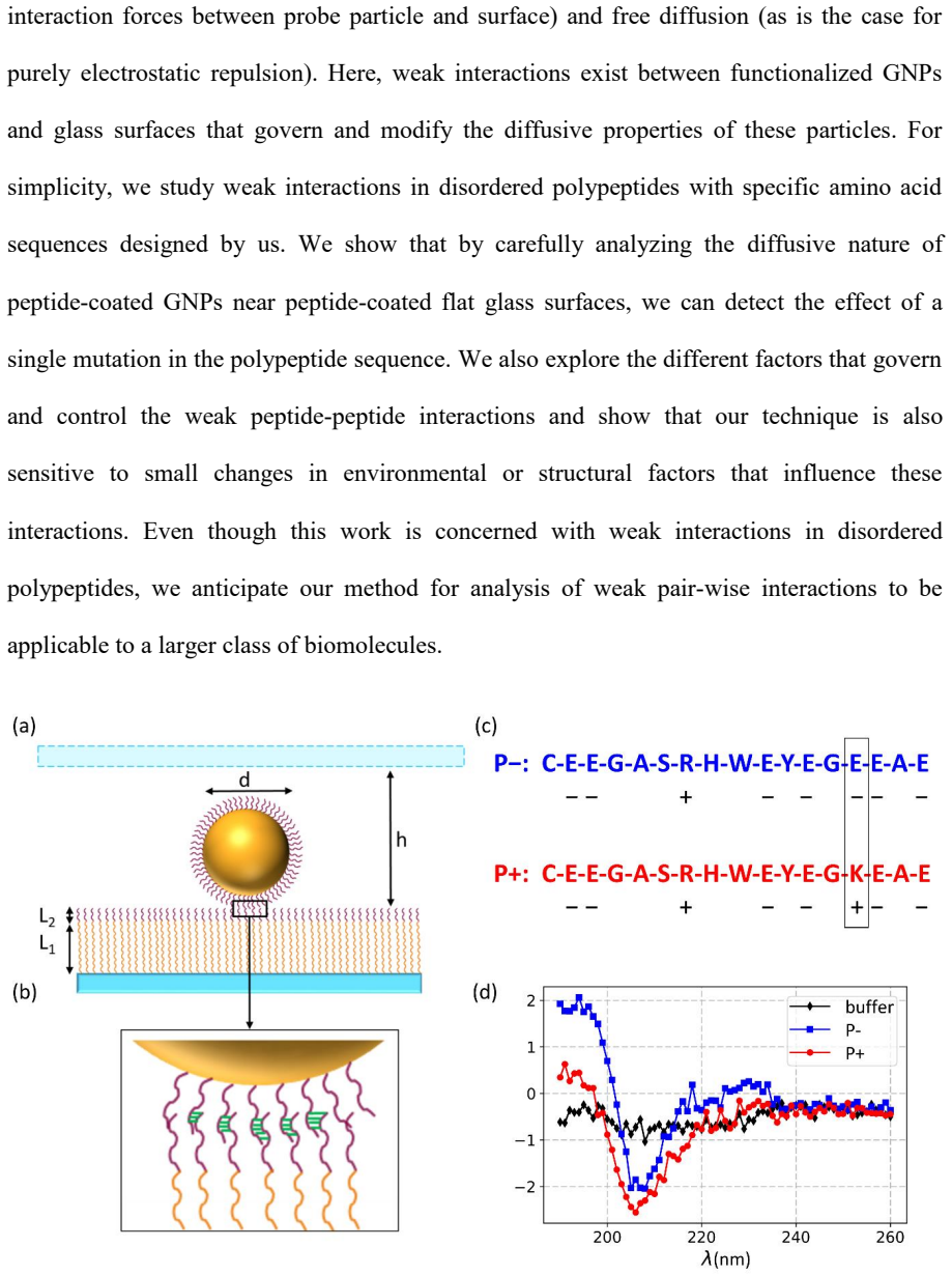
The central object is a multivalent probe: a 40 nm gold nanoparticle grafted with a disordered peptide at roughly 0.5 to 0.7 peptides per square nanometer, moving over a glass surface grafted with the same or a different peptide through a flexible 10 kDa PEG linker. Because a particle can touch many peptides at once, the effective lifetime of a weak bond is exponentially amplified, pulling the interaction above thermal noise while remaining sensitive to differences between sequences. The diagnostic machinery is single-particle tracking statistics: per-trajectory diffusion exponents $n$, transport coefficients $\tilde D$, the displacement distribution $G(\Delta x)$, and sticking-time distributions $P(t_{st})$. The bimodal shape of $P(n)$ and $P(\tilde D)$, not just the ensemble mean, is what separates a weakly interacting peptide from a stronger one.
What would settle it
Repeat the $P_-$ on $P_-$ versus $P_+$ on $P_+$ diffusion comparison with nanoparticles prepared at equal grafting density for both peptides and with identical PEG-linker geometry on the glass. If the bimodal subdiffusive population and the long sticking-time tail no longer track the E14K mutation once density is matched, the central attribution of the mobility change to peptide-peptide interaction strength fails.
Extended reading notes
Core claim
On its own terms, the paper establishes that the diffusive motion of peptide-coated 40 nm gold nanoparticles over peptide-coated glass can serve as a quantitative probe of weak transient peptide-peptide interactions. For the self-interaction comparison, the mutation E14K changes the total peptide charge from $-6.2e$ to $-4.2e$ at pH 7.5, and with it the mobility statistics: $P_-$ on $P_-$ shows single-peaked $P(n)$ near $n=1$ and a displacement distribution fitted by one Gaussian, whereas $P_+$ on $P_+$ shows two populations, one nearly free and one subdiffusive with $n$ near 0.15, giving a double-Gaussian $G(\Delta x)$ and two peaks in $P(\tilde D)$. The ensemble MSD remains linear in lag time for both, so the $P_+$ case is Fickian yet non-Gaussian. The authors interpret the bimodality as intermittent stick-and-hop binding by ionic bridges, amplified by the many simultaneous contacts between a particle and the surface. Salt addition strengthens both self-interactions and increases the subdiffusive fractions, and the two cross-interaction arrangements ($P_-$ on $P_+$ versus $P_+$ on $P_-$) are asymmetric in a way the authors attribute to grafting density, peptide conformation, and the flexibility of the PEG linker.
Load-bearing premise
The load-bearing premise is that the observed mobility difference between $P_-$ and $P_+$ comes from the E14K change in peptide-peptide interaction strength, not from the different grafting densities on the nanoparticles ($0.48$ versus $0.68\ \mathrm{nm}^{-2}$) or from the PEG linker and grafting geometry.
Editorial extensions
If this is right
- Sequence variants of disordered peptides can be ranked by their interaction strength using only diffusion measurements, without labels, force probes, or binding assays.
- Buffer salinity is a workable control knob: adding NaCl increases the subdiffusive fraction in a peptide-dependent way, so the assay can map how electrostatic screening changes weak interactions.
- The assay also reads cross-interactions between different peptides, and the asymmetry between the two grafting arrangements shows that molecular geometry and grafting density contribute to the measured interaction strength.
- Because the readout rests on standard dark-field imaging and particle tracking, it can be run in high-throughput formats such as microfluidics and extended to other biomolecules beyond synthetic peptides.
Reading between the lines
- The paper does not test it, but the multivalent-amplification picture predicts that changing the nanoparticle diameter should tune the accessible window of interaction energies: larger particles should show a larger subdiffusive fraction for the same peptide pair because they can form more simultaneous bonds.
- The E14K swap alters both net charge and the placement of charges; a natural extension the paper leaves implicit is to mutate charged residues at other positions, or make a double mutant with the same net charge, to separate charge-patterning effects from net-charge effects.
- Because the same weak, salt-sensitive interactions drive liquid-liquid phase separation in disordered proteins, the mobility readout could plausibly serve as a quick screen for sequences prone to phase separation, though the paper does not make that link.
Signed reviews
Editorial analysis
A structured set of objections, weighed in public.
Referee Report
Summary. The manuscript reports a single-particle tracking assay in which peptide-coated gold nanoparticles (GNPs) diffuse over peptide-coated glass surfaces, with the aim of probing weak, transient interactions between disordered peptides. The authors compare two 17-residue peptides, P− and P+, that differ by a single E14K mutation. They measure ensemble-averaged MSDs, distributions of diffusion exponents P(n), displacement distributions G(Δx), transport-coefficient distributions P(D̃), and sticking-time distributions, finding that P+ on P+ exhibits a bimodal diffusive population with a large subdiffusive fraction and a longer sticking-time tail than P− on P−. They also study the effect of added NaCl and cross-interactions (P+ on P− and vice versa). The central claim is that the assay is sensitive enough to detect the influence of a single amino acid mutation on transient peptide–peptide interactions.
Significance. If the central claim is validated, the technique would provide a relatively high-throughput, single-molecule-level probe for weak interactions in disordered peptides, with the notable strength of resolving subpopulations (e.g., Fickian yet non-Gaussian diffusion) that bulk measurements would mask. The paper's experimental design is generally careful: large trajectory counts, direct comparison of several independent observables, and a systematic salt-dependence study. The authors also provide explicit controls for surface homogeneity (AFM, spatial maps) and a control showing that surface peptide coverage strongly affects transport coefficients (Figure S3). These strengths are genuine and make the approach plausible. However, the load-bearing comparison that supports the single-mutation claim (P− on P− versus P+ on P+) is currently confounded by a difference in GNP grafting densities, which the manuscript itself reports. This issue must be resolved before the central claim can be accepted.
major comments (2)
- [Grafting density (main text near Figure S1; Methods: 'Measuring peptide grafting density')] The central self-interaction comparison P− on P− versus P+ on P+ is confounded by unequal GNP grafting densities: the manuscript reports 0.48 ± 0.01 nm−2 for P− and 0.68 ± 0.01 nm−2 for P+ (main text near Figure S1). The authors explicitly argue that 'having multiple bonds between the probe particle and the surface allows us to amplify the transient bond lifetime exponentially' (main text after Figure 1), and Figure S3 shows that surface peptide coverage is a first-order variable in this assay: the transport-coefficient distribution shifts substantially with grafting density. Thus a 42% higher graft density on P+ GNPs could, by itself, produce a stronger effective interaction, a longer sticking-time tail, and a larger subdiffusive population, even if the E14K mutation had no effect on per-bond affinity. The manuscript does not provide an equal-density control for the P− versus P+ comparison; density is invoked only to rationalize the cross-interaction asymmetry. A matched-density control (e.g., P− GNPs with a density brought to ~0.68 nm−2, or measurements over a range of P− densities) is essential to isolate the mutation effect. As written, the attribution of the bimodal P+ on P+ population to the E14K mutation (Figure 3 and surrounding text) is not uniquely supported.
- [Figure 3 and the 'nearly 50% subdiffusive' claim] The claim that 'nearly 50% of the particles are undergoing subdiffusion' for P+ on P+ (Figure 3b and text near it) is presented as a quantitative result, but it depends on two arbitrary thresholds: the immobile-exclusion cutoff (total displacement < 1.2 µm in the first 400 ms) and the definition of subdiffusion as n ≤ 0.5 (used for φs in Figure 6a). The fraction is also estimated from a single dataset with N = 806 trajectories, and the immobile fraction itself differs between P− on P− and P+ on P+ (Figure S4). The manuscript should provide the uncertainty in the 50% figure, show how it varies with the chosen thresholds, or at least state a plausible range. Without this, the quantitative characterization of the bimodal population is less reliable than the qualitative observation that a substantial subdiffusive population exists.
minor comments (4)
- [Methods: 'Preparation of peptide coated glass cover-slips'] The text mentions both 'silane-PEG-maleimide' and 'PEG-silane'; consider clarifying the molecular weights (10 kDa vs 5 kDa) at the points of first use to avoid confusion.
- [Figure 3 caption and text] The units for the transport coefficient D̃ are written as 'μm²/sⁿ' in the text but as 'μm²/sn' in some places; please standardize the notation.
- [Text near Equation for Kd] The definition of [GNP] (the 'measured concentration of GNPs diffusing on the surface') is not fully specified; since Kd is a derived quantity, please give the exact formula and mention that this is an operational definition.
- [Section on CTRW scaling] The fitted exponents α for the sticking-time distributions (2.4, 1.7, 1.0, 1.2) are outside the CTRW range (0 < α < 1), and the interpretation that the tracers 'diffused almost normally' could be stated more carefully given that two α values exceed 2; consider adding a brief comment on the possible influence of finite trajectory lengths.
Circularity Check
No circularity: the central claims are empirical comparisons of measured diffusion statistics; the Kd estimate is explicitly acknowledged as simplistic and is not used as a prediction.
full rationale
The paper's central claim, that the assay detects the effect of a single E14K mutation on transient peptide-peptide interactions, rests on direct comparisons of measured trajectory statistics: sticking-time distributions, P(n), G(Δx), and P(D̃) for P− on P− versus P+ on P+ (Figures 2 and 3). These quantities are measured, not derived from a model in which the mutation is an input; no parameter is fitted to the outcome and then renamed as a prediction. The Kd estimate in Figure 6b is computed from measured binding and unbinding rates and is explicitly described by the authors as 'extremely simplistic,' so it is an illustrative summary of the observed trend rather than a derivation that forces the conclusion. The reported difference in GNP grafting densities (0.48 ± 0.01 nm^-2 for P− versus 0.68 ± 0.01 nm^-2 for P+) is a potential experimental confound for the mutation comparison, and the paper uses grafting density and geometry to rationalize the asymmetry of the cross-interaction systems. However, this is an experimental-design and interpretation concern, not circular reasoning: the paper does not define the claimed interaction-strength difference in terms of grafting density, and the mobility differences are presented as direct empirical observations. Self-citations to the authors' prior neurofilament work (refs 12-14, 44-47) provide background about IDP weak interactions and ionic-bridging mechanisms, but they are not load-bearing for the present assay result. No uniqueness theorem is imported from the authors' prior work, no ansatz is smuggled in via citation, and no known result is renamed as a new principle. The derivation chain, such as it is, is self-contained as an empirical comparison, so no circularity is present.
Assumptions & free parameters
free parameters (3)
- Immobile exclusion threshold =
1.2 µm in 400 ms
- Sticking-time displacement cutoff =
0.6 µm
- Slow/fast transport coefficient threshold =
1 µm^2/s^n
assumptions (4)
- domain assumption P- and P+ remain fully disordered when grafted to GNPs and glass at pH 7.5.
- domain assumption Weak interactions between peptides occur through transient ionic bridges whose strength is modulated by monovalent salt.
- domain assumption Only a few peptides on each GNP can reach the surface, yet enough bonds form to amplify transient lifetime.
- domain assumption The peptide-coated glass surface is homogeneous and fully covered with binding sites.
Cite this review
Pith. "Pith review of Nanoparticle mobility over a surface as a probe for weak transient disordered peptide-peptide interactions." pith.science (2026). https://pith.science/paper/4UFAFQZH
@misc{pith2026190811117,
  author       = {Pith},
  title        = {Pith review of: Nanoparticle mobility over a surface as a probe for weak transient disordered peptide-peptide interactions},
  year         = {2026},
  howpublished = {\url{https://pith.science/paper/4UFAFQZH}},
  note         = {Machine review of arXiv:1908.11117}
}
read the original abstract
Weak interactions form the core basis of a vast number of biological processes, in particular, those involving intrinsically disordered proteins. Here, we establish a new technique capable of probing these weak interactions between synthetic unfolded polypeptides using a convenient yet efficient, quantitative method based on single particle tracking of peptide-coated gold nanoparticles over peptide-coated surfaces. We demonstrate that our technique is sensitive enough to observe the influence of a single amino acid mutation on the transient peptide-peptide interactions. Furthermore, the effects of buffer salinity, expected to alter weak electrostatic interactions, are also readily detected and examined in detail. The method presented here has the potential to evaluate in a high throughput manner, weak interactions for a wide range of disordered proteins, polypeptides, and other biomolecules.
Figures


Figures from the paper (2 more)


Reference graph
Works this paper leans on
-
[1]
Visualizing an Ultra-Weak Protein-Protein Interaction in Phosphorylation Signaling
(1) Xing, Q.; Huang, P.; Yang, J.; Sun, J.-Q.; Gong, Z.; Dong, X.; Guo, D.-C.; Chen, S.- M.; Yang, Y.-H.; Wang, Y.; et al. Visualizing an Ultra-Weak Protein-Protein Interaction in Phosphorylation Signaling. Angew. Chemie - Int. Ed. 2014, 53 (43), 11501–11505. https://doi.org/10.1002/anie.201405976. (2) Hahn, S.; Kim, D. Transient Protein-Protein Interacti...
-
[54]
https://doi.org/10.1007/s11671-010-9809-5. (29) Stout, A. L. Detection and Characterization of Individual Intermolecular Bonds Using Optical Tweezers. Biophys. J. 2001, 80 (6), 2976–2986. https://doi.org/10.1016/S0006- 3495(01)76263-7. (30) Heider, S.; Reimhult, E.; Metzner, C. Real-Time Analysis of Protein and Protein Mixture Interaction with Lipid Bilay...
-
[848]
(25) Hao, T.; Peng, W.; Wang, Q.; Wang, B.; Sun, J
https://doi.org/10.15252/msb.20156351. (25) Hao, T.; Peng, W.; Wang, Q.; Wang, B.; Sun, J. Reconstruction and Application of Protein–Protein Interaction Network. Int. J. Mol. Sci. 2016, 17 (6),
-
[907]
https://doi.org/10.3390/ijms17060907. (26) Henderson, R. M. Structural Dynamics of Single Molecules Studied with High-Speed Atomic Force Microscopy. Expert Opin. Drug Discov. 2015, 10 (3), 221–229. https://doi.org/10.1517/17460441.2015.998195. (27) Lee, C.-K.; Wang, Y.-M.; Huang, L.-S.; Lin, S. Atomic Force Microscopy: 28 Determination of Unbinding Force,...
arXiv 2015
-
[2016]
(53) Allan, D.; Caswell, T.; Keim, N.; van der Wel, C
https://doi.org/10.1021/jacs.6b09542. (53) Allan, D.; Caswell, T.; Keim, N.; van der Wel, C. Trackpy v0.3.0. Zenodo 2015, 34028. (54) Crocker, J. C.; Grier, D. G. Methods of Digital Video Microscopy for Colloidal Studies. J. Colloid Interface Sci. 1996, 179, 298–310. https://doi.org/10.1006/jcis.1996.0217. 1 Supporting information Nanoparticle mobility ov...
arXiv 2015
-
[2019]
(52) Yoo, J.; Lee, T.; Choi, B.; Shon, M
https://doi.org/10.1038/s42254-019-0077-8. (52) Yoo, J.; Lee, T.; Choi, B.; Shon, M. J.; Yoon, T. Observing Extremely Weak Protein − Protein Interactions with Conventional Single-Molecule Fluorescence Microscopy
Reviewed August 14, 2026 · model on record in the stance chip above.
Discussion (0). Continue with ORCID to comment.