REVIEW 4 major objections 5 minor 44 references
AFP-Net: Realtime Anchor-Free Polyp Detection in Colonoscopy
T0 review · 4 major / 5 minor · reviewed 2026-08-14 · deepseek-v4-flash
Pith's one-line read The paper claims that an anchor-free, center-point detector with a cosine ground-truth projection can detect colon polyps at 99.36% precision and 96.44% recall while running at 52.6 frames per second.
desk verdict Plausible anchor-free polyp detector with a solid ablation study, but the evaluation table is internally inconsistent and the SOTA claim doesn't hold as printed. read the letter →
The pith
A machine-rendered reading of the paper's core claim, the machinery that carries it, and where it could break.
The reading
What carries the argument
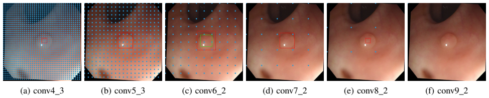
The load-bearing object is the cosine ground-truth projection: for a ground truth assigned to its best feature level $i$, the non-negative label region at another level $l$ is scaled by $\phi_l=\max(\cos(\lambda d\pi/2k),0)$, where $d=|i-l|$, $k$ is the number of feature levels, and $\lambda=2.5$. This factor decides which center points are positive, ignored, or negative at each feature-pyramid level, so that neighboring scales still supervise keypoints near the object center instead of labeling them negative. Around it sits the Context Enhancement Module, which splits channels into three branches of dilated convolutions and concatenates them to enlarge the receptive field, and the multitask loss that combines Smooth L1 box regression, focal loss on negative samples, and Gaussian-weighted cross-entropy on positive samples.
What would settle it
Re-run AFP-Net and the compared detectors on a colonoscopy test set that none of them trained on, count a detection as correct only when its predicted box overlaps the ground truth by at least 0.5 IoU, and report scores across multiple train/test splits; if AFP-Net no longer leads in F1 and F2, or the gap falls within run-to-run variation, the state-of-the-art claim would be refuted.
Extended reading notes
Core claim
On its own terms, the paper's central discovery is that an anchor-free detector can match or beat anchor-based polyp detectors if the label assignment is redesigned rather than copied. Removing anchors eliminates the need for IoU thresholds and anchor hyperparameters, but it also creates 'unnecessary negative' keypoints: near-center points on non-best feature levels are labeled negative even though the enlarged receptive field could cover the object. AFP-Net answers this with a cosine ground-truth projection, which shrinks the non-negative region on each neighboring feature level by a cosine factor based on distance from the best level, turning those points into useful training signals. Combined with a Context Enhancement Module that widens the receptive field and a loss using focal loss on negatives and a Gaussian distance penalty on positives, the model reaches the reported precision/recall numbers and runs in real time.
Load-bearing premise
The load-bearing premise is that the reported evaluation is a fair, unbiased measure, yet the test set is labeled 'CVC-Clinic-train,' the true-positive rule is only that the predicted center falls inside the ground-truth box, and no error bars or split variance are reported.
Editorial extensions
If this is right
- Anchor-based detectors are not required for accurate polyp detection: AFP-Net's center-point head outperforms its direct anchor-based counterpart, SSD-baseline, which shares the same backbone, pyramid, and context module.
- Cosine ground-truth projection is what recovers recall after anchors are removed: dropping it (Experiment 4 vs 6) lowers recall by a considerable margin in the ablation.
- Context enrichment is a necessary ingredient: the paper reports that removing the Context Enhancement Module degrades both precision and recall.
- The full pipeline operates at 52.6 FPS on an RTX 2080 Ti, satisfying real-time colonoscopy video requirements while keeping the reported accuracy.
- Because no anchors or IoU assignment thresholds need tuning, the detector can be adapted to new endoscopic datasets without per-dataset anchor design.
Reading between the lines
- The same cosine label-spreading trick could transfer to other sparse-object detection tasks in medical imaging, such as lesions in CT or capsule endoscopy, where small objects and class imbalance make anchor-free recall loss especially costly.
- The reported true-positive rule -- predicted center inside the ground-truth box -- is more permissive than an IoU threshold like 0.5; re-scoring with standard IoU would likely lower absolute numbers and might change the ranking against anchor-based methods.
- The test column labeled 'CVC-Clinic-train' raises the possibility that compared models trained on the same frames; a held-out split with no overlap could narrow or reverse the claimed advantage.
- Since the detector already runs at 52.6 FPS, a natural extension is to couple it with temporal tracking across video frames, which could suppress the fold-shaped false positives shown in the paper's failure cases.
Editorial analysis
A structured set of objections, weighed in public.
Referee Report
Summary. The paper proposes AFP-Net, an anchor-free polyp detector for colonoscopy. The architecture uses a VGG16 backbone, a feature pyramid, a Context Enhancement Module (CEM), a cosine ground-truth projection for multi-scale label assignment, focal loss with a Gaussian penalty, and anchor-free box regression. The authors report state-of-the-art precision/recall/F1/F2 on CVC-Clinic and ETIS-LARIB test sets, with 52.6 FPS inference speed, and support the claims with an ablation study and comparisons against anchor-based and anchor-free baselines.
Significance. If the reported results are reproducible, the paper makes a useful contribution: it is among the first anchor-free detectors for polyp detection, provides detailed ablations of each component, and directly compares with a matched SSD-style anchor-based baseline, showing a real-time speed advantage. The systematic ablation (Table I) and the inclusion of inference-time measurements are strengths. However, the central state-of-the-art claim depends entirely on the comparison in Table III, and that table has internal inconsistencies and ambiguous test-set descriptions. The evaluation protocol, especially the centroid-only true-positive criterion and the absence of error bars, is too weak to support the claimed margins over the closest baselines as currently written.
major comments (4)
- [Section IV.E / Table III] The rows labeled 'CVC-Clinic-train' in Table III are internally inconsistent. Using the paper's own definitions in Section IV.C, each ground-truth box is either a true positive or a false negative, so TP+FN equals the total number of ground-truth boxes in the test set and cannot depend on the method. The table reports TP+FN = 646 for AFP-Net (623+23), CenterNet (603+43), and SSD-baseline (618+28), but TP+FN = 531 for Faster-RCNN (523+8). The 115-instance gap means Faster-RCNN's numbers were produced on a different image set, a different annotation set, or under a different counting rule. Since the paper's central claim that AFP-Net 'outperforms all previous approaches' is supported only by this table, the claim is not established as printed.
- [Section IV.A / Section IV.E / Table III] The test-set description is ambiguous and potentially contradictory. Section IV.A states that all 612 CVC-Clinic images are used as the testing set, while Section IV.E says the test was run on 'CVC-Clinic training dataset' and Table III uses the column header 'CVC-Clinic-train'. Because the CVC-Clinic images are also said to be identical to the GIANA polyp-segmentation test set, and no image IDs or split files are provided, a reader cannot determine whether the compared methods were trained on the same images, whether there is overlap between training and testing, or whether the numbers in Table III were all obtained under the same protocol. The authors should specify the exact split, release or cite image IDs, and ensure that the evaluation set is clearly labeled and consistently used for all methods.
- [Section IV.C / Table III] The true-positive criterion is too loose to support the precision/recall comparisons. Section IV.C defines a TP as any detection whose centroid falls inside a ground-truth box, with no IoU threshold, and counts only one TP per ground truth even if multiple boxes overlap. This criterion does not penalize poor localization, and combined with the low NMS threshold of 0.1, it can inflate precision and recall relative to standard detection metrics. The differences against the closest baselines are small (for example, F1 97.88 vs. 97.17 for SSD-baseline on CVC-Clinic-train, and F1 84.63 vs. 83.3 for FRCNNPL on ETIS-LARIB), so a stricter metric or at least error bars across multiple runs is needed to establish that the improvement is meaningful. No error bars, variance estimates, or multiple-seed experiments are reported anywhere.
- [Section IV.E / Table III] The comparison with prior work mixes numbers obtained under different conditions. Some rows are taken from previously published papers on datasets that are no longer available (e.g., Y-Net and RYCO on ASU-MAYO*), some report only N/A for TP/FP/FN, and the hardware and input sizes differ across methods. It is not stated whether the prior methods were re-run under the same protocol as AFP-Net or whether the published numbers are quoted as-is. This weakens the 'outperforms all previous approaches' conclusion, especially because the reader cannot verify whether the compared methods used the same test split and the same true-positive definition.
minor comments (5)
- [Section IV.D / Figure 5] The text says 'we show the ROC curve with respect to precision and recall,' but Figure 5 is a precision-recall curve; the wording should be corrected.
- [Section III.A] There is a missing period in 'Our network uses VGG16 [34] as the backbone Our framework selects k = 6 feature maps'; the sentence break should be fixed.
- [Section III.C] The phrase 'we have m2 ... center points' is typeset ambiguously; it should be 'm^2 center points' or 'm-by-m center points' for clarity.
- [Section IV.A / Section IV.C] Section IV.A describes GIANA as a MICCAI2017 challenge while Section IV.C refers to the 'MICCAI2015 [5] challenge' protocol; the distinction between the challenge year and the cited protocol should be clarified to avoid confusion.
- [General] No code, trained models, or split files are provided, and the manuscript does not state whether they will be released. Given the ambiguities in the evaluation, releasing the exact test split and the evaluation script would substantially improve reproducibility.
Circularity Check
No material circularity: AFP-Net's novelty claims are empirical design choices evaluated on held-out data, not derivations that reduce to their own inputs.
full rationale
This is an empirical machine-learning paper. The proposed components (anchor-free label assignment, Context Enhancement Module, cosine ground-truth projection, Gaussian penalty, focal loss) are heuristic design choices with hand-set hyperparameters, and their contributions are assessed through ablation studies on a separate test set (CVC-Clinic and ETIS-LARIB, with training on GIANA). There is no self-definitional derivation: the cosine projection formula (Eq. 2-3) is a penalization heuristic, not derived from the metric it is claimed to improve. The F1/F2 claims are empirical measurements, not predictions forced by fitted parameters. The paper cites several of the authors' own prior works, but none of these citations carries the load of the central claim; the central comparison is against external baselines and the paper reports its own training/evaluation setup. The serious concern in Table III—the inconsistent TP+FN totals across methods on rows labeled 'CVC-Clinic-train' (646 for AFP-Net/CenterNet/SSD-baseline vs. 531 for Faster-RCNN)—is an evaluation-protocol or reporting error, not circularity: it undermines the fairness of the comparison but does not make the derivation equivalent to its inputs. Under the stated rules, this does not qualify as a circularity finding, so the score is kept at the low end (1, reflecting minor self-citation presence without load-bearing circularity).
Assumptions & free parameters
free parameters (6)
- epsilon_p =
0.75
- epsilon_n =
1.25
- lambda =
2.5
- alpha =
1
- beta =
0.45
- NMS_threshold =
0.1
assumptions (4)
- domain assumption GIANA training set is representative of polyp appearance in CVC-Clinic and ETIS-LARIB
- domain assumption ImageNet pre-training transfers to colonoscopy images
- domain assumption Centroid-in-box true-positive criterion is a valid measure of detection accuracy
- domain assumption Ground truth annotations in all datasets are accurate
Cite this review
Pith. "Pith review of AFP-Net: Realtime Anchor-Free Polyp Detection in Colonoscopy." pith.science (2026). https://pith.science/paper/N4WNLNY4
@misc{pith2026190902477,
  author       = {Pith},
  title        = {Pith review of: AFP-Net: Realtime Anchor-Free Polyp Detection in Colonoscopy},
  year         = {2026},
  howpublished = {\url{https://pith.science/paper/N4WNLNY4}},
  note         = {Machine review of arXiv:1909.02477}
}
read the original abstract
Colorectal cancer (CRC) is a common and lethal disease. Globally, CRC is the third most commonly diagnosed cancer in males and the second in females. For colorectal cancer, the best screening test available is the colonoscopy. During a colonoscopic procedure, a tiny camera at the tip of the endoscope generates a video of the internal mucosa of the colon. The video data are displayed on a monitor for the physician to examine the lining of the entire colon and check for colorectal polyps. Detection and removal of colorectal polyps are associated with a reduction in mortality from colorectal cancer. However, the miss rate of polyp detection during colonoscopy procedure is often high even for very experienced physicians. The reason lies in the high variation of polyp in terms of shape, size, textural, color and illumination. Though challenging, with the great advances in object detection techniques, automated polyp detection still demonstrates a great potential in reducing the false negative rate while maintaining a high precision. In this paper, we propose a novel anchor free polyp detector that can localize polyps without using predefined anchor boxes. To further strengthen the model, we leverage a Context Enhancement Module and Cosine Ground truth Projection. Our approach can respond in real time while achieving state-of-the-art performance with 99.36% precision and 96.44% recall.
Figures


Figures from the paper (3 more)


Reference graph
Works this paper leans on
-
[1]
M. F. Alcantara, Y . Cao, C. Liu, B. Liu, M. Brunette, N. Zhang, T. Sun, P. Zhang, Q. Chen, Y . Li, C. M. Albarracin, J. Peinado, E. S. Garavito, L. L. Garcia, and W. H. Curioso. Improving tuberculosis diagnostics using deep learning and mobile health technologies among resource-poor communities in per. Smart Health , 1-2(Supplement C):66 – 76, 2017. Conn...
work page 2017
-
[2]
H. Ali Qadir, Y . Shin, J. Solhusvik, J. Bergsland, L. Aabakken, and I. Balasingham. Polyp detection and segmentation using mask r-cnn: Does a deeper feature extractor cnn always perform better? pages 1–6, 05 2019
work page 2019
-
[3]
S. Ameling, S. Wirth, D. Paulus, G. Lacey, and F. Vilario. Texture-based polyp detection in colonoscopy. pages 346–350, 01 2009
work page 2009
- [4]
-
[5]
J. Bernal, N. Tajbakhsh, F. Javier Sanchez, B. J. Matuszewski, H. Chen, L. Yu, Q. Angermann, O. Romain, B. Rustad, I. Balasing- ham, K. Pogorelov, S. Choi, Q. Debard, L. Maier-Hein, S. Speidel, D. Stoyanov, P. Brandao, H. Crdova, C. Snchez-Montes, and A. His- tace. Comparative validation of polyp detection methods in video colonoscopy: Results from the mi...
work page 2015
-
[6]
M. D. Bloice, P. M. Roth, and A. Holzinger. Biomedical image augmentation using Augmentor. Bioinformatics, 04 2019
work page 2019
-
[7]
F. Bray, J. Ferlay, I. Soerjomataram, R. L. Siegel, L. A. Torre, and A. Jemal. Global cancer statistics 2018: Globocan estimates of incidence and mortality worldwide for 36 cancers in 185 countries. CA: A Cancer Journal for Clinicians , 68(6):394–424, 2018
work page 2018
-
[8]
Y . Cao, C. Liu, B. Liu, M. J. Brunette, N. Zhang, T. Sun, P. Zhang, J. Peinado, E. S. Garavito, L. L. Garcia, and W. H. Curioso. Improving tuberculosis diagnostics using deep learning and mobile health tech- nologies among resource-poor and marginalized communities. In 2016 IEEE First International Conference on Connected Health: Applications, Systems an...
work page 2016
Show all 44 references
-
[9]
K. Duan, S. Bai, L. Xie, H. Qi, Q. Huang, and Q. Tian. Centernet: Keypoint triplets for object detection. CoRR, abs/1904.08189, 2019
1904 arXiv
-
[10]
Eggert, S
C. Eggert, S. Brehm, A. Winschel, D. Zecha, and R. Lienhart. A closer look: Small object detection in faster r-cnn. In Multimedia and Expo (ICME), 2017 IEEE International Conference on, pages 421–426. IEEE, 2017
2017
-
[11]
Y . Gao, N. Zhang, H. Wang, X. Ding, X. Ye, G. Chen, and Y . Cao. ihear food: Eating detection using commodity bluetooth headsets. In 2016 IEEE First International Conference on Connected Health: Applications, Systems and Engineering Technologies (CHASE) , pages 163–172, June 2016
2016
-
[12]
R. B. Girshick. Fast R-CNN. CoRR, abs/1504.08083, 2015
2015 arXiv
-
[13]
K. He, G. Gkioxari, P. Doll ´ar, and R. Girshick. Mask r-cnn. In Computer Vision (ICCV), 2017 IEEE International Conference on , pages 2980–
2017
-
[14]
K. He, X. Zhang, S. Ren, and J. Sun. Deep residual learning for image recognition. In Proceedings of the IEEE conference on computer vision and pattern recognition , pages 770–778, 2016
2016
-
[15]
B. Y . Hsueh, W. Li, and I. Wu. Stochastic gradient descent with hyperbolic-tangent decay. CoRR, abs/1806.01593, 2018
2018 arXiv
-
[16]
Hwang, J
S. Hwang, J. Oh, W. Tavanapong, J. Wong, and P. C. de Groen. Polyp detection in colonoscopy video using elliptical shape feature. In 2007 IEEE International Conference on Image Processing , volume 2, pages II – 465–II – 468, Sep. 2007
2007
-
[17]
T. Kong, F. Sun, H. Liu, Y . Jiang, and J. Shi. Foveabox: Beyond anchor- based object detector. arXiv preprint arXiv:1904.03797 , 2019
1904 arXiv
-
[18]
Law and J
H. Law and J. Deng. Cornernet: Detecting objects as paired keypoints. CoRR, abs/1808.01244, 2018
2018 arXiv
-
[19]
Leufkens, M
A. Leufkens, M. V . Oijen, F. Vleggaar, and P. Siersema. Factors influencing the miss rate of polyps in a back-to-back colonoscopy study. Endoscopy, 44(05):470?475, 2012
2012
-
[20]
J. Li, Y . Wang, C. Wang, Y . Tai, J. Qian, J. Yang, C. Wang, J. Li, and F. Huang. DSFD: dual shot face detector. CoRR, abs/1810.10220, 2018
2018 arXiv
-
[21]
P. Li, Y . Luo, N. Zhang, and Y . Cao. Heterospark: A heterogeneous cpu/gpu spark platform for machine learning algorithms. In 2015 IEEE International Conference on Networking, Architecture and Storage (NAS), pages 347–348, Aug 2015
2015
-
[22]
T. Lin, P. Doll ´ar, R. B. Girshick, K. He, B. Hariharan, and S. J. Belongie. Feature pyramid networks for object detection. CoRR, abs/1612.03144, 2016
2016 arXiv
-
[23]
T.-Y . Lin, P. Doll´ar, R. Girshick, K. He, B. Hariharan, and S. Belongie. Feature pyramid networks for object detection. In CVPR, volume 1, page 4, 2017
2017
-
[24]
T.-Y . Lin, P. Goyal, R. Girshick, K. He, and P. Doll ´ar. Focal loss for dense object detection. arXiv preprint arXiv:1708.02002 , 2017
2017 arXiv
-
[25]
W. Liu, D. Anguelov, D. Erhan, C. Szegedy, S. E. Reed, C. Fu, and A. C. Berg. SSD: single shot multibox detector. CoRR, abs/1512.02325, 2015
2015 arXiv
-
[26]
X. Mo, K. Tao, Q. Wang, and G. Wang. An Efficient Approach for Polyps Detection in Endoscopic Videos Based on Faster R-CNN. arXiv e-prints, page arXiv:1809.01263, Sep 2018
2018 arXiv
-
[27]
A. K. Mohammed, S. Yildirim, I. Farup, M. Pedersen, and Ø. Hovde. Y-net: A deep convolutional neural network for polyp detection. CoRR, abs/1806.01907, 2018
2018 arXiv
-
[28]
Najibi, P
M. Najibi, P. Samangouei, R. Chellappa, and L. S. Davis. SSH: single stage headless face detector. CoRR, abs/1708.03979, 2017
2017 arXiv
-
[29]
Redmon, S
J. Redmon, S. K. Divvala, R. B. Girshick, and A. Farhadi. You only look once: Unified, real-time object detection. CoRR, abs/1506.02640, 2015
2015 arXiv
-
[30]
S. Ren, K. He, R. B. Girshick, and J. Sun. Faster R-CNN: towards real-time object detection with region proposal networks. CoRR, abs/1506.01497, 2015
2015 arXiv
-
[31]
J. S. Silva, A. Histace, O. Romain, X. Dray, and B. Granado. Toward embedded detection of polyps in wce images for early diagnosis of colorectal cancer. International journal of computer assisted radiology and surgery, 9, 09 2013
2013
-
[32]
Y . Shin, H. Ali Qadir, L. Aabakken, J. Bergsland, and I. Balasingham. Automatic colon polyp detection using region based deep cnn and post learning approaches. IEEE Access, PP:1–1, 07 2018
2018
-
[33]
R. L. Siegel, K. D. Miller, S. A. Fedewa, D. J. Ahnen, R. G. S. Meester, A. Barzi, and A. Jemal. Colorectal cancer statistics, 2017. CA: A Cancer Journal for Clinicians , 67(3):177–193, 2017
2017
-
[34]
Simonyan and A
K. Simonyan and A. Zisserman. Very deep convolutional networks for large-scale image recognition. arXiv 1409.1556, 09 2014
2014 arXiv
-
[35]
Sornapudi, F
S. Sornapudi, F. Meng, and S. Yi. Region-based automated localiza- tion of colonoscopy and wireless capsule endoscopy polyps. Applied Sciences, 9, 06 2019
2019
-
[36]
X. Sun, N. Zhang, Q. Chen, Y . Cao, and B. Liu. People re-identification by multi-branch cnn with multi-scale features. In 2019 26th IEEE International Conference on Image Processing (ICIP) . IEEE, 2019
2019
-
[37]
Szegedy, Wei Liu, Yangqing Jia, P
C. Szegedy, Wei Liu, Yangqing Jia, P. Sermanet, S. Reed, D. Anguelov, D. Erhan, V . Vanhoucke, and A. Rabinovich. Going deeper with convolutions. In 2015 IEEE Conference on Computer Vision and Pattern Recognition (CVPR), pages 1–9, June 2015
2015
-
[38]
Tajbakhsh, S
N. Tajbakhsh, S. Gurudu, and J. Liang. Automated polyp detection in colonoscopy videos using shape and context information. IEEE transactions on medical imaging , 35, 10 2015
2015
-
[39]
Z. Tian, C. Shen, H. Chen, and T. He. Fcos: Fully convolutional one- stage object detection. arXiv preprint arXiv:1904.01355 , 2019
1904 arXiv
-
[40]
J. Wang, K. Chen, S. Yang, C. C. Loy, and D. Lin. Region proposal by guided anchoring. arXiv preprint arXiv:1901.03278 , 2019
1901 arXiv
-
[41]
Zhang, Y
N. Zhang, Y . Cao, B. Liu, and Y . Luo. Improved multimodal represen- tation learning with skip connections. In Proceedings of the 2017 ACM on Multimedia Conference , MM ’17, pages 654–662, New York, NY , USA, 2017. ACM
2017
-
[42]
Zhang, D
N. Zhang, D. Wang, X. Sun, P. Zhang, C. Zhang, Y . Cao, and B. Liu. 3d anchor-free lesion detector on computed tomography scans, 2019
2019
-
[43]
Zhang, Y
R. Zhang, Y . Zheng, C. C. Poon, D. Shen, and J. Y . Lau. Polyp detection during colonoscopy using a regression-based convolutional neural network with a tracker. Pattern Recognition, 83:209 – 219, 2018
2018
-
[44]
X. Zhou, D. Wang, and P. Kr ¨ahenb¨uhl. Objects as points. CoRR, abs/1904.07850, 2019
1904 arXiv
Reviewed August 14, 2026 · model on record in the stance chip above.
Discussion (0). Continue with ORCID to comment.