REVIEW 4 major objections 5 minor 48 references
S3TU-Net: Structured Convolution and Superpixel Transformer for Lung Nodule Segmentation
T0 review · 4 major / 5 minor · reviewed 2026-08-12 · deepseek-v4-flash
Pith's one-line read The paper claims that S3TU-Net, a U-Net hybrid with structured convolutions, a superpixel-based transformer, and spatial-shift skip links, achieves 89.04% Dice on LIDC-IDRI and 86.40% on a private adenocarcinoma dataset, outperforming…
desk verdict Plausible incremental architecture; reported 4.5-point Dice gain not yet trustworthy because of split leakage risk and metric inconsistencies. read the letter →
The pith
A machine-rendered reading of the paper's core claim, the machinery that carries it, and where it could break.
The reading
What carries the argument
The load-bearing machinery is a stack of three modular innovations inside a U-Net skeleton. (1) DWF-Conv and D2BR-Conv replace standard convolution blocks: DWF-Conv inserts a large-kernel attention (LKA) module with depthwise and dilated convolutions plus scaled ReLU activations, and D2BR-Conv inserts DropBlock and batch normalization after each 3x3 convolution. (2) RM-SViT divides the encoded feature tensor into non-overlapping patches, initializes 'super tokens' by averaging features in an 8x8 grid, iteratively refines the token-superpixel association via a softmax similarity (Eq. 2), then applies multi-branch self-attention with a residual connection. (3) The S2-MLP Link expands channels threefold, spatially shifts two of the three branches in orthogonal directions, and recombines them with split attention. The paper's argument is that these modules respectively widen local receptive fields, model global context cheaply, and improve multi-scale fusion, and that the ensemble is what lifts segmentation metrics.
What would settle it
A controlled re-evaluation would split LIDC-IDRI by patient (or by nodule) rather than by slice, train S3TU-Net and at least the two strongest baselines (USG-Net and SA-UNet) under identical preprocessing, loss, and training budget, and report mean Dice over multiple seeds. If the per-nodule split shrinks S3TU-Net's DSC margin to near the reported 4.52% gap or turns it negative, the central claim as stated would be refuted. Additionally, checking whether the reported mIoU exceeds the reported DSC (90.70% vs 89.04%) is consistent with the stated IoU definition would settle concerns about metric integrity.
Extended reading notes
Core claim
The central discovery is that a U-shaped encoder-decoder can be made substantially more accurate for lung nodule segmentation by replacing plain convolutions with two specialized blocks, inserting a superpixel-based vision transformer between encoder and decoder, and adding spatial-shift MLP links at the skip connections. The DWF-Conv block uses large-kernel attention and scaled activations to broaden receptive fields, while the D2BR-Conv block applies DropBlock to regularize training. The RM-SViT module reduces the token sequence into learnable 'super tokens' via iterative sparse association, applies multi-branch self-attention, and fuses the result back with a residual connection. The S2-MLP Link performs channel-wise spatial shifts and split attention to fuse encoder and decoder features. Taken together, these components produce the reported 89.04% DSC on LIDC-IDRI and 86.40% on the EPDB adenocarcinoma dataset, with ablation experiments attributing the largest single gain to the structured convolution blocks.
Load-bearing premise
The reported advantage assumes the LIDC-IDRI 9:1 split and the comparison protocols are fair, meaning slices from the same nodule are not shared between training and testing and all baselines were measured under identical preprocessing and metric definitions; if either condition fails, the 4.5-point DSC lead could evaporate.
Editorial extensions
If this is right
- On the LIDC-IDRI benchmark, the full model improves DSC by 4.52% over USG-Net and 4.69% over SA-UNet, the two best prior baselines, and raises sensitivity by 3.16% over SA-UNet.
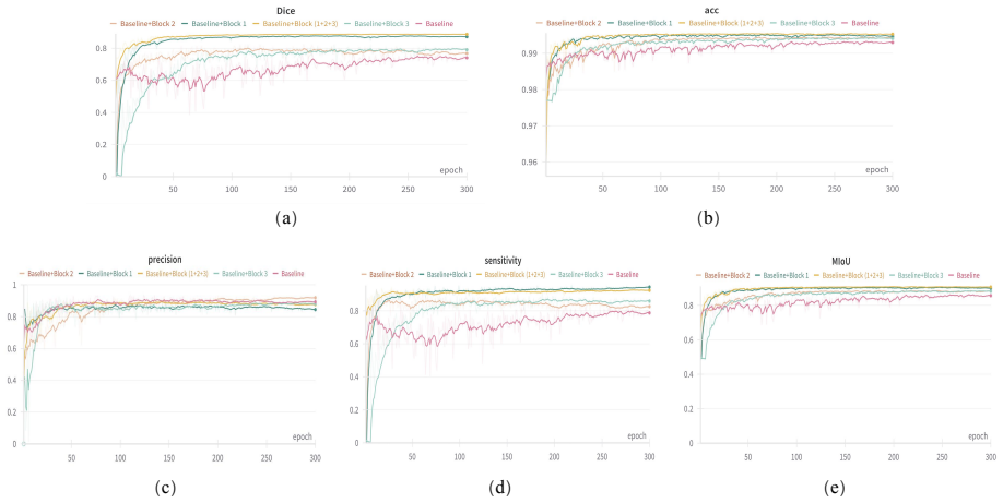
- Ablation shows the structured convolution blocks (Block1) contribute the largest gain, adding 9.41% DSC over the U-Net baseline, which supports the design choice of specialized early-stage convolution.
- The RM-SViT module achieves its best result with a single iteration and an 8x8 grid on 128x128 inputs, indicating that cheap superpixel attention suffices for this task size.
- The EPDB result (86.40% DSC across AAH/AIS/MIA/IAC subtypes) suggests the model transfers to adenocarcinoma staging-relevant nodule shapes without retraining.
Reading between the lines
- Because superpixel tokenization compresses the sequence length, the RM-SViT design may scale to 3D volumes or high-resolution images better than global ViTs, though the paper only demonstrates 2D 128x128 slices.
- The slice-level data split, if not per-nodule, is a common source of inflated Dice; a per-patient split on LIDC-IDRI would give a more clinically meaningful estimate and would also let readers judge whether the claimed gain over baselines persists.
- The reported mIoU higher than DSC is atypical for binary segmentation unless the IoU is computed on a probabilistic or multi-class basis; clarifying this definition would strengthen the comparison.
- If the gains replicate under a fair protocol, the same three-module recipe (structured convolution, superpixel transformer, spatial-shift MLP skip) could be transferred to other small-structure segmentation tasks like skin lesions or prostate MRI.
Signed reviews
Editorial analysis
A structured set of objections, weighed in public.
Referee Report
Summary. The paper proposes S3TU-Net, a U-shaped CNN-Transformer hybrid for lung nodule segmentation in CT images. The architecture combines two structured convolutional blocks (DWF-Conv, D2BR-Conv), a superpixel-based residual multi-branch vision transformer (RM-SViT), and a spatial-shift MLP skip-connection module (S2-MLP Link). The authors report a DSC of 89.04%, precision of 90.73%, mIoU of 90.70%, and sensitivity of 93.70% on LIDC-IDRI, claim a 4.52% DSC improvement over the best compared method, and report a DSC of 86.40% on a private EPDB dataset. The paper also includes ablation studies and comparisons against seven published segmentation networks.
Significance. If the reported results hold under a clean evaluation protocol, the architecture is a credible contribution to lung nodule segmentation: it combines several recent design ideas (large-kernel attention, DropBlock, spatial-shift MLPs, and superpixel transformers) in a U-Net framework and demonstrates generalization on an independent private dataset. The ablation study on EPDB is a useful strength. However, the paper does not release code or trained models, and the central quantitative claims currently rest on an evaluation protocol with several unresolved issues: inconsistent headline numbers, an undefined IoU metric that is reported above DSC, a slice-level data split that may leak nodule information, and test-set-based hyperparameter selection. For these reasons, the significance depends on whether the authors can repair the evaluation and confirm the reported margins.
major comments (4)
- [Section IV-B, Table V, Abstract] The numerical results are internally inconsistent. The abstract and Table V report DSC 89.04%, mIoU 90.70%, precision 90.73%, and sensitivity 93.70%, while the text of Section IV-B states that S3TU-Net achieves a maximum DSC of 88.87%, MIoU of 91.14%, sensitivity of 93.48%, and precision of 91.97%. These are different numbers, and the subsequent claims of improvements by 4.52% in DSC and 3.16% in sensitivity are computed from the Table V values. The authors must state which numbers are the official results, correct the inconsistency, and recompute all comparative claims consistently.
- [Section III-C, Section III-A] The LIDC-IDRI split is described only at the slice level: 5,717 training images and 757 test images from 6,474 total slices, with the same section reporting 1,303 nodules. The paper never states that the split is patient-disjoint or nodule-disjoint. Because adjacent CT slices of the same nodule are near-duplicates, a slice-level split can place nearly identical images in both training and test sets, inflating DSC and sensitivity through memorization. The authors must clarify the split granularity, and if it is slice-level, they must repeat the main comparison under a patient-level or nodule-level split and report whether the claimed 4.52% DSC advantage persists.
- [Section III-B, Eqs. (15)-(17), Table V] The metric definitions are incomplete and appear to produce impossible values. Equations (15)-(17) define DSC, sensitivity, and precision using raw probabilities, but no definition is given for IoU/mIoU, and no thresholding or binarization procedure is stated. In Table V, S3TU-Net reports mIoU 90.70% alongside DSC 89.04%; for any fixed pair of binary masks, IoU = DSC/(2-DSC), which would give at most about 80.3% foreground IoU for DSC 89.04%. If the reported mIoU is instead a mean of per-class IoUs (including background), that definition must be stated explicitly and must be used for all compared methods. The caption of Table III also labels results as '(Max)', indicating best-of-run selection; the authors should report mean and standard deviation over runs, or at least justify why maximum statistics are a valid basis for comparison.
- [Section IV-A.2, Fig. 9] The choice of the superpixel grid size (8x8) and the iteration count (1) is made by comparing performance in Fig. 9, which appears to be evaluated on the LIDC-IDRI test set. Selecting hyperparameters using the test set, and then reporting test-set performance with those hyperparameters, introduces selection bias and makes the reported margins over baselines (which are not given the same tuned hyperparameters) difficult to interpret. The authors should either tune on a held-out validation split and report test-set results, or disclose the selection procedure and quantify its effect.
minor comments (5)
- [Eqs. (7)-(8)] The spatial shift formulas in Equations (7) and (8) are formatted incorrectly: the piecewise cases are written as assignments with conditions placed on the right-hand side, making the intended operations unclear. Please rewrite them as standard piecewise definitions.
- [References] Several citations are mismatched: reference [41] is listed as a ResNeSt paper but is cited for LIDC-IDRI in Section III-A, and reference [45] is listed as a roof-extraction paper but is cited for Attention-UNet in Table V. The LIDC-IDRI dataset and the Attention-UNet method need correct citations. References [15] and [19] also appear to duplicate the same roof-extraction paper.
- [Fig. 7 caption] The caption of Fig. 7 refers to 'stage IV lung adenocarcinoma (AAH/MIA/IAC/AIS)', but AAH, AIS, MIA, and IAC are pathological subtypes, not stages; please correct the terminology.
- [Section IV-A.1] The text states that 'the DSC and sensitivity are increased by 9.41%' for Baseline+Block1 compared with Baseline; from Table III, DSC increases by 9.41 percentage points (77.44 to 86.85) but sensitivity increases by 8.41 percentage points (86.94 to 95.35), so the sentence should be corrected.
- [Section II-B] There is a typo in the description of DropBlock parameters: 'block size and y.block size' should presumably be 'block size and drop block size' or similar.
Circularity Check
No significant circularity: the paper is an empirical architecture-and-evaluation study with no derivation chain reducing to its inputs; the external EPDB validation is independent.
full rationale
S3TU-Net is presented as a U-Net hybrid with structured convolution blocks, an RM-SViT transformer, and an S2-MLP Link. The module equations (Eqs. 1-14) are self-contained definitions of the proposed operators, not fitted quantities from which the reported DSC/mIoU numbers are derived. The headline LIDC-IDRI results (DSC 89.04%, etc.) are experimental measurements on a held-out test portion, and the EPDB private dataset provides an external generalization check, so the central claims are not equivalent to their inputs by construction. The ablation study selects a superpixel grid size of 8×8 and one iteration based on observed performance; this is model selection rather than a fitted parameter renamed as a prediction, and if the selection and the slice-level 9:1 split were performed on the same test data, that is a leakage/correctness risk, not a circularity step. The mIoU-above-DSC values and the '(Max)' statistics indicate metric-definition and best-of-run reporting concerns, but they do not make the derivation circular. No load-bearing self-citation chain or imported uniqueness theorem appears; references to prior work by potential co-authors (e.g., [10]) are contextual and are not used to justify the central result. The future-work section notes annotation challenges but does not assert a circular limitation. Accordingly, the circularity score is 0.
Assumptions & free parameters
free parameters (6)
- Superpixel grid size =
8x8
- RM-SViT iteration count =
1
- DropBlock block size =
7
- Initial learning rate =
0.001
- Batch size and epoch count =
16, 300
- Base channel width C
assumptions (5)
- domain assumption Radiologist annotations in LIDC-IDRI (nodules marked by at least two of four radiologists) are treated as ground truth.
- domain assumption Slices are independent samples; no nodule- or patient-level split is enforced.
- ad hoc to paper DSC, sensitivity, and precision computed with raw probabilities (Eqs. 15-17) are comparable to standard thresholded metrics.
- ad hoc to paper Reporting maximum metrics over the training run is a valid summary.
- domain assumption Grid-averaged superpixel initialization and iterative softmax association preserve nodule-relevant information.
invented entities (4)
-
DWF-Conv block
-
D2BR-Conv block
-
S2-MLP Link
-
RM-SViT module
Cite this review
Pith. "Pith review of S3TU-Net: Structured Convolution and Superpixel Transformer for Lung Nodule Segmentation." pith.science (2026). https://pith.science/paper/Y5FXCUOD
@misc{pith2026241112547,
  author       = {Pith},
  title        = {Pith review of: S3TU-Net: Structured Convolution and Superpixel Transformer for Lung Nodule Segmentation},
  year         = {2026},
  howpublished = {\url{https://pith.science/paper/Y5FXCUOD}},
  note         = {Machine review of arXiv:2411.12547}
}
read the original abstract
The irregular and challenging characteristics of lung adenocarcinoma nodules in computed tomography (CT) images complicate staging diagnosis, making accurate segmentation critical for clinicians to extract detailed lesion information. In this study, we propose a segmentation model, S3TU-Net, which integrates multi-dimensional spatial connectors and a superpixel-based visual transformer. S3TU-Net is built on a multi-view CNN-Transformer hybrid architecture, incorporating superpixel algorithms, structured weighting, and spatial shifting techniques to achieve superior segmentation performance. The model leverages structured convolution blocks (DWF-Conv/D2BR-Conv) to extract multi-scale local features while mitigating overfitting. To enhance multi-scale feature fusion, we introduce the S2-MLP Link, integrating spatial shifting and attention mechanisms at the skip connections. Additionally, the residual-based superpixel visual transformer (RM-SViT) effectively merges global and local features by employing sparse correlation learning and multi-branch attention to capture long-range dependencies, with residual connections enhancing stability and computational efficiency. Experimental results on the LIDC-IDRI dataset demonstrate that S3TU-Net achieves a DSC, precision, and IoU of 89.04%, 90.73%, and 90.70%, respectively. Compared to recent methods, S3TU-Net improves DSC by 4.52% and sensitivity by 3.16%, with other metrics showing an approximate 2% increase. In addition to comparison and ablation studies, we validated the generalization ability of our model on the EPDB private dataset, achieving a DSC of 86.40%.
Figures


Figures from the paper (6 more)




Reference graph
Works this paper leans on
-
[45]
A bag of tricks for fine-grained roof extraction,
J. Hu, Z. Huang, F. Shen, D. He, and Q. Xian, “A bag of tricks for fine-grained roof extraction,” 2023
work page 2023
-
[1]
Lung cancer prediction using electronic claims records: A transformer-based approach,
H.-Y . Chen, H.-M. Wang, C.-H. Lin, R. Yang, and C.- C. Lee, “Lung cancer prediction using electronic claims records: A transformer-based approach,” IEEE Journal of Biomedical and Health Informatics , 2023
work page 2023
-
[2]
Modality- specific segmentation network for lung tumor segmenta- tion in pet-ct images,
D. Xiang, B. Zhang, Y . Lu, and S. Deng, “Modality- specific segmentation network for lung tumor segmenta- tion in pet-ct images,” IEEE Journal of Biomedical and Health Informatics, vol. 27, no. 3, pp. 1237–1248, 2022
work page 2022
-
[3]
Z. Li, J. Zhang, T. Tan, X. Teng, X. Sun, H. Zhao, L. Liu, Y . Xiao, B. Lee, Y . Liet al., “Deep learning methods for lung cancer segmentation in whole-slide histopathology images—the acdc@ lunghp challenge 2019,” IEEE Jour- nal of Biomedical and Health Informatics , vol. 25, no. 2, pp. 429–440, 2020
work page 2019
-
[4]
Spectrum of lung adenocarcinoma,
B. D. Hutchinson, G. S. Shroff, M. T. Truong, and J. P. Ko, “Spectrum of lung adenocarcinoma,” in Seminars in Ultrasound, CT and MRI, vol. 40, no. 3. Elsevier, 2019, pp. 255–264
work page 2019
-
[5]
D. J. Myers and J. M. Wallen, “Lung adenocarcinoma,” in StatPearls [Internet]. StatPearls Publishing, 2023
work page 2023
-
[6]
A. C. Borczuk, “Prognostic considerations of the new world health organization classification of lung adeno- carcinoma,” European Respiratory Review , vol. 25, no. 142, pp. 364–371, 2016
work page 2016
-
[7]
Who classification of tumours of the lung, pleura, thymus and heart,
W. H. Organization et al., “Who classification of tumours of the lung, pleura, thymus and heart,” WHO/IARC Classification of Tumours, , vol. 7, 2015
work page 2015
Show all 48 references
-
[8]
Role of pet/ct in management of early lung adenocarcinoma,
X. Shao, R. Niu, Z. Jiang, X. Shao, and Y . Wang, “Role of pet/ct in management of early lung adenocarcinoma,” American Journal of Roentgenology , vol. 214, no. 2, pp. 437–445, 2020
2020
-
[9]
Lung ade- nocarcinomas: correlation of computed tomography and pathology findings,
J. Cohen, E. Reymond, A. Jankowski, E. Brambilla, F. Arbib, S. Lantuejoul, and G. Ferretti, “Lung ade- nocarcinomas: correlation of computed tomography and pathology findings,” Diagnostic and interventional imag- ing, vol. 97, no. 10, pp. 955–963, 2016
2016
-
[10]
Integrating lung parenchyma segmentation and nodule detection with deep multi-task learning,
W. Liu, X. Liu, H. Li, M. Li, X. Zhao, and Z. Zhu, “Integrating lung parenchyma segmentation and nodule detection with deep multi-task learning,” IEEE Journal of Biomedical and Health Informatics , vol. 25, no. 8, pp. 3073–3081, 2021
2021
-
[11]
Advancing pose-guided image synthesis with progressive conditional diffusion models,
F. Shen, H. Ye, J. Zhang, C. Wang, X. Han, and Y . Wei, “Advancing pose-guided image synthesis with progressive conditional diffusion models,” in The Twelfth International Conference on Learning Representations , 2023
2023
-
[12]
Boosting consistency in story visualization with rich-contextual conditional diffusion models,
F. Shen, H. Ye, S. Liu, J. Zhang, C. Wang, X. Han, and W. Yang, “Boosting consistency in story visualization with rich-contextual conditional diffusion models,” arXiv preprint arXiv:2407.02482, 2024
2024 arXiv
-
[13]
Pulmonary nodule detection in medical images: a survey,
J. Zhang, Y . Xia, H. Cui, and Y . Zhang, “Pulmonary nodule detection in medical images: a survey,” Biomedi- cal Signal Processing and Control, vol. 43, pp. 138–147, 2018
2018
-
[14]
Deep feature transfer learning in combination with traditional features predicts survival among patients with lung adenocarcinoma,
R. Paul, S. H. Hawkins, Y . Balagurunathan, M. B. Schabath, R. J. Gillies, L. O. Hall, and D. B. Goldgof, “Deep feature transfer learning in combination with traditional features predicts survival among patients with lung adenocarcinoma,” Tomography, vol. 2, no. 4, p. 388, 2016
2016
-
[15]
A bag of tricks for fine-grained roof extraction,
J. Hu, Z. Huang, F. Shen, D. He, and Q. Xian, “A bag of tricks for fine-grained roof extraction,” in IGARSS 2023-2023 IEEE International Geoscience and Remote Sensing Symposium. IEEE, 2023
2023
-
[16]
A fast weak-supervised pulmonary nodule segmentation method based on modified self-adaptive fcm algorithm,
H. Liu, F. Geng, Q. Guo, C. Zhang, and C. Zhang, “A fast weak-supervised pulmonary nodule segmentation method based on modified self-adaptive fcm algorithm,” Soft Computing, vol. 22, pp. 3983–3995, 2018
2018
-
[17]
Au- tomated pulmonary nodule detection in ct images using deep convolutional neural networks,
H. Xie, D. Yang, N. Sun, Z. Chen, and Y . Zhang, “Au- tomated pulmonary nodule detection in ct images using deep convolutional neural networks,”Pattern recognition, vol. 85, pp. 109–119, 2019
2019
-
[18]
Pulmonary lung nodule detection from computed tomography images using two-stage convolutional neural network,
S. Jain, P. Choudhari, and M. Gour, “Pulmonary lung nodule detection from computed tomography images using two-stage convolutional neural network,”The Com- puter Journal, vol. 66, no. 4, pp. 785–795, 2023
2023
-
[19]
A rubust method for roof extraction and height estimation,
J. Hu, Z. Huang, F. Shen, D. He, and Q. Xian, “A rubust method for roof extraction and height estimation,” in IGARSS 2023-2023 IEEE International Geoscience and Remote Sensing Symposium . IEEE, 2023
2023
-
[20]
Segmentation of the pulmonary nodule and the attached vessels in the ct scan of the chest using morphological features and topological skeleton of the nodule,
M. B. Tavakoli, M. Orooji, M. Teimouri, and R. Sha- habifar, “Segmentation of the pulmonary nodule and the attached vessels in the ct scan of the chest using morphological features and topological skeleton of the nodule,” IET Image Processing, vol. 14, no. 8, pp. 1520– 1528, 2020
2020
-
[21]
Clustering by transmission learning from data density to label manifold with statistical diffusion,
Y . Zhang, F.-l. Chung, and S. Wang, “Clustering by transmission learning from data density to label manifold with statistical diffusion,” Knowledge-Based Systems , vol. 193, p. 105330, 2020
2020
-
[22]
U-net: Con- volutional networks for biomedical image segmenta- tion,
O. Ronneberger, P. Fischer, and T. Brox, “U-net: Con- volutional networks for biomedical image segmenta- tion,” in Medical image computing and computer-assisted intervention–MICCAI 2015: 18th international confer- ence, Munich, Germany, October 5-9, 2015, proceedings, part III...
2015
-
[23]
Review of semantic segmentation of medical images using mod- ified architectures of unet,
M. Krithika Alias AnbuDevi and K. Suganthi, “Review of semantic segmentation of medical images using mod- ified architectures of unet,” Diagnostics, vol. 12, no. 12, p. 3064, 2022
2022
-
[24]
A cascaded dual-pathway residual network for lung nodule segmentation in ct images,
H. Liu, H. Cao, E. Song, G. Ma, X. Xu, R. Jin, Y . Jin, and C.-C. Hung, “A cascaded dual-pathway residual network for lung nodule segmentation in ct images,” Physica Medica, vol. 63, pp. 112–121, 2019
2019
-
[25]
A novel deep learning network and its application for pulmonary nodule segmentation,
D. Lu, J. Chu, R. Zhao, Y . Zhang, and G. Tian, “A novel deep learning network and its application for pulmonary nodule segmentation,” Computational Intelligence and Neuroscience, vol. 2022, no. 1, p. 7124902, 2022
2022
-
[26]
Pulmonary nodules segmentation based on crf 3d-unet structure,
T. Hou, J. Zhao, Y . Qiang, S. Wang, and P. Wang, “Pulmonary nodules segmentation based on crf 3d-unet structure,” Comput Eng Des , vol. 41, no. 6, pp. 1663– 1669, 2020
2020
-
[27]
Vits vs. cnns for 3d medical image segmentation: Are transformers all you need?
X. Arregui Garc ´ıa, “Vits vs. cnns for 3d medical image segmentation: Are transformers all you need?” Master’s thesis, 2023
2023
-
[28]
Attention is all you need,
A. Vaswani, “Attention is all you need,” Advances in Neural Information Processing Systems , 2017
2017
-
[29]
Enhancing landslide segmen- tation with guide attention mechanism and fast fourier transformer,
K. Yan, F. Shen, and Z. Li, “Enhancing landslide segmen- tation with guide attention mechanism and fast fourier transformer,” in International Conference on Intelligent Computing. Springer, 2024, pp. 296–307
2024
-
[30]
An image is worth 16x16 words: Trans- formers for image recognition at scale,
A. Dosovitskiy, “An image is worth 16x16 words: Trans- formers for image recognition at scale,” arXiv preprint arXiv:2010.11929, 2020
2010 arXiv
-
[31]
Swin transformer: Hierarchical vi- sion transformer using shifted windows,
Z. Liu, Y . Lin, Y . Cao, H. Hu, Y . Wei, Z. Zhang, S. Lin, and B. Guo, “Swin transformer: Hierarchical vi- sion transformer using shifted windows,” in Proceedings of the IEEE/CVF international conference on computer vision, 2021, pp. 10 012–10 022
2021
-
[32]
Dropout: a simple way to pre- vent neural networks from overfitting,
N. Srivastava, G. Hinton, A. Krizhevsky, I. Sutskever, and R. Salakhutdinov, “Dropout: a simple way to pre- vent neural networks from overfitting,” The journal of machine learning research, vol. 15, no. 1, pp. 1929–1958, 2014
1929
-
[33]
Dropblock: A regu- larization method for convolutional networks,
G. Ghiasi, T.-Y . Lin, and Q. V . Le, “Dropblock: A regu- larization method for convolutional networks,” Advances in neural information processing systems , vol. 31, 2018
2018
-
[34]
Visual attention network,
M.-H. Guo, C.-Z. Lu, Z.-N. Liu, M.-M. Cheng, and S.- M. Hu, “Visual attention network,” Computational Visual Media, vol. 9, no. 4, pp. 733–752, 2023
2023
-
[35]
Freeu: Free lunch in diffusion u-net,
C. Si, Z. Huang, Y . Jiang, and Z. Liu, “Freeu: Free lunch in diffusion u-net,” in Proceedings of the IEEE/CVF Conference on Computer Vision and Pattern Recognition, 2024, pp. 4733–4743
2024
-
[36]
Squeeze-and-excitation networks,
J. Hu, L. Shen, and G. Sun, “Squeeze-and-excitation networks,” in Proceedings of the IEEE conference on computer vision and pattern recognition, 2018, pp. 7132– 7141
2018
-
[37]
Malunet: A multi-attention and light-weight unet for skin lesion segmentation,
J. Ruan, S. Xiang, M. Xie, T. Liu, and Y . Fu, “Malunet: A multi-attention and light-weight unet for skin lesion segmentation,” in 2022 IEEE International Conference on Bioinformatics and Biomedicine (BIBM) . IEEE, 2022, pp. 1150–1156
2022
-
[38]
S2-mlp: Spatial-shift mlp architecture for vision,
T. Yu, X. Li, Y . Cai, M. Sun, and P. Li, “S2-mlp: Spatial-shift mlp architecture for vision,” in Proceedings of the IEEE/CVF winter conference on applications of computer vision, 2022, pp. 297–306
2022
-
[39]
Mlp-mixer: An all-mlp architecture for vision,
I. O. Tolstikhin, N. Houlsby, A. Kolesnikov, L. Beyer, X. Zhai, T. Unterthiner, J. Yung, A. Steiner, D. Keysers, J. Uszkoreit et al. , “Mlp-mixer: An all-mlp architecture for vision,” Advances in neural information processing systems, vol. 34, pp. 24 261–24 272, 2021
2021
-
[40]
Multilayer perceptron (mlp),
H. Taud and J.-F. Mas, “Multilayer perceptron (mlp),” Geomatic approaches for modeling land change scenar- ios, pp. 451–455, 2018
2018
-
[41]
Resnest: Split-attention networks,
H. Zhang, C. Wu, Z. Zhang, Y . Zhu, H. Lin, Z. Zhang, Y . Sun, T. He, J. Mueller, R. Manmatha et al. , “Resnest: Split-attention networks,” in Proceedings of the IEEE/CVF conference on computer vision and pattern recognition, 2022, pp. 2736–2746
2022
-
[42]
Y-net: joint segmentation and classifi- cation for diagnosis of breast biopsy images,
S. Mehta, E. Mercan, J. Bartlett, D. Weaver, J. G. Elmore, and L. Shapiro, “Y-net: joint segmentation and classifi- cation for diagnosis of breast biopsy images,” in Medical Image Computing and Computer Assisted Intervention– MICCAI 2018: 21st International Conference, Granada...
2018
-
[43]
Unet++: A nested u-net architecture for medical image segmentation,
Z. Zhou, M. M. Rahman Siddiquee, N. Tajbakhsh, and J. Liang, “Unet++: A nested u-net architecture for medical image segmentation,” in Deep Learning in Medical Image Analysis and Multimodal Learning for Clinical Decision Support: 4th International Workshop, DLMIA 2018, and 8th ...
2018
-
[44]
Recurrent residual convolutional neural network based on u-net (r2u-net) for medical image segmentation,
M. Z. Alom, M. Hasan, C. Yakopcic, T. M. Taha, and V . K. Asari, “Recurrent residual convolutional neural network based on u-net (r2u-net) for medical image segmentation,” arXiv preprint arXiv:1802.06955 , 2018
2018 arXiv
-
[46]
Unet 3+: A full- scale connected unet for medical image segmentation,
H. Huang, L. Lin, R. Tong, H. Hu, Q. Zhang, Y . Iwamoto, X. Han, Y .-W. Chen, and J. Wu, “Unet 3+: A full- scale connected unet for medical image segmentation,” in ICASSP 2020-2020 IEEE international conference on acoustics, speech and signal processing (ICASSP) . IEEE, 2020, ...
2020
-
[47]
Sa-unet: Spatial attention u-net for retinal vessel segmentation,
C. Guo, M. Szemenyei, Y . Yi, W. Wang, B. Chen, and C. Fan, “Sa-unet: Spatial attention u-net for retinal vessel segmentation,” in 2020 25th international conference on pattern recognition (ICPR). IEEE, 2021, pp. 1236–1242
2020
-
[48]
Uncertainty- guided lung nodule segmentation with feature-aware attention,
H. Yang, L. Shen, M. Zhang, and Q. Wang, “Uncertainty- guided lung nodule segmentation with feature-aware attention,” in International Conference on Medical Im- age Computing and Computer-Assisted Intervention . Springer, 2022, pp. 44–54
2022
Reviewed August 12, 2026 · model on record in the stance chip above.
Discussion (0). Continue with ORCID to comment.