REVIEW 4 major objections 6 minor 33 references
Automating Sonologists USG Commands with AI and Voice Interface
T0 review · 4 major / 6 minor · reviewed 2026-08-12 · deepseek-v4-flash
Pith's one-line read This paper claims that a voice-controlled AI pipeline can segment organs and classify liver fibrosis in real-time ultrasound, reporting about 98 percent accuracy for fibrosis classification.
desk verdict A plausible voice-controlled ultrasound integration, but the organ-segmentation claim is validated only by model confidence, not ground-truth accuracy. read the letter →
The pith
A machine-rendered reading of the paper's core claim, the machinery that carries it, and where it could break.
The reading
What carries the argument
The load-bearing machinery is the pairing of Mask R-CNN, a region-based convolutional network that outputs pixel-level masks and confidence scores for each detected organ, with a MobileNet backbone that was fine-tuned on a five-class liver histopathology dataset and topped with dense layers for classification. A voice layer uses speech recognition to map commands into a queue that freezes video, restarts it, or triggers prediction. The system runs these in threads so that command execution does not lag the video playback.
What would settle it
Hold out ultrasound videos recorded on a different scanner or at a different site, run the trained organ-segmentation and liver-classification models on them, and measure mean mask intersection-over-union and classification accuracy; if either collapses relative to the reported 40-95 percent confidence range or 98 percent accuracy, the central claim fails.
Extended reading notes
Core claim
The central discovery is that the components can be chained end to end: a live ultrasound video feed is buffered and processed by Mask R-CNN for organ segmentation; when the liver is seen and the predict command is given, a MobileNet-based classifier labels the frame into one of five histopathology classes; and all actions are triggered by a speech-recognition layer that executes freeze, continue, and predict commands. In the authors' tests, the classifier reached 98 percent overall accuracy and 97.78 percent test accuracy, with the abstract-level statement of 98.6 percent for the fibrosis-optimized module, while organ masks carried confidence scores from 40 to 95 percent, and clear images exceeded 90 percent. The paper argues this shows the system can assist sonologists by reducing manual interaction and providing early pathological clues.
Load-bearing premise
The load-bearing premise is that a Mask R-CNN model initialized with weights from large natural-image datasets and fine-tuned on a small set of ultrasound frames from one hospital will segment organs accurately enough on unseen patients, machines, and image qualities.
Editorial extensions
If this is right
- Hands-free operation: a sonologist can freeze, continue, and ask for prediction without touching the scanner, which the paper says reduces workflow interruptions.
- Real-time organ awareness: the segmentation module draws masks and confidence scores for liver, kidney, and prostate as the live feed plays, potentially reducing manual measurement time.
- Early liver-fibrosis signal: the classifier assigns a frame to one of five histopathology classes, and the paper argues this helps flag fibrosis early and speeds diagnosis.
- Consistency over manual reading: automated predictions remove observer variability in basic interpretation, according to the authors' comparison with traditional methods.
Reading between the lines
- The paper does not quantify how the segmentation model behaves on scans from other hospitals; a direct test would be to rerun the trained weights on a multi-scanner ultrasound set and compare mask accuracy with the reported 40-95 percent confidence range.
- The confidence scores could be repurposed as a quality gate: frames below a threshold could trigger a re-scan. The authors do not propose this, but their own reported spread makes it plausible.
- Expanding the voice vocabulary from freeze, continue, and predict to measurement commands would be a small engineering step with large clinical payoff; this is my inference, not a paper claim.
Signed reviews
Editorial analysis
A structured set of objections, weighed in public.
Referee Report
Summary. The manuscript describes an ultrasound imaging system that combines real-time organ detection and segmentation using Mask R-CNN (Detectron2), a liver histopathology classifier based on MobileNet, and a voice-command interface for hands-free control. The authors report command recognition accuracy above 90%, organ segmentation confidence scores between 40% and 95%, and liver histopathology classification accuracy of 98% (with 97.78% test accuracy and a 98.6% figure in the abstract). The paper includes system architecture, implementation details, and a brief evaluation using a recorded ultrasound video and a histopathology dataset. The central claim is that the integrated system improves workflow and diagnostic support for sonologists, but the evidence presented is largely based on self-reported model confidence and internally inconsistent accuracy figures, without external validation or standard segmentation metrics.
Significance. If the system performed as claimed, it would represent a useful engineering contribution to ultrasound workflow automation, combining voice control with real-time organ segmentation and liver fibrosis classification. The paper clearly describes a plausible system architecture and uses standard deep learning components (Mask R-CNN, MobileNet, Google Speech-to-Text). However, the current evaluation is not sufficient to establish the central claims: organ segmentation is validated only by model confidence rather than ground-truth metrics, the histopathology accuracy figures are inconsistent and lack dataset details, and the voice interface is evaluated without a defined protocol. The authors also explicitly acknowledge narrow training datasets and image-quality sensitivity in Section 6.2, yet the abstract promises a clinically generalizable system. With substantial additional experiments and transparent reporting, the contribution could be valuable, but as presented the evidence is too weak to support the stated conclusions.
major comments (4)
- [Section 5.1.2, Section 3.4] The organ detection and segmentation claim is unsupported because the paper reports only model confidence scores ('more than 40% and less than 95% average confidence scores') rather than any ground-truth segmentation accuracy. Confidence from Mask R-CNN is the model's estimated probability of a correct prediction, which can be high even for anatomically incorrect masks. No Dice coefficient, IoU, boundary error, or clinician-verified manual annotation comparison is provided, and there is no per-organ breakdown or test set from a different machine or hospital. Section 3.4 states that training used only ultrasound data from Ramachandra hospital, and Section 6.2 admits 'narrow training datasets' and sensitivity to image quality. Because the voice commands ('freeze', 'predict') and the histopathology module rely on correct organ localization, this missing validation is load-bearing for the paper's central claim.
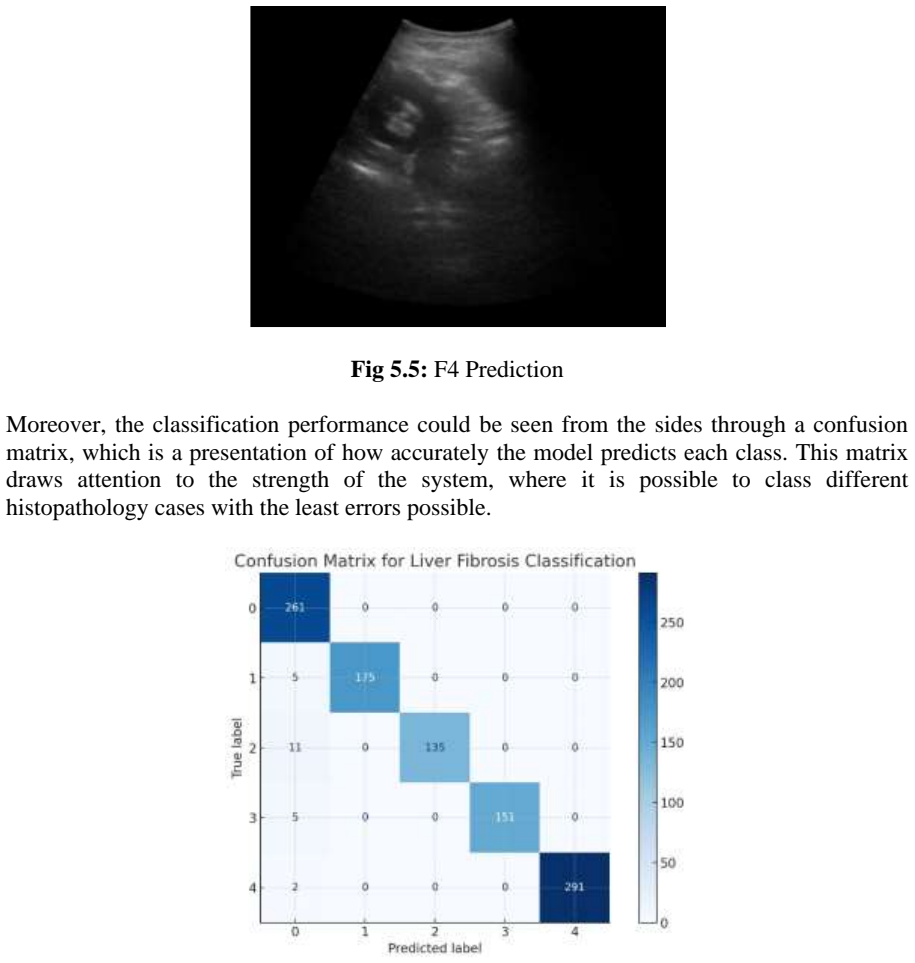
- [Section 5.1.3, Abstract] The liver histopathology accuracy results are internally inconsistent and not auditable. The abstract reports 'an impressive accuracy of 98.6%', Section 5.1.3 reports 'a total accuracy of 98%', and the same section reports 'test accuracy being 97.78%'. The paper does not specify the dataset size, class-wise sample counts beyond two classes (Class 0 with 261 entries and Class 4 with 293), the train/validation/test split, or how the 98.6% figure was obtained. Without these details, the flagship accuracy cannot be reconstructed or independently verified. The statement 'This large difference shows the capacity of the model to adapt well to new data' is also unclear, since the difference between 98% and 97.78% is small.
- [Section 5.1.1, Section 6.2] The voice interface evaluation is insufficient to support the claim of 'over 90% accuracy'. No sample size, number of speakers, command set, environment conditions, or confusion matrix is provided, and the only quantitative statement is a single percentage. Section 6.2 acknowledges failures under background noise, accents, overlapping speech, and rapid commands, but these are not quantified. Since the paper relies on Google's Speech-to-Text API and does not describe any custom noise-robustness evaluation, the clinical usability of the voice interface in real ultrasound environments is not established.
- [Section 5.2.1, Section 6.2] The case-study evaluation in Section 5.2.1 uses a single previously recorded ultrasound video to emulate clinical use. This is not a substitute for a prospective or multi-patient evaluation, and it does not address the acknowledged generalizability limitation from Section 3.4 (single-hospital data) and Section 6.2 (narrow training datasets). The paper's conclusion that the system is 'easily amenable for incorporation into clinical usage' is therefore an extrapolation beyond the evidence. I would ask the authors to either remove this claim or provide a systematic evaluation with multiple patients, ultrasound machines, and operators.
minor comments (6)
- [Section 3.3] The sentence 'Key technologies used include Google's Speech-to-Text API for systems that are not connected to the internet' is contradictory, since an API typically requires an internet connection; please clarify whether an offline speech recognition engine was used.
- [Section 3.4] Mask R-CNN performs instance segmentation, not semantic segmentation as stated; please correct the terminology.
- [Section 5.1.1] The phrase 'registering over 90% accuracy with little cases of misinterpretation' is grammatically awkward; consider rewriting as 'over 90% accuracy with few misinterpretation cases.'
- [Section 8 (References)] References [1] through [7] are given without complete bibliographic details (journal volumes, pages, or DOI), and several appear to be generic placeholders rather than verifiable publications. Please provide full citations or remove uncited sources.
- [Section 5.1.3] The phrase 'This large difference shows the capacity of the model to adapt well to new data without being over-fitted or any other learning effects limiting it' is unclear; please specify which numbers are being compared and what statistical conclusion follows.
- [Section 5.2.1] The case study reports segmentation scores on a recorded video but does not state how many frames were evaluated or how representative the video is; please add this context.
Circularity Check
No circular derivation; reported metrics are empirical claims, not results forced by construction or by self-citation.
full rationale
This paper does not present a formal derivation chain; it reports measured performance of a voice interface, a Detectron2 Mask R-CNN organ segmentation module, and a MobileNet-based liver histopathology classifier. The histopathology accuracies (abstract 98.6%, body 98% total, 97.78% test) are empirical results on a held-out test split, and although dataset size and split details are omitted, nothing in the text defines the reported accuracy in terms of the model's own training objective or fitted parameters. The organ segmentation evaluation is weaker: Section 5.1.2 reports only the model's internal confidence scores (40-95%) and no Dice/IoU or ground-truth comparison, so the efficacy claim is under-supported; however, this is a validation gap, not circularity, because the confidence score is not being renamed as an accuracy computed from labels. The references include Detectron2 and MobileNets as external method citations, and no self-citation is load-bearing. Section 6.2 explicitly acknowledges narrow training data and image-quality sensitivity, which further indicates the claims are empirical and limited rather than circular. Under the stated rules requiring a specific reduction of a claim to its inputs, no circular step can be identified.
Assumptions & free parameters
free parameters (2)
- Trained organ segmentation model weights =
not provided
- Trained histopathology classifier weights =
not provided
assumptions (3)
- domain assumption COCO-pretrained features transfer to ultrasound organ segmentation.
- domain assumption The annotated datasets are correctly labeled and representative.
- ad hoc to paper Google Speech-to-Text API is reliable in clinical settings.
Cite this review
Pith. "Pith review of Automating Sonologists USG Commands with AI and Voice Interface." pith.science (2026). https://pith.science/paper/UIBGJKHZ
@misc{pith2026241113006,
  author       = {Pith},
  title        = {Pith review of: Automating Sonologists USG Commands with AI and Voice Interface},
  year         = {2026},
  howpublished = {\url{https://pith.science/paper/UIBGJKHZ}},
  note         = {Machine review of arXiv:2411.13006}
}
read the original abstract
This research presents an advanced AI-powered ultrasound imaging system that incorporates real-time image processing, organ tracking, and voice commands to enhance the efficiency and accuracy of diagnoses in clinical practice. Traditional ultrasound diagnostics often require significant time and introduce a degree of subjectivity due to user interaction. The goal of this innovative solution is to provide Sonologists with a more predictable and productive imaging procedure utilizing artificial intelligence, computer vision, and voice technology. The functionality of the system employs computer vision and deep learning algorithms, specifically adopting the Mask R-CNN model from Detectron2 for semantic segmentation of organs and key landmarks. This automation improves diagnostic accuracy by enabling the extraction of valuable information with minimal human input. Additionally, it includes a voice recognition feature that allows for hands-free operation, enabling users to control the system with commands such as freeze or liver, all while maintaining their focus on the patient. The architecture comprises video processing and real-time segmentation modules that prepare the system to perform essential imaging functions, such as freezing and zooming in on frames. The liver histopathology module, optimized for detecting fibrosis, achieved an impressive accuracy of 98.6%. Furthermore, the organ segmentation module produces output confidence levels between 50% and 95%, demonstrating its efficacy in organ detection.
Figures


Figures from the paper (2 more)

Reference graph
Works this paper leans on
-
[1]
Introduction In modern healthcare, diagnostic imaging plays a pivotal role in identifying, diagnosing, and monitoring a wide array of conditions. Ultrasound imaging is one of the most common diagnostic methods used across nearly every medical department today. Its popularity stems from its real -time imaging capability, non -invasive nature, and easy acce...
-
[2]
The authors used ResNet and DenseNet along with KNN
Automatic Recognition of Abdominal Organs in Ultrasound Images The review work in this article is geared towards the application of deep learning techniques for recognizing abdominal organs automatically in ultrasound images. The authors used ResNet and DenseNet along with KNN. Their method uses feature vector-based techniques on the ultrasound images ins...
-
[3]
The authors underline the potential of AI to increase the accuracy and speed of diagnostic processes
Towards Clinical Application of AI in Ultrasound Imaging The potential uses of artificial intelligence (AI) in ultrasound imaging in the clinical field are assessed in this paper th rough a review of recent developments in deep learning models for pre-processing, segmentation and detection tasks. The authors underline the potential of AI to increase the a...
-
[4]
Voice-Controlled Medical Ultrasound Systems This paper proposes a novel ultrasound imaging and scanning system which can be operated using voice control and aims to optimize medical imaging work flow. The system includes speech recognition, thus, it is possible to operate the system without hands, in which case a clinician can assign tags to the images an...
-
[5]
Automated Measurement and Calcification Detectionin Carotid Ultrasound The present work describes a fully automated method to detect and quantify calcifications in carotid ultrasound images, employing advanced image segmentation techniques along with deep learning models. This system improves the diagnostic care of vascular calcifications, prompting a sig...
-
[6]
Deep Learning-Based Tumor Detection in Breast Ultrasound Imaging The research concern of Johnson et al. finds application of advanced computing techniques such as deep convolution neural networks in real -time detection of tumours in breast ultrasounds. Using the CNN architecture, the authors illustrate that the model can effectively find tumors, hence co...
-
[7]
AI-Based Liver Segmentation in Ultrasound Imaging The research described in this paper looks at the use of AI technology in liver segmentationtasks from ultrasound images with a specific focus on the use of convolutional neural networks (CNN) for the automated and accurate delineation of liver boundaries. The authors state that the method supported with A...
-
[8]
Integrated Voice and Image Processing in Ultrasound Diagnostics The authors of this manuscript present a machine learning model that comb ines speech recognition and image analysis to improve the efficiency of ultrasound diagnosis. The purpose of the system is to help the clinicians arrive at faster and more accurate diagnostic conclusions by eliminating ...
Show all 33 references
-
[9]
freeze”, “deep freeze
Methodology 3.1 Definition The USG Voice Automation System is a cutting -edge technology that transforms ultrasound imaging by allowing hands -free control through voice commands. This innovative system enables radiologists to efficiently manage ultrasound images and carry out...
-
[10]
FREEZE,"
Implementation 4.1 System Integration Cohesive functionality in the system was achieved through the integration of several components. The first step involved incorporating the organ segmentation and detection model developed using the Detectron 2 framework. This model was als...
-
[11]
The system operated with very low latency, thus ensuring a near real -time interaction and workflow
Results 5.1 Performance Evaluation 5.1.1 Assessing the Performance of the Voice User Interface The voice user interface embedded into the system worked perfectly, supporting the execution of the commands ‘freeze, ‘predict’ or ‘continue’ among others effortlessly. The system op...
-
[12]
The voice interface was effective, where more than 90% command recognit ion accuracy was achieved with little or no delay
Discussion 6.1 Explanation of the Findings and Their Importance The findings gained from the execution and assessment of the system , reinforces its claim of being a valuable repertoire to sonologists and pathologists. The voice interface was effective, where more than 90% com...
-
[13]
It is observed that in cases of poor quality or high levels of noise images, segmentation accuracy and confidence scores may fall significantly
Image Quality Limitations: The performance of the organ detection model is drawn to a great extent from the quality and definition of the ultrasound images. It is observed that in cases of poor quality or high levels of noise images, segmentation accuracy and confidence scores...
-
[14]
This impacts how well this system might perform in new clinical situations not encountered during training
Narrow Training Datasets: The model was built using a dimensions space that does not cover every possible aspect of any given organ features, deformities, ethnicities or diseases. This impacts how well this system might perform in new clinical situations not encountered during...
-
[15]
Issues with Speech Recognition: The voice interface worked remarkably well apart from the fact that it had problems in instances of high background noise, presence of accents, when people spoke at the same time or when commands were given in qui ck succession. There was also a...
-
[16]
Hardware Specifications: Real-time segmentation and classification of the system requires sophisticated computational power, which limits the implementation in such circumstances where the resources are few. 6.3 Potential Improvements and Future Work Within the existing limita...
-
[17]
Harnessing Latest Datasets: Employing the latest available annotated images and histopathology slides to include more peculiari ties and demographics’ rare diseases, would greatly improve the ability of the system to perform in different clinical environments
-
[18]
Voice Interface Improvement: Enhanced microphone technology employing noise suppression algorithms and voice recognitio n systems capable of understanding different accents and fast speech while reducing turnaround time would improve the device’s efficiency in clinical setting...
-
[19]
Morphological Changes’ Detection: When kidneys are encountered, a calcification model can be deployed to predict those as well
-
[20]
Deployment of the Edge AI: Fine-tuning the model for edge -computing-capable devices would allow the application to be used in low -resource settings and allow the processing of images without the need for expensive computer systems. The above limitations, the latest datasets,...
-
[21]
The organ detection model built in the Detectron2 framework produced results that have some signs of segmentation of the liver with confidence levels of between 40% and 95%
Conclusion 7.1 Summary of Findings The project involves the development and testing of a system that integrates AI -based organ segmentation, liver histopathology classification, as well as voice -command ultras ound imaging. The organ detection model built in the Detectron2 f...
-
[22]
Automatic Recognition of Abdominal Organs in Ultrasound Images
Smith, J., Kumar, R., and Chen, Y. Automatic Recognition of Abdominal Organs in Ultrasound Images. arXiv Preprint, 2021
2021
-
[23]
Towards Clinical Application of AI in Ultrasound Imaging
Brown, L., Williams, P., and Davis, T. Towards Clinical Application of AI in Ultrasound Imaging. MDPI Journal of Imaging, 2021
2021
-
[24]
Voice -Controlled Medical Ultrasound Systems
Nguyen, T., Patel, A., and Lee, K. Voice -Controlled Medical Ultrasound Systems. IEEE Transactions on Medical Devices, 2022
2022
-
[25]
Automated Measurement and Calcification Detection in Carotid Ultrasound
Garcia, M., Zhou, L., and Taylor, F. Automated Measurement and Calcification Detection in Carotid Ultrasound. Journal of Cardiovascular Imaging, 2022
2022
-
[26]
Deep Learning -Based Tumor Detection in Breast Ultrasound Imaging
Johnson, P., Singh, V., and Wang, Z. Deep Learning -Based Tumor Detection in Breast Ultrasound Imaging. IEEE Transactions on Biomedical Engineering, 2022
2022
-
[27]
AI -Based Liver Segmentation in Ultrasound Imaging
Martinez, R., O’Connor, S., and Chang, D. AI -Based Liver Segmentation in Ultrasound Imaging. Journal of Medical Ultrasound, 2023
2023
-
[28]
Integrated Voice and Image Processing in Ultraso und Diagnostics
Kim, H., Lopez, J., and Zhao, Q. Integrated Voice and Image Processing in Ultraso und Diagnostics. Springer Proceedings on Intelligent Healthcare Systems, 2023
2023
-
[29]
Detectron 2: A PyTorch-based modular object detection library
Wu, Y., Kirillov, A., Massa, F., Lo, W.-Y., and Girshick, R. Detectron 2: A PyTorch-based modular object detection library. Facebook AI Research, 2019
2019
-
[30]
G., Zhu, M., Chen, B., Kalenichenko, D., Wang, W., Weyand, T., Andreetto, M., and Adam, H
Howard, A. G., Zhu, M., Chen, B., Kalenichenko, D., Wang, W., Weyand, T., Andreetto, M., and Adam, H. Mobile Nets: Efficient Convolutional Neural Networks for Mobile Vision Applications. arXiv Preprint, 2017
2017
-
[31]
Dimitriou, N., Arandjelovi´c, O., and Caie, P. D. Deep learning for whole slide image analysis: From datasets to algorithms. Journal of Pathology Informatics, 2019
2019
-
[32]
Liver Histopathology Fibrosis Ultrasound Images
Gupta, V. Liver Histopathology Fibrosis Ultrasound Images. Kaggle Dataset
-
[33]
Learned Annotation for Medical Datasets: A Comprehensive Guide
Sanders, A. Learned Annotation for Medical Datasets: A Comprehensive Guide
Reviewed August 12, 2026 · model on record in the stance chip above.
Discussion (0). Continue with ORCID to comment.