REVIEW 4 major objections 5 minor 69 references
Virtual Staining of Label-Free Tissue in Imaging Mass Spectrometry
T0 review · 4 major / 5 minor · reviewed 2026-08-12 · deepseek-v4-flash
Pith's one-line read A diffusion model turns low-resolution mass spectrometry images of label-free kidney tissue into realistic PAS-stained histology, letting pathologists identify glomeruli and tubules without chemical staining.
desk verdict A genuinely new application of diffusion-based virtual staining to IMS data, with a load-bearing statistical flaw in the equivalence claim that needs fixing before publication. read the letter →
The pith
A machine-rendered reading of the paper's core claim, the machinery that carries it, and where it could break.
The reading
What carries the argument
The engine is a Brownian Bridge Diffusion Model (BBDM) for image-to-image translation, conditioned on the IMS ion images; its forward process interpolates from the stained ground-truth image to the IMS input, and the reverse process denoises the IMS input back into a stain-like image using an attention-based U-Net that estimates the posterior mean. Because the final reverse-diffusion steps inject large noise variance, the authors add a 'mean sampling' strategy that stops adding random noise after an exit point, cutting run-to-run variance while preserving perceptual similarity; they also evaluate a 'skip sampling' variant. The model is trained on 712 registered IMS–PAS patches from four patients and tested on 36 patches from a fifth.
What would settle it
Retrain the model on the same data with the PAS ground truth deliberately shifted by 5–10 µm relative to the IMS images; if the model reproduces the shifted structures in its virtual stains, then the reported concordance is substantially an artifact of registration accuracy rather than of molecular-to-morphology mapping. Alternatively, feed the trained model spatially scrambled or noise-only IMS channels; if it still outputs realistic PAS images, the generated histology is coming from the model's prior rather than from the mass-spectrometry signal.
Extended reading notes
Core claim
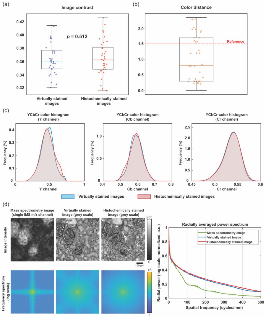
The central discovery is that a Brownian-bridge diffusion model can jointly perform ten-fold super-resolution and cross-modal translation: it takes 1,453 low-resolution ion images (10 µm pixels, each an m/z channel) of unlabeled kidney tissue and outputs 1 µm-pixel brightfield images that mimic PAS histochemistry. On 36 held-out fields of view from a patient not used in training, the generated images showed no statistically significant contrast difference from the true stains (p=0.512), CIE-94 color distances mostly below 1.5, matching radial power spectra, and pathologist-annotated structures that agreed between virtual and histochemical stains. The authors attribute this success to the rich molecular information carried by the mass-spectrometry channels and to diffusion models' ability to model complex distributions at extreme super-resolution factors.
Load-bearing premise
The whole pipeline rests on the assumption that the PAS-stained ground-truth image and the label-free IMS ion image of the same section are aligned at pixel scale, and the registration is done through an intermediate autofluorescence image plus a manual affine fit with 8–12 fiducial markers, so any residual misalignment becomes a systematic error the model can learn as if it were tissue structure.
Editorial extensions
If this is right
- IMS users can obtain histology-equivalent PAS images immediately after a scan, without chemical staining, brightfield imaging, or registration.
- The label-free tissue remains available for genomics, epigenetics, or further mass spectrometry, since the staining is purely digital.
- The method can augment existing IMS datasets: stored ion images can be re-run through the trained model to produce virtual stains.
- More mass-spectrometry channels improve virtual staining fidelity, so higher-multiplex IMS acquisitions yield better histology predictions.
- The mean sampling strategy makes diffusion-based virtual staining repeatable enough for digital pathology workflows.
Reading between the lines
- Editorial inference: the same trained model could be applied to archival IMS datasets from other institutions without retraining, provided m/z calibration and pixel size match; this would be a cheap test of cross-laboratory robustness.
- Editorial inference: the channel-reduction result hints that specific m/z channels carry most of the morphology-relevant signal, so analyzing which channels the model relies on most could reveal molecular correlates of PAS staining and possibly new biomarkers.
- Editorial inference: if the co-registration assumption is imperfect, some 'tissue structure' the model generates may actually be learned misalignment; comparing virtual stains trained on deliberately misregistered pairs would expose how much of the concordance depends on registration accuracy.
- Editorial extension: the same Brownian-bridge setup should transfer to other stains (for example H&E) and other organs; the paper claims generality, but the direct test of training and blinded pathologist reading on a second stain or organ has not been performed here.
Editorial analysis
A structured set of objections, weighed in public.
Referee Report
Summary. The manuscript proposes a Brownian-bridge diffusion model that maps low-resolution (10 µm pixel) MALDI IMS ion images of label-free human kidney tissue to super-resolved (1 µm pixel) virtual Periodic Acid-Schiff (PAS) stained brightfield images. The model is trained on 712 image-pair patches from four patients and tested on 36 fields of view from a held-out fifth patient. The authors report that a board-certified pathologist could identify glomeruli and proximal/distal tubules in the virtual stains, and they support the claim of close matching with quantitative metrics: image contrast, CIE-94 color distance, YCbCr histograms, and radial power spectra. They also propose a deterministic 'mean sampling' strategy to reduce diffusion-output variance and a channel-reduction analysis to show the value of multiplexed IMS input. The core idea is to use rich molecular IMS information to synthesize histology-like contrast without chemical staining.
Significance. If the results hold, the paper would contribute a practically valuable method for molecular histology, enabling histological interpretation of IMS data without tissue staining and without inference-time registration. The strengths include the use of a held-out patient for blind testing, comparison against actual histochemical ground truth, qualitative pathologist annotation, and a quantitative channel-ablation study. The proposed mean-sampling strategy addresses a real reproducibility concern in diffusion-based image translation. However, the quantitative foundation is currently weakened by an invalid statistical-equivalence argument and by validation limited to one test patient. These issues are fixable and do not undermine the plausibility of the method, but they must be addressed before the central claims can be accepted.
major comments (4)
- [Results, Fig. 3(a); Statistical analysis] The claim of 'statistical equivalence' between contrast of VS and HS images is not supported by the reported two-tailed paired t-test (p=0.512). A t-test tests the null hypothesis of zero mean difference; failing to reject that null does not establish equivalence. With 36 FOVs, a non-significant p-value can simply reflect large variance or low power. The manuscript should report a confidence interval for the mean contrast difference or perform a two one-sided tests (TOST) procedure with a pre-specified equivalence margin. This is load-bearing because the abstract's 'closely match their histochemically stained counterparts' is quantitatively supported substantially by this contrast claim.
- [Methods: Multimodal image registration; Data division and preparation] The training premise is that the IMS ion images and PAS-stained brightfield images are accurately co-registered to sub-cellular accuracy. The Methods describe elastix rigid/affine registration and a manual affine fit using 8-12 fiducial markers on laser ablation marks. Any residual misalignment between the low-resolution IMS grid and the high-resolution PAS image becomes a systematic error that the model may learn as tissue structure. The manuscript does not quantify registration error or report a sensitivity analysis to misalignment. Because the central application claim includes automatic registration of the virtual stains, the accuracy of the training-pair registration should be documented.
- [Results: blind testing and Figure 2] The blind test comprises 36 FOVs from a single patient. The text states that concordance was demonstrated 'across multiple FOVs of tissue' and refers to 'robustness and generalizability,' but a single-patient test set does not support cross-patient generalizability claims. The manuscript should either temper the generalization claim or provide results on multiple held-out patients, with per-patient variability reported.
- [Figure 2 and Results: pathologist annotations] The pathologist concordance is presented only as qualitative annotation images and the statement 'very good concordance.' No quantitative agreement measure (e.g., counts of annotated structures, detection rates, Dice overlap, or kappa) is reported. This is a central evidence item for the claimed 'high concordance in identifying key renal pathology structures,' and it should be quantified.
minor comments (5)
- [Statistical analysis] The sentence 'A p-value greater than 0.05 indicates no statistically significant difference' is acceptable, but the preceding use of 'statistically equivalent' in the same section and in Figure 3(a) is terminologically incorrect. The manuscript should distinguish 'no significant difference' from 'equivalence.'
- [Figure 4 and Statistical analysis] The one-tailed t-tests comparing models with different channel counts are applied to multiple model pairs and two metrics without multiple-comparison correction. This should be stated, or adjusted p-values should be reported.
- [Introduction and Discussion] The Discussion states that GANs 'might struggle' at extreme super-resolution factors and that diffusion models are 'superior,' but no GAN or other non-generative baseline is evaluated in this work. The statement should be framed as a hypothesis or supported by the cited literature rather than by the present experiments.
- [Discussion: inference-time registration] The paper says the method 'eliminates image registration steps.' This is true for inference on new IMS data, but training required registration of IMS and PAS images. The wording should be clarified to avoid overstatement.
- [Additional implementation details] No statement of code or data availability is provided. For a deep-learning method paper, sharing the trained model or inference code would substantially aid reproducibility; please add an availability statement.
Circularity Check
No circularity: blind-test evaluation against held-out histochemical ground truth keeps the prediction claim independent of fitted inputs and self-citations.
full rationale
The paper's central claim is an empirical supervised-translation result: a Brownian Bridge diffusion model is trained on paired low-resolution IMS ion images and high-resolution PAS-stained brightfield images from four patients and evaluated on 36 FOVs from a held-out fifth patient. The evaluation metrics (image contrast, CIE-94 color distance, YCbCr histograms, radial power spectra, PSNR, LPIPS, and pathologist concordance) are all computed against the histochemically stained ground-truth images, which are external to the model and not constructed from the model's fitted parameters. No derived quantity in the paper is defined in terms of the quantity it is claimed to predict; the contrast metric, PSNR, LPIPS, and FID are standard definitions applied to model outputs versus ground truth. The tuned exit point t_e in the noise-sampling optimization affects the repeatability/variance claim and is not used as evidence that the virtual stain matches histochemistry. The notable self-citation (ref 30, an overlapping-author arXiv preprint) supports the general statement that diffusion models outperform GANs in multiplexed image-restoration tasks, which is background for the architecture choice and is not load-bearing for the central blind-test result; rejecting that citation would not collapse the paper's prediction claim. The 'statistical equivalence' wording based on a two-tailed paired t-test (p=0.512) is a statistical inference concern (failure to reject a difference is not the same as demonstrating equivalence), but that is a validity issue, not a circularity issue, because the p-value is computed from an external comparison rather than from the model's own definitions. Therefore no circular step reduces the paper's predictions to its inputs.
Assumptions & free parameters
free parameters (2)
- Diffusion reverse-process exit point t_e =
~10
- Trained network weights and U-Net/Brownian bridge hyperparameters =
Not released
assumptions (4)
- domain assumption IMS ion images at 10 um pixel size contain sufficient molecular information to predict 1 um PAS-stained cellular morphology.
- domain assumption Co-registration accuracy between IMS and PAS images is high enough for pixel-level supervised training.
- domain assumption A non-significant paired t-test (p=0.512) supports the claim of contrast equivalence.
- domain assumption The single-patient 36-FOV test set supports the claimed generalizability.
Cite this review
Pith. "Pith review of Virtual Staining of Label-Free Tissue in Imaging Mass Spectrometry." pith.science (2026). https://pith.science/paper/QOAGZ2XP
@misc{pith2026241113120,
  author       = {Pith},
  title        = {Pith review of: Virtual Staining of Label-Free Tissue in Imaging Mass Spectrometry},
  year         = {2026},
  howpublished = {\url{https://pith.science/paper/QOAGZ2XP}},
  note         = {Machine review of arXiv:2411.13120}
}
read the original abstract
Imaging mass spectrometry (IMS) is a powerful tool for untargeted, highly multiplexed molecular mapping of tissue in biomedical research. IMS offers a means of mapping the spatial distributions of molecular species in biological tissue with unparalleled chemical specificity and sensitivity. However, most IMS platforms are not able to achieve microscopy-level spatial resolution and lack cellular morphological contrast, necessitating subsequent histochemical staining, microscopic imaging and advanced image registration steps to enable molecular distributions to be linked to specific tissue features and cell types. Here, we present a virtual histological staining approach that enhances spatial resolution and digitally introduces cellular morphological contrast into mass spectrometry images of label-free human tissue using a diffusion model. Blind testing on human kidney tissue demonstrated that the virtually stained images of label-free samples closely match their histochemically stained counterparts (with Periodic Acid-Schiff staining), showing high concordance in identifying key renal pathology structures despite utilizing IMS data with 10-fold larger pixel size. Additionally, our approach employs an optimized noise sampling technique during the diffusion model's inference process to reduce variance in the generated images, yielding reliable and repeatable virtual staining. We believe this virtual staining method will significantly expand the applicability of IMS in life sciences and open new avenues for mass spectrometry-based biomedical research.
Figures

Reference graph
Works this paper leans on
-
[1]
Caprioli, R. M., Farmer, T. B. & Gile, J. Molecular Imaging of Biological Samples: Localization of Peptides and Proteins Using MALDI-TOF MS. Anal. Chem. 69, 4751– 4760 (1997)
work page 1997
-
[2]
Colley, M. E., Esselman, A. B., Scott, C. F. & Spraggins, J. M. High-Specificity Imaging Mass Spectrometry. Annu. Rev. Anal. Chem. 17, 1–24 (2024)
work page 2024
-
[3]
Djambazova, K. V ., van Ardenne, J. M. & Spraggins, J. M. Advances in imaging mass spectrometry for biomedical and clinical research. TrAC Trends Anal. Chem. 117344 (2023)
work page 2023
-
[4]
Krestensen, K. K., Heeren, R. M. & Balluff, B. State -of-the-art mass spectrometry imaging applications in biomedical research. Analyst 148, 6161–6187 (2023)
work page 2023
-
[5]
Spraggins, J. M. et al. High-Performance Molecular Imaging with MALDI Trapped Ion- Mobility Time-of-Flight (timsTOF) Mass Spectrometry. Anal. Chem. 91, 14552–14560 (2019)
work page 2019
-
[6]
Neumann, E. K. et al. Spatial Metabolomics of the Human Kidney using MALDI Trapped Ion Mobility Imaging Mass Spectrometry. Anal. Chem. 92, 13084 –13091 (2020)
work page 2020
-
[7]
Taylor, M. J., Lukowski, J. K. & Anderton, C. R. Spatially Resolved Mass Spectrometry at the Single Cell: Recent Innovations in Proteomics and Metabolomics. J. Am. Soc. Mass Spectrom. 32, 872–894 (2021)
work page 2021
-
[8]
Spivey, E. C., McMillen, J. C., Ryan, D. J., Spraggins, J. M. & Caprioli, R. M. Combining MALDI ‐2 and transmission geometry laser optics to achieve high sensitivity for ultra‐high spatial resolution surface analysis. J. Mass Spectrom. 54, 366– 370 (2019)
work page 2019
Show all 69 references
-
[9]
Niehaus, M., Soltwisch, J., Belov, M. E. & Dreisewerd, K. Transmission-mode MALDI- 2 mass spectrometry imaging of cells and tissues at subcellular resolution. Nat. Methods 16, 925–931 (2019)
2019
-
[10]
& Soltwisch, J
Bien, T., Bessler, S., Dreisewerd, K. & Soltwisch, J. Transmission -Mode MALDI Mass Spectrometry Imaging of Single Cells: Optimizing Sample Preparation Protocols. Anal. Chem. 93, 4513–4520 (2021)
2021
-
[11]
M., Burnum, K
Puolitaival, S. M., Burnum, K. E., Cornett, D. S. & Caprioli, R. M. Solvent -free matrix dry-coating for MALDI imaging of phospholipids. J. Am. Soc. Mass Spectrom. 19, 882–886 (2008)
2008
-
[12]
Anderson, D. M. G. et al. A method to prevent protein delocalization in imaging mass spectrometry of non-adherent tissues: application to small vertebrate lens imaging. Anal. Bioanal. Chem. 407, 2311–2320 (2015)
2015
-
[13]
& Caprioli, R
Van de Plas, R., Yang, J., Spraggins, J. & Caprioli, R. M. Image fusion of mass spectrometry and microscopy: a multimodality paradigm for molecular tissue mapping. Nat. Methods 12, 366–372 (2015). 22
2015
-
[14]
Tideman, L. E. et al. Automated biomarker candidate discovery in imaging mass spectrometry data through spatially localized Shapley additive explanations. Anal. Chim. Acta 1177, 338522 (2021)
2021
-
[15]
Verbeeck, N. et al. Connecting imaging mass spectrometry and magnetic resonance imaging-based anatomical atlases for automated anatomical interpretation and differential analysis. Biochim. Biophys. Acta BBA-Proteins Proteomics 1865, 967–977 (2017)
2017
-
[16]
Patterson, N. H. et al. Next Generation Histology -Directed Imaging Mass Spectrometry Driven by Autofluorescence Microscopy. Anal. Chem. 90, 12404–12413 (2018)
2018
-
[17]
Abdelmoula, W. M. et al. Automatic 3D Nonlinear Registration of Mass Spectrometry Imaging and Magnetic Resonance Imaging Data. Anal. Chem. 91, 6206– 6216 (2019)
2019
-
[18]
Jones, M. A. et al. Discovering New Lipidomic Features Using Cell Type Specific Fluorophore Expression to Provide Spatial and Biological Specificity in a Multimodal Workflow with MALDI Imaging Mass Spectrometry. Anal. Chem. 92, 7079 –7086 (2020)
2020
-
[19]
Bai, B. et al. Deep learning -enabled virtual histological staining of biological samples. Light Sci. Appl. 12, 57 (2023)
2023
-
[20]
Rivenson, Y . et al. Virtual histological staining of unlabelled tissue - autofluorescence images via deep learning. Nat. Biomed. Eng. 3, 466–477 (2019)
2019
-
[21]
J., Boppart, S
Borhani, N., Bower, A. J., Boppart, S. A. & Psaltis, D. Digital staining through the application of deep neural networks to multimodal multi -photon microscopy. Biomed. Opt. Express 10, 1339–1350 (2019)
2019
-
[22]
Zhang, Y . et al. Digital synthesis of histological stains using micro -structured and multiplexed virtual staining of label-free tissue. Light Sci. Appl. 9, 1–13 (2020)
2020
-
[23]
Li, J. et al. Biopsy-free in vivo virtual histology of skin using deep learning. Light Sci. Appl. 10, 1–22 (2021)
2021
-
[24]
Pradhan, P. et al. Computational tissue staining of non-linear multimodal imaging using supervised and unsupervised deep learning. Biomed. Opt. Express 12, 2280–2298 (2021)
2021
-
[25]
Bai, B. et al. Label-Free Virtual HER2 Immunohistochemical Staining of Breast Tissue using Deep Learning. BME Front. 2022, 9786242 (2022)
2022
-
[26]
Abraham, T. M. et al. Label-and slide -free tissue histology using 3D epi -mode quantitative phase imaging and virtual hematoxylin and eosin staining. Optica 10, 1605– 1618 (2023)
2023
-
[27]
Cao, R. et al. Label-free intraoperative histology of bone tissue via deep-learning- assisted ultraviolet photoacoustic microscopy. Nat. Biomed. Eng. 7, 124–134 (2023)
2023
-
[28]
Li, Y . et al. Virtual histological staining of unlabeled autopsy tissue. Nat. Commun. 15, 1684 (2024). 23
2024
-
[29]
Yang, X. et al. Virtual birefringence imaging and histological staining of amyloid deposits in label-free tissue using autofluorescence microscopy and deep learning. Nat. Commun. 15, 7978 (2024)
2024
-
[30]
Zhang, Y . et al. Super-resolved virtual staining of label -free tissue using diffusion models. arXiv.org https://arxiv.org/abs/2410.20073v1 (2024)
2024 arXiv
-
[31]
Winter, D. et al. Mask-guided cross -image attention for zero -shot in -silico histopathologic image generation with a diffusion model. Preprint at http://arxiv.org/abs/2407.11664 (2024)
2024 arXiv
-
[32]
Li, Z. et al. His-MMDM: Multi -domain and Multi -omics Translation of Histopathology Images with Diffusion Models. medRxiv 2024–07 (2024)
2024
-
[33]
& Lai, Y
Li, B., Xue, K., Liu, B. & Lai, Y . -K. Bbdm: Image -to-image translation with brownian bridge diffusion models. in Proceedings of the IEEE/CVF conference on computer vision and pattern Recognition 1952–1961 (2023)
2023
-
[34]
& Yor, M
Revuz, D. & Yor, M. Continuous Martingales and Brownian Motion . vol. 293 (Springer Science & Business Media, 2013)
2013
-
[35]
& Brox, T
Ronneberger, O., Fischer, P. & Brox, T. U -net: Convolutional networks for biomedical image segmentation. in International Conference on Medical image computing and computer-assisted intervention 234–241 (Springer, 2015)
2015
-
[36]
Esselman, A. B. et al. Microscopy-Directed Imaging Mass Spectrometry for Rapid High Spatial Resolution Molecular Imaging of Glomeruli. J. Am. Soc. Mass Spectrom. 34, 1305–1314 (2023)
2023
-
[37]
& Madabhushi, A
Janowczyk, A., Zuo, R., Gilmore, H., Feldman, M. & Madabhushi, A. HistoQC: An Open-Source Quality Control Tool for Digital Pathology Slides. JCO Clin. Cancer Inform. 1–7 (2019) doi:10.1200/CCI.18.00157
2019 doi
-
[38]
& Ozcan, A
Zhang, Y ., Wu, Y ., Zhang, Y . & Ozcan, A. Color calibration and fusion of lens-free and mobile -phone microscopy images for high -resolution and accurate color reproduction. Sci. Rep. 6, 27811 (2016)
2016
-
[39]
Rivenson, Y . et al. Deep learning microscopy. Optica 4, 1437–1443 (2017)
2017
-
[40]
S., Rivenson, Y ., Wu, Y
de Haan, K., Ballard, Z. S., Rivenson, Y ., Wu, Y . & Ozcan, A. Resolution enhancement in scanning electron microscopy using deep learning. Sci. Rep. 9, 1 –7 (2019)
2019
-
[41]
A., Shechtman, E
Zhang, R., Isola, P., Efros, A. A., Shechtman, E. & Wang, O. The unreasonable effectiveness of deep features as a perceptual metric. in Proceedings of the IEEE conference on computer vision and pattern recognition 586–595 (2018)
2018
-
[42]
& Abbeel, P
Ho, J., Jain, A. & Abbeel, P. Denoising diffusion probabilistic models. Adv. Neural Inf. Process. Syst. 33, 6840–6851 (2020)
2020
-
[43]
Nichol, A. Q. & Dhariwal, P. Improved denoising diffusion probabilistic models. in International conference on machine learning 8162–8171 (PMLR, 2021). 24
2021
-
[44]
& Ommer, B
Rombach, R., Blattmann, A., Lorenz, D., Esser, P. & Ommer, B. High -resolution image synthesis with latent diffusion models. in Proceedings of the IEEE/CVF conference on computer vision and pattern recognition 10684–10695 (2022)
2022
-
[45]
Goodfellow, I. et al. Generative Adversarial Nets. in Advances in Neural Information Processing Systems (eds. Ghahramani, Z., Welling, M., Cortes, C., Lawrence, N. & Weinberger, K. Q.) vol. 27 (Curran Associates, Inc., 2014)
2014
-
[46]
Conditional generative adversarial nets
Mirza, M. Conditional generative adversarial nets. ArXiv Prepr. ArXiv14111784 (2014)
2014
-
[47]
-Y ., Zhou, T
Isola, P., Zhu, J. -Y ., Zhou, T. & Efros, A. A. Image -to-image translation with conditional adversarial networks. in Proceedings of the IEEE conference on computer vision and pattern recognition 1125–1134 (2017)
2017
-
[48]
Wang, H. et al. Deep learning enables cross -modality super -resolution in fluorescence microscopy. Nat. Methods 16, 103–110 (2019)
2019
-
[49]
& Ozcan, A
de Haan, K., Rivenson, Y ., Wu, Y . & Ozcan, A. Deep -learning-based image reconstruction and enhancement in optical microscopy. Proc. IEEE 108, 30–50 (2019)
2019
-
[50]
Saharia, C. et al. Image super -resolution via iterative refinement. IEEE Trans. Pattern Anal. Mach. Intell. 45, 4713–4726 (2022)
2022
-
[51]
Gao, S. et al. Implicit diffusion models for continuous super -resolution. in Proceedings of the IEEE/CVF conference on computer vision and pattern recognition 10021–10030 (2023)
2023
-
[52]
Huang, L. et al. Multi-scale Conditional Generative Modeling for Microscopic Image Restoration. Preprint at http://arxiv.org/abs/2407.05259 (2024)
2024 arXiv
-
[53]
& Zhou, M
Wang, Z., Zheng, H., He, P., Chen, W. & Zhou, M. Diffusion -gan: Training gans with diffusion. ArXiv Prepr. ArXiv220602262 (2022)
2022
-
[54]
Neumann, E. et al. PAS Staining of Fresh Frozen or Paraffin Embedded Human Kidney Tissue. (2023)
2023
-
[55]
Klein, S., Staring, M., Murphy, K., Viergever, M. A. & Pluim, J. P. Elastix: a toolbox for intensity-based medical image registration. IEEE Trans. Med. Imaging 29, 196–205 (2009)
2009
-
[56]
NHPatterson/wsireg
Patterson, H. NHPatterson/wsireg. (2022)
2022
-
[57]
NHPatterson/napari-imsmicrolink
Patterson, H. NHPatterson/napari-imsmicrolink. (2022)
2022
-
[58]
Shi, W. et al. Real-time single image and video super-resolution using an efficient sub-pixel convolutional neural network. in Proceedings of the IEEE conference on computer vision and pattern recognition 1874–1883 (2016)
2016
-
[59]
Attention is all you need
Vaswani, A. Attention is all you need. Adv. Neural Inf. Process. Syst. (2017)
2017
-
[60]
& Gimpel, K
Hendrycks, D. & Gimpel, K. Gaussian error linear units (gelus). ArXiv Prepr. ArXiv160608415 (2016)
2016
-
[61]
& Doya, K
Elfwing, S., Uchibe, E. & Doya, K. Sigmoid -weighted linear units for neural network function approximation in reinforcement learning. Neural Netw. 107, 3 –11 (2018). 25
2018
-
[62]
Decoupled weight decay regularization
Loshchilov, I. Decoupled weight decay regularization. ArXiv Prepr. ArXiv171105101 (2017)
2017
-
[63]
https://cie.co.at/publications/industrial-colour-difference-evaluation
Industrial colour -difference evaluation | CIE. https://cie.co.at/publications/industrial-colour-difference-evaluation
-
[64]
https://www.mathworks.com/help/images/ref/imcolordiff.html
imcolordiff - Color difference based on CIE94 or CIE2000 standard - MATLAB. https://www.mathworks.com/help/images/ref/imcolordiff.html
-
[65]
& Zisserman, A
Simonyan, K. & Zisserman, A. Very Deep Convolutional Networks for Large-Scale Image Recognition. Preprint at http://arxiv.org/abs/1409.1556 (2015)
2015 arXiv
-
[66]
& Wojna, Z
Szegedy, C., Vanhoucke, V ., Ioffe, S., Shlens, J. & Wojna, Z. Rethinking the inception architecture for computer vision. in Proceedings of the IEEE conference on computer vision and pattern recognition 2818–2826 (2016)
2016
-
[67]
& Hochreiter, S
Heusel, M., Ramsauer, H., Unterthiner, T., Nessler, B. & Hochreiter, S. Gans trained by a two time -scale update rule converge to a local nash equilibrium. Adv. Neural Inf. Process. Syst. 30, (2017)
2017
-
[68]
completely blind
Mittal, A., Soundararajan, R. & Bovik, A. C. Making a “completely blind” image quality analyzer. IEEE Signal Process. Lett. 20, 209–212 (2012)
2012
-
[69]
https://www.mathworks.com/help/images/ref/niqe.html
niqe - Naturalness Image Quality Evaluator (NIQE) no -reference image quality score - MATLAB. https://www.mathworks.com/help/images/ref/niqe.html. 26 Figure 1. Diffusion model -based virtual staining of label-free IMS-measured ion images . (a) The workflow for histochemical st...
Reviewed August 12, 2026 · model on record in the stance chip above.
Discussion (0). Continue with ORCID to comment.