REVIEW 4 major objections 6 minor 84 references
A Contrast-Agnostic Method for Ultra-High Resolution Claustrum Segmentation
T0 review · 4 major / 6 minor · reviewed 2026-08-12 · deepseek-v4-flash
Pith's one-line read The paper claims to provide the first automatic claustrum segmentation method that is accurate at ultra-high resolution and robust to changes in contrast and resolution, trained on synthetic images and manual labels from…
desk verdict A practical, honestly-reported SynthSeg adaptation for claustrum segmentation with released code; the in vivo accuracy claim leans on an unvalidated QC proxy, but the authors say so themselves. read the letter →
The pith
A machine-rendered reading of the paper's core claim, the machinery that carries it, and where it could break.
The reading
What carries the argument
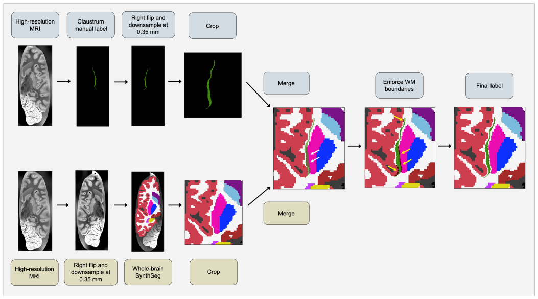
The load-bearing mechanism is label-conditioned synthetic image generation: a 3D U-Net is trained on pairs of heavily augmented label maps and intensity images synthesized from those labels with randomly sampled contrast and resolution, including anisotropic downsampling from 0.35 mm up to 5 mm and added voxel-wise Gaussian noise. Because the training intensities are synthetic, the network learns shape and context rather than scanner-specific intensity statistics, and the same weights can be applied to ex vivo and in vivo images of different contrasts. A contrast-insensitive affine registration into a standard space is used only to locate a cropping field of view around the claustrum; it plays no role in assigning labels.
What would settle it
Manually label a held-out set of in vivo T1-weighted scans at about 1 mm resolution (say 20-30 subjects) and run the released model; if the mean Dice against these manual labels is far below the reported quality-control estimate (~0.57) and below the reported cross-modal agreement (~0.7), then the in vivo accuracy claim fails.
Extended reading notes
Core claim
The central claim is that this is the first accurate automatic method for ultra-high-resolution claustrum segmentation that is robust to changes in contrast and resolution. The method uses a segmentation framework that requires only label maps for training: intensity images are synthesized on the fly with randomized contrast, bias field, noise, smoothing, and downsampling, so the network does not learn a fixed acquisition protocol. The authors manually labeled the claustrum in 18 ultra-high-resolution hemispheres, generated dense whole-field labels by adding surrounding structures, trained a 3D U-Net at 0.35 mm isotropic resolution, and then applied the same model to in vivo T1-weighted, T2-weighted, proton-density, and quantitative T1 scans at standard resolutions. They report that performance degrades gracefully when inputs are downsampled, remains stable in test-retest settings, and does not fail catastrophically across 581 subjects in an independent T1-weighted dataset.
Load-bearing premise
The in vivo performance claim rests on a quality-control score that assumes the nonlinear registration of a test brain into a standard space is accurate enough that looking like one of the 18 training labels after alignment means the segmentation is correct.
Editorial extensions
If this is right
- A single trained model can segment the full dorsal and ventral claustrum in standard ~1 mm T1-weighted in vivo scans, including scans from different field strengths and manufacturers.
- The same model transfers to T2-weighted, proton-density, and quantitative T1 images, so multimodal studies can use one segmentation pipeline without retraining.
- Test-retest Dice of 0.781 supports use in longitudinal and clinical studies where scans are acquired weeks apart.
- Released code and integration into a widely used neuroimaging software package would let other groups segment the claustrum and correct putamen overlabeling errors without building their own method.
Reading between the lines
- If the registration-to-reference-label quality-control strategy is sound, the same approach could screen segmentations of other small, low-contrast structures whose manual labels are scarce, without requiring manual ground truth on every test image.
- The resolution robustness down to about 1.4 mm suggests the method could be applied retrospectively to legacy datasets that lack ultra-high-resolution acquisitions, enabling large-scale claustrum morphometry.
- Because the in vivo evaluation relies on similarity to training labels in a standard space rather than manual labels on the test scans, an independent test with manual in vivo ground truth would clarify how much of the reported generalization comes from the network itself versus the evaluation proxy.
Editorial analysis
A structured set of objections, weighed in public.
Referee Report
Summary. The paper presents a SynthSeg-based 3D U-Net for claustrum segmentation, trained on manual labels from 18 ultra-high-resolution hemispheres (mostly ex vivo) and evaluated with 6-fold cross-validation (average Dice 0.632), a downsampling simulation, and in vivo experiments on the IXI, Miriad, and FSM datasets. The authors claim this is the first accurate, contrast- and resolution-agnostic method for ultra-high-resolution claustrum segmentation, and they release the method on GitHub and in FreeSurfer.
Significance. If the central claims held, this would be a practically valuable tool for claustrum research, especially because existing automatic methods are limited and the method is released with FreeSurfer integration and public in vivo evaluations. The paper also deserves credit for using a contrast-synthesis training strategy, for testing on multiple independent datasets, and for reporting test-retest and cross-modality consistency. However, the evidence for the 'accurate' part of the claim is incomplete: the CV Dice is below the reported inter-rater Dice, the in vivo accuracy argument depends on an unvalidated QC proxy, the resolution-robustness experiment stays within the training augmentation range, and the IXI volume inflation is unexplained. The study is therefore a solid engineering contribution but needs additional validation before the abstract-level claim is justified.
major comments (4)
- [Sec. 3.4 and Sec. 5 (IXI experiments and epoch selection)] The in vivo accuracy claim rests on the QC score defined in Sec. 3.4, yet the paper itself states that QC 'should not be interpreted as a direct measure of segmentation accuracy' and that even a perfect segmentation will not reach QC = 1. Because the QC score is the maximum Dice against the same 18 manual labels used for training, after nonlinear registration to MNI152, it can reward segmentations that fall at the population-typical claustrum location rather than at the individual's true claustrum, and no experiment in the paper shows that QC correlates with native-space Dice. Since the final epoch is selected on a 20-subject IXI subset using this score and the IXI 'never critically failed' conclusion is based on QC plus visual inspection of extremes, the load-bearing 'accurate in vivo' part of the abstract claim is not yet supported. Please validate QC against native-space Dice on the 18 CV cases (or another labeled set) and report the relationship, or explicitly downgrade the in vivo claim to robustness/plausibility.
- [Sec. 3.3 and Fig. 9] The resolution-robustness experiment downsamples the 18 high-resolution hemispheres to 0.4-3.5 mm and reports graceful Dice degradation, but the training procedure already simulates downsampling with resolutions sampled from U(0.35,3.5) isotropically and U(0.35,5) anisotropically. The tested range is therefore almost entirely inside the training augmentation envelope, and the simulated images inherit the high SNR and ex vivo contrast of the source scans rather than the noise and partial-volume properties of native ~1 mm in vivo acquisitions. This experiment supports robustness within the training distribution but does not by itself establish resolution-agnostic accuracy at typical in vivo resolutions; please either test on native-resolution in vivo images with manual labels or restrict the claim accordingly.
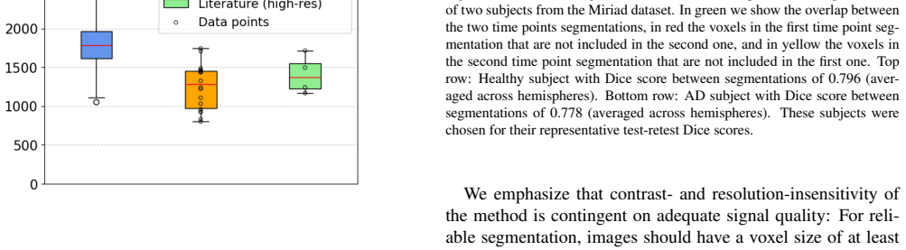
- [Sec. 5 (IXI volumes)] The IXI automatic claustrum volumes average 1,793.92 ± 259.16 mm3 versus 1,253.05 ± 283.79 mm3 for the manual labels, a ~43% inflation that the paper leaves unexplained ('It is not clear why the IXI volumes are so much higher'). This unexplained systematic bias undermines the use of the method for quantitative in vivo volumetry and should be addressed (e.g., by validating against manual labels on native-resolution scans or by modeling partial-volume effects) or explicitly listed as a limitation of quantitative accuracy.
- [Sec. 5 and Table 2] The primary ultra-high-resolution accuracy evidence is CV Dice 0.632 ± 0.061, which is substantially below the inter-rater Dice 0.805 ± 0.018 reported for the seven shared samples. The discussion acknowledges this gap but does not establish that 0.632 constitutes 'accurate' segmentation rather than moderate agreement; since the abstract's first claim is accuracy at ultra-high resolution, please provide a more direct argument (e.g., error analysis, comparison with a baseline on the same labels, or a stated acceptability threshold) or soften the claim.
minor comments (6)
- [Abstract and Section 1] The URL 'https://github.com/chiara-mauri/claustrum segmentation' contains a space; please provide the correct link.
- [Fig. 3 caption] The caption says 'ev vivo' and should read 'ex vivo'.
- [Sec. 5] The text refers to 'Vichow-Robins spaces' and should read 'Virchow-Robin spaces'.
- [Appendix A] The inline equations for Dice, IoU, TPR, FDR, and volumetric similarity are garbled by line wrapping; please typeset them as display equations.
- [Table 1] The '?' entries for sample 15 are not explained; please add a footnote stating that postmortem interval and brain weight were unavailable.
- [Throughout] The notation FoV/FOV is used inconsistently (e.g., Sec. 3.2 'FoV' versus Fig. 4 'FOV'); please unify.
Circularity Check
Partial circularity: the in vivo IXI evidence uses a QC score defined against the training labels, and the final epoch is selected on IXI and then reported on the same IXI dataset; CV and cross-modal results remain independent.
-
fitted input called prediction
[Sec. 3.4 (epoch selection) and Sec. 5 (IXI QC results)]
"We based the epoch selection on T1-weighted in vivo images, in particular on a set of 20 subjects from the IXI dataset. On this validation set, we ran the automatic segmentation for each epoch and used the nonlinear registration to MNI152 to compute the QC metric as discussed above. We then chose the epoch that had the highest mean QC score on the validation subjects. ... We obtained an average QC score of 0.570 ± 0.060 for the IXI dataset."
The final model's epoch is selected by maximizing the mean QC on 20 IXI subjects, and the reported IXI QC statistics are computed on the IXI dataset including those same validation subjects. Thus the 'IXI performance' is not an independent prediction: the same statistic used for model selection is later reported as evidence of robustness. The inflation is limited because only one of 100 epochs is chosen, but the evaluation is not held-out.
-
other
[Sec. 3.4 (QC definition) and Sec. 5 (IXI conclusions)]
"We then compute the Dice score between the registered segmentation and each of the 18 manual labels in MNI space. The maximum Dice score across these comparisons is used as our quality control (QC) measure. ... A high QC score indicates that the segmentation closely resembles at least one manual label after alignment to MNI space, and is thus likely to be accurate."
The 18 manual labels used as the QC reference are exactly the labels used to train the network ('These data were used to obtain claustrum manual labels and subsequently train a SynthSeg segmentation method'). Therefore a high QC score mainly shows that the automatic output resembles the training set after registration; it is not independent evidence of correct native-space segmentation. The paper itself warns that QC 'should not be interpreted as a direct measure of segmentation accuracy', yet it uses QC to conclude that the method 'did not severely fail on any subject' in IXI. This makes the in vivo accuracy claim partly circular, although test-retest and cross-modality Dice provide independent support.
full rationale
The derivation of the segmentation itself is not circular: the CV Dice (0.632) is measured on held-out folds of the 18 labeled hemispheres, and the final network is a standard SynthSeg U-Net trained on label maps with on-the-fly synthetic intensities, not on the QC score. The circularity concerns are confined to the in vivo evaluation. First, the IXI QC evidence is not independent: the QC metric is defined as the maximum Dice against the same 18 manual labels used for training, so a high score can reflect training-set resemblance rather than native-space accuracy; the paper itself warns QC is not a direct accuracy measure. Second, the final epoch is selected by maximizing mean QC on 20 IXI subjects, and the reported IXI QC average includes those subjects, so part of the reported IXI performance is a selection artifact. The resolution sweep from 0.4 to 3.5 mm also lies inside the SynthSeg training augmentation range U(0.35,3.5)/U(0.35,5), so it is an in-distribution check rather than an extrapolation test. However, the test-retest Dice (0.781), cross-modality Dice (0.696-0.809), comparison to Casamitjana et al., and the CV metrics provide independent evidence not forced by the QC construction. These independent components keep the circularity partial, not total.
Assumptions & free parameters
free parameters (5)
- Target label resolution =
0.35 mm isotropic
- Random voxel noise sigma range =
U(0, 100)
- Probability of adding noise =
0.95
- Atlas threshold for FoV cropping =
0.001
- Crop FoV size =
56 mm (training), 60 mm (testing)
assumptions (6)
- domain assumption Manual labels from two raters are accurate ground truth for claustrum boundaries
- domain assumption SmartInterpol produces accurate labels for slices between manually traced sections
- domain assumption Whole-brain SynthSeg segmentations provide sufficiently accurate labels for surrounding structures
- domain assumption SynthMorph registration to MNI152 is accurate for high-resolution ex vivo hemispheres and for in vivo scans
- domain assumption QC score (max Dice against the 18 training labels in MNI space) is a valid proxy for segmentation quality on unseen data
- domain assumption Synthetic intensity images with random contrast/resolution capture the variability of real MRI
Cite this review
Pith. "Pith review of A Contrast-Agnostic Method for Ultra-High Resolution Claustrum Segmentation." pith.science (2026). https://pith.science/paper/GBC3YJAQ
@misc{pith2026241115388,
  author       = {Pith},
  title        = {Pith review of: A Contrast-Agnostic Method for Ultra-High Resolution Claustrum Segmentation},
  year         = {2026},
  howpublished = {\url{https://pith.science/paper/GBC3YJAQ}},
  note         = {Machine review of arXiv:2411.15388}
}
read the original abstract
The claustrum is a band-like gray matter structure located between putamen and insula whose exact functions are still actively researched. Its sheet-like structure makes it barely visible in in vivo Magnetic Resonance Imaging (MRI) scans at typical resolutions and neuroimaging tools for its study, including methods for automatic segmentation, are currently very limited. In this paper, we propose a contrast- and resolution-agnostic method for claustrum segmentation at ultra-high resolution (0.35 mm isotropic); the method is based on the SynthSeg segmentation framework (Billot et al., 2023), which leverages the use of synthetic training intensity images to achieve excellent generalization. In particular, SynthSeg requires only label maps to be trained, since corresponding intensity images are synthesized on the fly with random contrast and resolution. We trained a deep learning network for automatic claustrum segmentation, using claustrum manual labels obtained from 18 ultra-high resolution MRI scans (mostly ex vivo). We demonstrated the method to work on these 18 high resolution cases (Dice score = 0.632, mean surface distance = 0.458 mm, and volumetric similarity = 0.867 using 6-fold Cross Validation (CV)), and also on in vivo T1-weighted MRI scans at typical resolutions (~1 mm isotropic). We also demonstrated that the method is robust in a test-retest setting and when applied to multimodal imaging (T2-weighted, Proton Density and quantitative T1 scans). To the best of our knowledge this is the first accurate method for automatic ultra-high resolution claustrum segmentation, which is robust against changes in contrast and resolution. The method is released at https://github.com/chiara-mauri/claustrum_segmentation and as part of the neuroimaging package Freesurfer (Fischl, 2012).
Figures


Figures from the paper (13 more)




Reference graph
Works this paper leans on
-
[1]
A., Shah, S
Albishri, A. A., Shah, S. J. H., Kang, S. S., and Lee, Y. (2022). AM - UN et: automated mini 3D end-to-end U -net based network for brain claustrum segmentation. Multimedia Tools and Applications , 81(25):36171--36194
2022
-
[2]
Arrigo, A., Calamuneri, A., Milardi, D., Mormina, E., Gaeta, M., Corallo, F., Lo Buono, V., Chillemi, G., Marino, S., Cacciola, A., et al. (2019). Claustral structural connectivity and cognitive impairment in drug na \" ve P arkinson’s disease. Brain Imaging and Behavior , 13:933--944
2019
-
[3]
K., McGrath, T
Atilgan, H., Doody, M., Oliver, D. K., McGrath, T. M., Shelton, A. M., Echeverria-Altuna, I., Tracey, I., Vyazovskiy, V. V., Manohar, S. G., and Packer, A. M. (2022). Human lesions and animal studies link the claustrum to perception, salience, sleep and pain. Brain , 145(5):1610--1623
2022
-
[4]
J., Pozner, G., Tasaka, G.-i., Goll, Y., Refaeli, R., Zviran, O., et al
Atlan, G., Terem, A., Peretz-Rivlin, N., Sehrawat, K., Gonzales, B. J., Pozner, G., Tasaka, G.-i., Goll, Y., Refaeli, R., Zviran, O., et al. (2018). The claustrum supports resilience to distraction. Current Biology , 28(17):2752--2762
2018
-
[5]
Atzeni, A., Jansen, M., Ourselin, S., and Iglesias, J. E. (2018). A probabilistic model combining deep learning and multi-atlas segmentation for semi-automated labelling of histology. In Medical Image Computing and Computer Assisted Intervention--MICCAI 2018: 21st International Conference, Granada, Spain, September 16-20, 2018, Proceedings, Part II 11 , p...
work page 2018
-
[6]
Baizer, J. S., Sherwood, C. C., Noonan, M., and Hof, P. R. (2014). Comparative organization of the claustrum: what does structure tell us about function? Frontiers in systems neuroscience , 8:117
work page 2014
-
[7]
Banati, R. B., Goerres, G. W., Tjoa, C., Aggleton, J. P., and Grasby, P. (2000). The functional anatomy of visual-tactile integration in man: a study using positron emission tomography. Neuropsychologia , 38(2):115--124
work page 2000
-
[8]
Benarroch, E. E. (2021). What is the role of the claustrum in cortical function and neurologic disease? Neurology , 96(3):110--113
work page 2021
Show all 84 references
-
[9]
Berman, S., Schurr, R., Atlan, G., Citri, A., and Mezer, A. A. (2020). Automatic segmentation of the dorsal claustrum in humans using in vivo high-resolution MRI . Cerebral Cortex Communications , 1(1):tgaa062
2020
-
[10]
Bernstein, H.-G., Ortmann, A., Dobrowolny, H., Steiner, J., Brisch, R., Gos, T., and Bogerts, B. (2016). Bilaterally reduced claustral volumes in schizophrenia and major depressive disorder: a morphometric postmortem study. European archives of psychiatry and clinical neurosci...
2016
-
[11]
N., Puonti, O., Thielscher, A., Van Leemput, K., Fischl, B., Dalca, A
Billot, B., Greve, D. N., Puonti, O., Thielscher, A., Van Leemput, K., Fischl, B., Dalca, A. V., Iglesias, J. E., et al. (2023a). Synth S eg: S egmentation of brain MRI scans of any contrast and resolution without retraining. Medical image analysis , 86:102789
2023
-
[12]
E., Das, S., and Iglesias, J
Billot, B., Magdamo, C., Cheng, Y., Arnold, S. E., Das, S., and Iglesias, J. E. (2023b). Robust machine learning segmentation for large-scale analysis of heterogeneous clinical brain MRI datasets. Proceedings of the National Academy of Sciences , 120(9):e2216399120
2023
-
[13]
P., Mathur, B
Brown, S. P., Mathur, B. N., Olsen, S. R., Luppi, P.-H., Bickford, M. E., and Citri, A. (2017). New breakthroughs in understanding the role of functional interactions between the neocortex and the claustrum. Journal of Neuroscience , 37(45):10877--10881
2017
-
[14]
D., McGeown, W
Bruen, P. D., McGeown, W. J., Shanks, M. F., and Venneri, A. (2008). Neuroanatomical correlates of neuropsychiatric symptoms in A lzheimer's disease. Brain , 131(9):2455--2463
2008
-
[15]
M., Lehmann, P., de Rochefort, L., Besson, P., Massire, A., Ridley, B., Girard, N., Guye, M., et al
Brun, G., Testud, B., Girard, O. M., Lehmann, P., de Rochefort, L., Besson, P., Massire, A., Ridley, B., Girard, N., Guye, M., et al. (2022). Automatic segmentation of deep grey nuclei using a high-resolution 7T magnetic resonance imaging atlas— Q uantification of T1 values in...
2022
-
[16]
Calarco, N., Kashyap, S., and Uludağ, K. (2023). Establishing an MRI reference for the human claustrum. Poster presented at OHBM 2023, Montreal
2023
-
[17]
Calarco, N., Kedo, O., Bludau, S., Herold, C., Uludag, K., and Amunts, K. (2024). Cytoarchitectonic mapping and probabilistic atlas of the human claustrum. Poster presented at OHBM 2024, Seoul
2024
-
[18]
Casamitjana, A., Mancini, M., Robinson, E., Peter, L., Annunziata, R., Althonayan, J., Crampsie, S., Blackburn, E., Billot, B., Atzeni, A., et al. (2024). A next-generation, histological atlas of the human brain and its application to automated brain MRI segmentation. bioRxiv ...
2024
-
[19]
G., Gerner, G
Cascella, N. G., Gerner, G. J., Fieldstone, S. C., Sawa, A., and Schretlen, D. J. (2011). The insula--claustrum region and delusions in schizophrenia. Schizophrenia Research , 133(1-3):77--81
2011
-
[20]
Cascella, N. G. and Sawa, A. (2014). The claustrum in schizophrenia. In The claustrum , pages 237--243. Elsevier
2014
-
[21]
M., Krueger, F., Cristofori, I., and Grafman, J
Chau, A., Salazar, A. M., Krueger, F., Cristofori, I., and Grafman, J. (2015). The effect of claustrum lesions on human consciousness and recovery of function. Consciousness and Cognition , 36:256--264
2015
-
[22]
Coates, A., Linhardt, D., Windischberger, C., Ischebeck, A., and Zaretskaya, N. (2023). High-resolution 7T f MRI reveals the visual sensory zone of the human claustrum. bioRxiv , pages 2023--09
2023
-
[23]
and Zaretskaya, N
Coates, A. and Zaretskaya, N. (2024). High-resolution dataset of manual claustrum segmentation. Data in Brief , 54:110253
2024
-
[24]
M., et al
Costantini, I., Morgan, L., Yang, J., Balbastre, Y., Varadarajan, D., Pesce, L., Scardigli, M., Mazzamuto, G., Gavryusev, V., Castelli, F. M., et al. (2023). A cellular resolution atlas of B roca’s area. Science Advances , 9(41):eadg3844
2023
-
[25]
Crick, F. C. and Koch, C. (2005). What is the function of the claustrum? Philosophical Transactions of the Royal Society B: Biological Sciences , 360(1458):1271--1279
2005
-
[26]
Davis, W. (2008). The claustrum in autism and typically developing male children: a quantitative MRI study . Brigham Young University
2008
-
[27]
C., Guttmann, C
Dewey, J., Hana, G., Russell, T., Price, J., McCaffrey, D., Harezlak, J., Sem, E., Anyanwu, J. C., Guttmann, C. R., Navia, B., et al. (2010). Reliability and validity of MRI -based automated volumetry software relative to auto-assisted manual measurement of subcortical structu...
2010
-
[28]
M., Jankowski, M
Dillingham, C. M., Jankowski, M. M., Chandra, R., Frost, B. E., and O’Mara, S. M. (2017). The claustrum: C onsiderations regarding its anatomy, functions and a programme for research. Brain and Neuroscience Advances , 1:2398212817718962
2017
-
[29]
J., Sunkin, S
Ding, S.-L., Royall, J. J., Sunkin, S. M., Ng, L., Facer, B. A., Lesnar, P., Guillozet-Bongaarts, A., McMurray, B., Szafer, A., Dolbeare, T. A., et al. (2016). Comprehensive cellular-resolution atlas of the adult human brain. Journal of comparative neurology , 524(16):3127--3481
2016
-
[30]
L., Mareyam, A., Horn, A., Polimeni, J
Edlow, B. L., Mareyam, A., Horn, A., Polimeni, J. R., Witzel, T., Tisdall, M. D., Augustinack, J. C., Stockmann, J. P., Diamond, B. R., Stevens, A., et al. (2019). 7 T esla MRI of the ex vivo human brain at 100 micron resolution. Scientific data , 6(1):244
2019
-
[31]
M., Collins, D
Ewert, S., Plettig, P., Li, N., Chakravarty, M. M., Collins, D. L., Herrington, T. M., K \"u hn, A. A., and Horn, A. (2018). Toward defining deep brain stimulation targets in MNI space: a subcortical atlas based on multimodal mri, histology and structural connectivity. Neuroim...
2018
-
[32]
Fischl, B. (2012). Free S urfer. Neuroimage , 62(2):774--781
2012
-
[33]
Goll, Y., Atlan, G., and Citri, A. (2015). Attention: the claustrum. Trends in neurosciences , 38(8):486--495
2015
-
[34]
Greve, D. N. and Fischl, B. (2024). The F ree S urfer M aintenance D ataset. OpenNeuro, doi:10.18112/openneuro.ds004958.v1.0.0
2024 doi
-
[35]
and Roland, P
Hadjikhani, N. and Roland, P. E. (1998). Cross-modal transfer of information between the tactile and the visual representations in the human brain: a positron emission tomographic study. Journal of Neuroscience , 18(3):1072--1084
1998
-
[36]
N., Iglesias, J
Hoffmann, M., Billot, B., Greve, D. N., Iglesias, J. E., Fischl, B., and Dalca, A. V. (2021). Synth M orph: learning contrast-invariant registration without acquired images. IEEE transactions on medical imaging , 41(3):543--558
2021
-
[37]
S., Dalca, A
Hoopes, A., Mora, J. S., Dalca, A. V., Fischl, B., and Hoffmann, M. (2022). Synth S trip: skull-stripping for any brain image. NeuroImage , 260:119474
2022
-
[38]
P., Klanderman, G
Huttenlocher, D. P., Klanderman, G. A., and Rucklidge, W. J. (1993). Comparing images using the H ausdorff distance. IEEE Transactions on pattern analysis and machine intelligence , 15(9):850--863
1993
-
[39]
E., Billot, B., Balbastre, Y., Magdamo, C., Arnold, S
Iglesias, J. E., Billot, B., Balbastre, Y., Magdamo, C., Arnold, S. E., Das, S., Edlow, B. L., Alexander, D. C., Golland, P., and Fischl, B. (2023). Synth SR : A public AI tool to turn heterogeneous clinical brain scans into high-resolution T1 -weighted images for 3D morphomet...
2023
-
[40]
E., Billot, B., Balbastre, Y., Tabari, A., Conklin, J., Gonz \'a lez, R
Iglesias, J. E., Billot, B., Balbastre, Y., Tabari, A., Conklin, J., Gonz \'a lez, R. G., Alexander, D. C., Golland, P., Edlow, B. L., Fischl, B., et al. (2021). Joint super-resolution and synthesis of 1 mm isotropic MP-RAGE volumes from clinical MRI exams with scans of differ...
2021
-
[41]
Kalaitzakis, M., Pearce, R., and Gentleman, S. (2009). Clinical correlates of pathology in the claustrum in P arkinson's disease and dementia with L ewy bodies. Neuroscience letters , 461(1):12--15
2009
-
[42]
S., Bodenheimer, J., and Butler, T
Kang, S. S., Bodenheimer, J., and Butler, T. (2020). A comprehensive protocol for manual segmentation of the human claustrum and its sub-regions using high-resolution MRI . arXiv preprint arXiv:2010.06423
2020 arXiv
-
[43]
Kapakin, S. (2011). The claustrum: three-dimensional reconstruction, photorealistic imaging, and stereotactic approach. Folia morphologica , 70(4):228--234
2011
-
[44]
R., Roy, S., Kovacs, B., Ulrich, C., Wald, T., Zenk, M., Vollmuth, P., Kleesiek, J., Isensee, F., et al
Kirchhoff, Y., Rokuss, M. R., Roy, S., Kovacs, B., Ulrich, C., Wald, T., Zenk, M., Vollmuth, P., Kleesiek, J., Isensee, F., et al. (2024). Skeleton recall loss for connectivity conserving and resource efficient segmentation of thin tubular structures. arXiv preprint arXiv:2404.03010
2024 arXiv
-
[45]
Z., Bartolomei, F., Beltagy, A., and Picard, F
Koubeissi, M. Z., Bartolomei, F., Beltagy, A., and Picard, F. (2014). Electrical stimulation of a small brain area reversibly disrupts consciousness. Epilepsy & Behavior , 37:32--35
2014
-
[46]
R., White, M
Krimmel, S. R., White, M. G., Panicker, M. H., Barrett, F. S., Mathur, B. N., and Seminowicz, D. A. (2019). Resting state functional connectivity and cognitive task-related activation of the human claustrum. Neuroimage , 196:59--67
2019
-
[47]
J., Shit, S., Sorg, C., Menze, B., and Hedderich, D
Li, H., Menegaux, A., Schmitz-Koep, B., Neubauer, A., B \"a uerlein, F. J., Shit, S., Sorg, C., Menze, B., and Hedderich, D. (2021). Automated claustrum segmentation in human brain MRI using deep learning. Human Brain Mapping , 42(18):5862--5872
2021
-
[48]
B., Tolsgaard, M
Lin, M., Weng, N., Mikolaj, K., Bashir, Z., Svendsen, M. B., Tolsgaard, M. G., Christensen, A. N., and Feragen, A. (2024). Shortcut learning in medical image segmentation. In International Conference on Medical Image Computing and Computer-Assisted Intervention , pages 623--63...
2024
-
[49]
L \"u sebrink, F., Sciarra, A., Mattern, H., Yakupov, R., and Speck, O. (2017). T1-weighted in vivo human whole brain MRI dataset with an ultrahigh isotropic resolution of 250 m. Scientific data , 4(1):1--12
2017
-
[50]
B., Stewart, B
Madden, M. B., Stewart, B. W., White, M. G., Krimmel, S. R., Qadir, H., Barrett, F. S., Seminowicz, D. A., and Mathur, B. N. (2022). A role for the claustrum in cognitive control. Trends in cognitive sciences , 26(12):1133--1152
2022
-
[51]
K., Majtanik, M., and Paxinos, G
Mai, J. K., Majtanik, M., and Paxinos, G. (2015). Atlas of the human brain . Academic Press
2015
-
[52]
B., Cash, D., Ridgway, G
Malone, I. B., Cash, D., Ridgway, G. R., MacManus, D. G., Ourselin, S., Fox, N. C., and Schott, J. M. (2013). MIRIAD — P ublic release of a multiple time point A lzheimer's MR imaging dataset. NeuroImage , 70:33--36
2013
-
[53]
P., Kober, T., Krueger, G., van der Zwaag, W., Van de Moortele, P.-F., and Gruetter, R
Marques, J. P., Kober, T., Krueger, G., van der Zwaag, W., Van de Moortele, P.-F., and Gruetter, R. (2010). MP2RAGE , a self bias-field corrected sequence for improved segmentation and T1 -mapping at high field. Neuroimage , 49(2):1271--1281
2010
-
[54]
Mathur, B. N. (2014). The claustrum in review. Frontiers in systems neuroscience , 8:48
2014
-
[55]
Meletti, S., Slonkova, J., Mareckova, I., Monti, G., Specchio, N., Hon, P., Giovannini, G., Marcian, V., Chiari, A., Krupa, P., et al. (2015). Claustrum damage and refractory status epilepticus following febrile illness. Neurology , 85(14):1224--1232
2015
-
[56]
Milardi, D., Bramanti, P., Milazzo, C., Finocchio, G., Arrigo, A., Santoro, G., Trimarchi, F., Quartarone, A., Anastasi, G., and Gaeta, M. (2015). Cortical and subcortical connections of the human claustrum revealed in vivo by constrained spherical deconvolution tractography. ...
2015
-
[57]
R., Eriksson, J., Larsson, A., and Nyberg, L
Naghavi, H. R., Eriksson, J., Larsson, A., and Nyberg, L. (2007). The claustrum/insula region integrates conceptually related sounds and pictures. Neuroscience letters , 422(1):77--80
2007
-
[58]
B., Wendt, J., Schmitz-Koep, B., Menegaux, A., Schinz, D., Menze, B., Zimmer, C., Sorg, C., and Hedderich, D
Neubauer, A., Li, H. B., Wendt, J., Schmitz-Koep, B., Menegaux, A., Schinz, D., Menze, B., Zimmer, C., Sorg, C., and Hedderich, D. M. (2022). Efficient claustrum segmentation in T2 -weighted neonatal brain mri using transfer learning from adult scans. Clinical Neuroradiology ,...
2022
-
[59]
N., Rizaeva, N
Nikolenko, V. N., Rizaeva, N. A., Beeraka, N. M., Oganesyan, M. V., Kudryashova, V. A., Dubovets, A. A., Borminskaya, I. D., Bulygin, K. V., Sinelnikov, M. Y., and Aliev, G. (2021). The mystery of claustral neural circuits and recent updates on its role in neurodegenerative pa...
2021
-
[60]
Patru, M. C. and Reser, D. H. (2015). A new perspective on delusional states--evidence for C laustrum involvement. Frontiers in psychiatry , 6:158
2015
-
[61]
A., Bogner, P., Doczi, T., Janszky, J., and Orsi, G
Perlaki, G., Horvath, R., Nagy, S. A., Bogner, P., Doczi, T., Janszky, J., and Orsi, G. (2017). Comparison of accuracy between FSL ’s FIRST and F reesurfer for caudate nucleus and putamen segmentation. Scientific reports , 7(1):2418
2017
-
[62]
G., and Pujol, J.-F
Redout \'e , J., Stol \'e ru, S., Gr \'e goire, M.-C., Costes, N., Cinotti, L., Lavenne, F., Le Bars, D., Forest, M. G., and Pujol, J.-F. (2000). Brain processing of visual sexual stimuli in human males. Human brain mapping , 11(3):162--177
2000
-
[63]
K., and Kayser, C
Remedios, R., Logothetis, N. K., and Kayser, C. (2010). Unimodal responses prevail within the multisensory claustrum. Journal of Neuroscience , 30(39):12902--12907
2010
-
[64]
K., and Kayser, C
Remedios, R., Logothetis, N. K., and Kayser, C. (2014). A role of the claustrum in auditory scene analysis by reflecting sensory change. Frontiers in systems neuroscience , 8:44
2014
-
[65]
J., Rosas, H
Reuter, M., Schmansky, N. J., Rosas, H. D., and Fischl, B. (2012). Within-subject template estimation for unbiased longitudinal image analysis. Neuroimage , 61(4):1402--1418
2012
-
[66]
Rodr \' guez-Vidal, L., Alcauter, S., and Barrios, F. A. (2024). The functional connectivity of the human claustrum, according to the H uman C onnectome P roject database. Plos one , 19(4):e0298349
2024
-
[67]
Ronneberger, O., Fischer, P., and Brox, T. (2015). U-net: C onvolutional networks for biomedical image segmentation. In Medical image computing and computer-assisted intervention--MICCAI 2015: 18th international conference, Munich, Germany, October 5-9, 2015, proceedings, part...
2015
-
[68]
Sener, R. (1993). The claustrum on MRI : normal anatomy, and the bright claustrum as a new sign in W ilson's disease. Pediatric radiology , 23:594--596
1993
-
[69]
Sener, R. N. (1998). Lesions affecting the claustrum. Computerized medical imaging and graphics , 22(1):57--61
1998
-
[70]
Silva, G., Jacob, S., Melo, C., Alves, D., and Costa, D. (2018). Claustrum sign in a child with refractory status epilepticus after febrile illness: why does it happen? Acta Neurologica Belgica , 118:303--305
2018
-
[71]
H., Pifl, C., Rajput, A
Sitte, H. H., Pifl, C., Rajput, A. H., H \"o rtnagl, H., Tong, J., Lloyd, G. K., Kish, S. J., and Hornykiewicz, O. (2017). Dopamine and noradrenaline, but not serotonin, in the human claustrum are greatly reduced in patients with P arkinson's disease: possible functional impli...
2017
-
[72]
B., Lee, A
Smith, J. B., Lee, A. K., and Jackson, J. (2020). The claustrum. Current Biology , 30(23):R1401--R1406
2020
-
[73]
B., Watson, G
Smith, J. B., Watson, G. D., Liang, Z., Liu, Y., Zhang, N., and Alloway, K. D. (2019). A role for the claustrum in salience processing? Frontiers in neuroanatomy , 13:64
2019
-
[74]
Smythies, J., Edelstein, L., and Ramachandran, V. (2012). Hypotheses relating to the function of the claustrum. Frontiers in integrative neuroscience , 6:53
2012
-
[75]
J., Peretz-Rivlin, N., Ashwal-Fluss, R., Bleistein, N., del Mar Reus-Garcia, M., Mukherjee, D., Groysman, M., and Citri, A
Terem, A., Gonzales, B. J., Peretz-Rivlin, N., Ashwal-Fluss, R., Bleistein, N., del Mar Reus-Garcia, M., Mukherjee, D., Groysman, M., and Citri, A. (2020). Claustral neurons projecting to frontal cortex mediate contextual association of reward. Current Biology , 30(18):3522--3532
2020
-
[76]
Tian, F., Tu, S., Qiu, J., Lv, J., Wei, D., Su, Y., and Zhang, Q. (2011). Neural correlates of mental preparation for successful insight problem solving. Behavioural brain research , 216(2):626--630
2011
-
[77]
M., Irimia, A., Goh, S
Torgerson, C. M., Irimia, A., Goh, S. M., and Van Horn, J. D. (2015). The DTI connectivity of the human claustrum. Human brain mapping , 36(3):827--838
2015
-
[78]
and Shanks, M
Venneri, A. and Shanks, M. (2014). The claustrum and A lzheimer’s disease. The claustrum , pages 263--275
2014
-
[79]
G., Schooler, L
Volz, K. G., Schooler, L. J., and von Cramon, D. Y. (2010). It just felt right: T he neural correlates of the fluency heuristic. Consciousness and Cognition , 19(3):829--837
2010
-
[80]
Wada, J. A. and Kudo, T. (1997). Involvement of the claustrum in the convulsive evolution of temporal limbic seizure in feline amygdaloid kindling. Electroencephalography and clinical neurophysiology , 103(2):249--256
1997
-
[81]
Y., Imaki, H., Wegiel, J., Frackowiak, J., Kolecka, B
Wegiel, J., Flory, M., Kuchna, I., Nowicki, K., Ma, S. Y., Imaki, H., Wegiel, J., Frackowiak, J., Kolecka, B. M., Wierzba-Bobrowicz, T., et al. (2015). Neuronal nucleus and cytoplasm volume deficit in children with autism and volume increase in adolescents and adults. Acta neu...
2015
-
[82]
G., Panicker, M., Mu, C., Carter, A
White, M. G., Panicker, M., Mu, C., Carter, A. M., Roberts, B. M., Dharmasri, P. A., and Mathur, B. N. (2018). Anterior cingulate cortex input to the claustrum is required for top-down action control. Cell reports , 22(1):84--95
2018
-
[83]
Yamamoto, R., Iseki, E., Murayama, N., Minegishi, M., Marui, W., Togo, T., Katsuse, O., Kosaka, K., Kato, M., Iwatsubo, T., et al. (2007). Correlation in lewy pathology between the claustrum and visual areas in brains of dementia with L ewy bodies. Neuroscience letters , 415(3...
2007
-
[84]
K., Saucier, D
Zhang, X., Hannesson, D. K., Saucier, D. M., Wallace, A. E., Howland, J., and Corcoran, M. E. (2001). Susceptibility to kindling and neuronal connections of the anterior claustrum. Journal of Neuroscience , 21(10):3674--3687
2001
Reviewed August 12, 2026 · model on record in the stance chip above.
Discussion (0). Continue with ORCID to comment.