REVIEW 3 major objections 5 minor 1 cited by
Benchmarking analytical electron ptychography methods for the low-dose imaging of beam-sensitive materials
T0 review · 3 major / 5 minor · reviewed 2026-08-10 · deepseek-v4-flash
Pith's one-line read The paper claims that the dose required for a fixed precision in analytical electron ptychography scales with the reconstructed frequency-space area, proportional to $q_A^2$, making sparse few-electron diffraction patterns viable for…
desk verdict A solid, honest benchmark of analytical ptychography with a modest new algorithm; the main caveat is that real-detector effects are deliberately left out. read the letter →
The pith
A machine-rendered reading of the paper's core claim, the machinery that carries it, and where it could break.
The reading
What carries the argument
The argument runs through the Wigner distribution formalism of analytical ptychography, in which the Fourier transform of the recorded diffraction patterns over scan positions is expressed as a product of a probe Wigner distribution, the specimen Wigner distribution, and the detector modulation transfer function. WDD performs a Wiener-filter deconvolution of this product to recover the transmission function; SBI-D and SBI-S use the weak-phase linearization to sum or deconvolve the two sidebands in the same distribution; iCoM computes the scan-position-wise center of mass and integrates it in Fourier space. Two contrast transfer functions carry the frequency weighting: the double-overlap phase contrast transfer function and the probe-autocorrelation optical transfer function. The new implementation element is the scan-frequency partitioning algorithm (SFPA), which replaces the full scan-space fast Fourier transform with an explicit summation over packets of scan positions and domains of reconstruction frequencies, lowering memory use, enabling parallelization, and decoupling the scan grid from the reconstruction grid. The dose law is carried by the Cramér–Rao lower bound formula and verified by Fourier ring correlations.
What would settle it
Measure the dose required to reach a fixed Fourier ring correlation threshold at a fixed spatial frequency in an experimental 4D-STEM dataset of a beam-sensitive specimen at two semi-convergence angles with matched overlap; the ratio of the required doses should be close to the ratio of the squared apertures, and a clear departure would falsify the central dose law.
Extended reading notes
Core claim
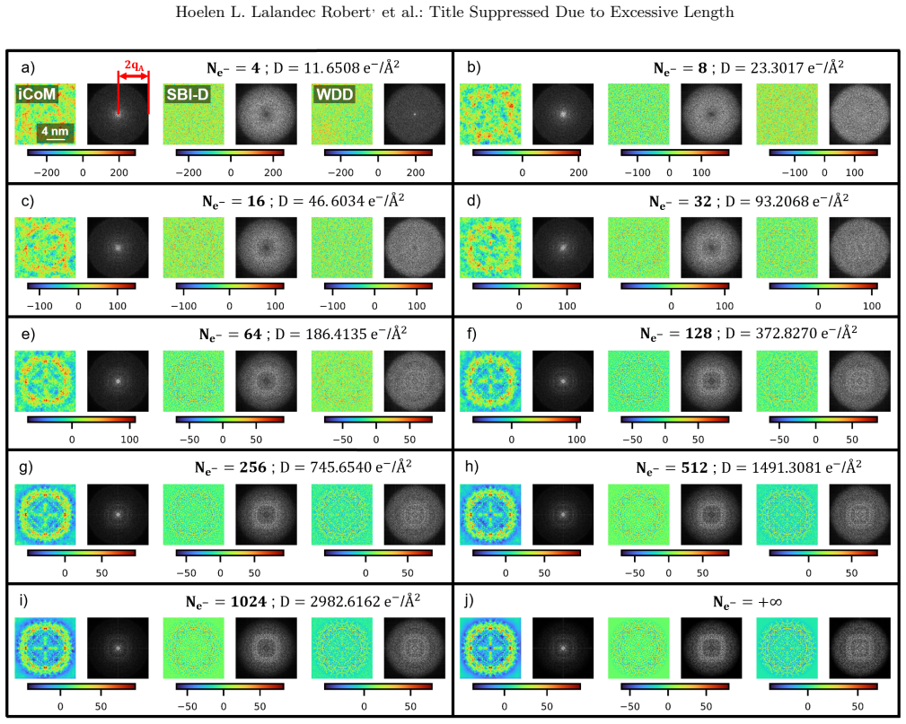
The central discovery is a quantitative dose law: the Cramér–Rao lower bound for real-space phase precision is $\mathrm{CRLB}_{\mathrm{RS}} = \sqrt{N_{\vec Q}/(2N_s N_{e^-})} \ge \sqrt{(2\pi q_A^2 - 1/(2S))/D}$, so for a target precision the dose $D$ must grow with the reconstructed frequency-space surface, which is a disk of radius $2q_A$ and therefore scales as $q_A^2$. Empirically, Fourier ring correlations comparing infinite-dose reconstructions with dose-limited ones show that WDD, SBI-D and iCoM have largely similar per-frequency dose efficiency on both model objects, and that specimen-rich frequencies are recovered much more efficiently than empty frequencies. Sparse diffraction patterns with very few counts still reconstruct, so count sparsity itself is not a limitation. The paper also finds that the weak-phase object approximation is violated for both model specimens, so the sideband method's contrast transfer function removes real object information as well as noise, while WDD, based on the more general phase object approximation, retains dark-field information and higher frequencies.
Load-bearing premise
The benchmark assumes that idealized multislice simulations without a detector MTF, without partial coherence, without inelastic scattering, and with Poisson-sampled intensities faithfully represent real low-dose experiments on beam-sensitive specimens; if real detector statistics or radiation-driven dynamics differ, the comparative dose-efficiency ranking could change.
Editorial extensions
If this is right
- If the dose law holds, then for a fixed overlap ratio the numerical aperture must be chosen against the specimen's critical dose: smaller apertures need proportionally less dose for a set precision, at the cost of resolution.
- Individual diffraction patterns can be extremely sparse (tens of electrons or fewer per pattern), so event-driven detectors with microsecond dwell times fit naturally with analytical ptychography.
- Because WDD, SBI-D and iCoM show comparable per-frequency dose efficiency in these benchmarks, method choice can be guided by noise shaping and artifact behavior rather than by dose economics.
- In the overfocused geometry the reconstruction window can exceed the scanned area and the SFPA can retrieve frequencies beyond the scan-grid Nyquist limit, but more electrons per pattern are needed than in the focused case.
- When proteins are embedded in amorphous ice, a small numerical aperture can keep the ice's frequency ring outside the reconstructed band, whereas a larger aperture lets the ice dominate the image.
Reading between the lines
- The $q_A^2$ dose law suggests a direct experimental check: measure the dose needed to cross a fixed Fourier ring correlation threshold at one spatial frequency for two apertures, and see whether it scales as the aperture ratio squared.
- Because the simulation omits detector MTF, partial coherence, inelastic scattering and multiple-counting effects, the equality of WDD, SBI-D and iCoM is a clean-optics benchmark; real detector statistics could change the absolute ranking.
- The SFPA's decoupling of scan and reconstruction grids opens a route to non-uniform or adaptive scan trajectories and on-the-fly reconstruction on low-memory devices, which is testable with live experimental data.
- WDD's specimen-dependent effective contrast transfer implies that dose-efficiency predictions for a new material may need specimen-specific Cramér–Rao calculations or multislice simulation rather than one universal transfer function.
Editorial analysis
A structured set of objections, weighed in public.
Referee Report
Summary. This paper benchmarks three analytical ptychography methods — Wigner distribution deconvolution (WDD), sideband-integration in its deconvolutive form (SBI-D), and integrated center-of-mass (iCoM) — for low-dose imaging of beam-sensitive materials. The benchmark is conducted by multislice simulations of monolayer MoS2 and apoferritin (in vacuum and in amorphous ice), with Poisson-limited sparse diffraction patterns and Fourier ring correlation (FRC) analysis across varying numerical apertures and defocus conditions. The paper also introduces a scan-frequency partitioning algorithm (SFPA) that replaces the full scan-to-frequency FFT with explicit term-by-term summations, allowing memory-efficient, parallelizable processing and flexible reconstruction grids. The central claims are that the per-frequency dose-efficiencies of WDD, SBI-D and iCoM are roughly equal in the studied idealized setting, that the dose required for a given precision scales with the reconstructed frequency surface (proportional to qA^2), and that analytical ptychography is therefore an attractive option for low-dose imaging.
Significance. If the conclusions hold, this work provides a useful reference for experimenters choosing among analytical ptychography methods at low dose, and the SFPA implementation addresses a real practical bottleneck in memory and parallelization. The paper is careful in several respects: the theory is presented in detail, the dose-limitation procedure explicitly models Poissonian sparse counting, multiple numerical apertures and two model objects are tested, and the limitations concerning detector MTF and multiple counting are openly acknowledged. The explicit derivation of the contrast transfer functions and the empirical FRC comparisons are valuable. However, the central equal-dose-efficiency claim is supported only by single noise realizations and by FRC referenced to each method's own infinite-dose output, which weakens the quantitative force of the benchmark. With added statistical robustness and a clearer separation of precision from accuracy, the paper would offer practical guidance that is currently somewhat conditional.
major comments (3)
- [Sec. 2.1.3, Sec. 2.1.4, Eq. (31), Figs. 5, 13, 17] The FRC curves are computed from a single Poisson realization at each dose level, and the conclusion that the three methods have 'more-or-less the same' dose-efficiency is based on visual comparison of these single-realization curves. Without repeated noise realizations or confidence bands, the observed differences among methods (e.g., the reportedly lower noise in SBI-D for Ne−≤64) could be realization-specific. Please add multiple realizations and report means/error bars, or, if this is not feasible, soften the equal-dose-efficiency claim so that it is not stated as a quantitative benchmark result.
- [Sec. 2.1.3, Eq. (31)] The FRC in Eq. (31) compares each dose-limited reconstruction with the same method's infinite-dose reconstruction, not with the ground-truth potential used in the simulation. This measures method-specific precision relative to the method's own converged output, and it does not penalize systematic biases such as the SBI-D dark halo or the iCoM low-frequency weighting discussed in Sec. 2.1.4. In particular, the phrase 'best achievable precision' is potentially misleading, since a method with a large systematic error can still have high FRC against its own noiseless reference. I recommend adding a complementary fidelity metric against the simulated ground truth (with an appropriate common frequency filter, if needed) or explicitly redefining the FRC-based claim as one about precision rather than accuracy.
- [Sec. 2.1.1, Appendix B] The simulations omit detector MTF and multiple-counting effects, and Appendix B states that those 'subtleties become important' for the sparse patterns used here. Since WDD exploits dark-field electrons at high scattering angles whereas SBI-D and iCoM weight frequencies differently, a realistic detector response could change the relative ranking of the methods. This is a self-identified limitation of the central comparative claim. Please either include a quantitative sensitivity check with a simplified MTF or multi-counting model, or explicitly restrict the benchmark conclusions to ideal single-counting detectors and revise the abstract/conclusion wording so that the practical guidance is not overstated.
minor comments (5)
- [Sec. 2.2.1] The text says the reconstruction results are 'displayed in figure 10', but Fig. 10 contains the FRC curves; the potential maps appear in Fig. 9. Please correct the cross-reference.
- [Sec. 2.1.1] There is a typo in 'acceleraton voltage'; it should read 'acceleration voltage'.
- [Introduction] The sentence 'They can be thus be considered as an extension...' contains a duplicated 'be'; please remove the second occurrence.
- [Sec. 1.5.3, Eq. (29)] The quantity S_rec is used in Eq. (29) but is defined only later in the text; please define the reconstructed real-space surface at first use.
- [Appendix B] The multiple-counting discussion is useful, but the statement that 'those subtleties become important' for sparse patterns is qualitative; a quantitative estimate or a reference to measured cluster sizes would make the severity of the limitation clearer.
Circularity Check
No significant circularity: the benchmark conclusions rest on independent multislice simulations, and the cited CRLB dose-scaling formula is used as an interpretive guide rather than as a fitted or self-constructed result.
full rationale
The paper's central claims, namely the relative dose-efficiency of iCoM, SBI-D and WDD and the approximately equal per-frequency precision, are obtained from forward multislice simulations of MoS2 and apoferritin with Poisson-sampled sparse counting; no parameter is fitted to produce those conclusions. The Fourier ring correlation comparisons are self-contained benchmarks against an infinite-dose reference computed from the same independent simulation chain. The dose-scaling statement in Section 2.3.4 restates equation 30, which is attributed to ref. [197] by overlapping author J. Verbeeck, but this is a parameter-free formula with stated ideal-illumination assumptions that the paper explicitly says are not met ('While it was derived in ideal illumination conditions which are not met here... this metric remains useful'). The 15/30/60 mrad simulations provide an independent empirical check that comparable FRC profiles are obtained at fixed Ne-, so the qA^2 scaling is not imposed by a fit or by definition. Self-citations for event-driven detection, live-processing implementations, apoferritin simulation setup and low-dose SBI aberration correction (refs. 48, 100, 101, 110, 130, 148) support methodology and context but do not carry the benchmark conclusion. The paper's own stated limitations - no explicit MTF, no absorption/partial coherence, and deferral of multiple counting to future work (Sections 1.1.1, 2.1.1 and Appendix B) - are correctness and external-validity risks for real detectors, not evidence of circularity. Overall, the derivation chain is not equivalent to its inputs, so no circular step is identified.
Assumptions & free parameters
free parameters (1)
- Wiener filter parameter epsilon =
1e-6
assumptions (5)
- domain assumption Phase object approximation (POA): specimen can be described by a single multiplicative transmission function exp(i sigma mu(r)) with no amplitude variation through the thickness.
- domain assumption Weak phase object approximation (WPOA) for SBI and the PCTF: transmission function is linearized as T = 1 + i sigma mu, requiring phase shifts well below 1 rad.
- domain assumption Simulation fidelity: multislice propagation with Lobato-Van Dyck potentials plus frozen phonon (MoS2) or Debye-Waller damping (apoferritin) accurately approximates electron scattering.
- domain assumption Ideal detector model: no MTF other than pixelation, full coherence, no absorption, no multiple counting, and Poisson/multinomial count statistics represent a Timepix3-like detector at low voltage.
- domain assumption FRC reference validity: each method's own infinite-dose reconstruction is treated as the best achievable image for that method.
Cite this review
Pith. "Pith review of Benchmarking analytical electron ptychography methods for the low-dose imaging of beam-sensitive materials." pith.science (2026). https://pith.science/paper/6M5KUBKF
@misc{pith2026250108874,
  author       = {Pith},
  title        = {Pith review of: Benchmarking analytical electron ptychography methods for the low-dose imaging of beam-sensitive materials},
  year         = {2026},
  howpublished = {\url{https://pith.science/paper/6M5KUBKF}},
  note         = {Machine review of arXiv:2501.08874}
}
abstract
This publication presents an investigation of the performance of different analytical electron ptychography methods for low-dose imaging. In particular, benchmarking is performed for two model-objects, monolayer MoS$_2$ and apoferritin, by means of multislice simulations. Specific attention is given to cases where the individual diffraction patterns remain sparse. After a first rigorous introduction to the theoretical foundations of the methods, an implementation based on the scan-frequency partitioning of calculation steps is described, permitting a significant reduction of memory needs and high sampling flexibility. By analyzing the role of contrast transfer and illumination conditions, this work provides insights into the trade-off between resolution, signal-to-noise ratio and probe focus, as is necessary for the optimization of practical experiments. Furthermore, important differences between the different methods are demonstrated. Overall, the results obtained for the two model-objects demonstrate that analytical ptychography is an attractive option for the low-dose imaging of beam-sensitive materials.
Figures


Figures from the paper (19 more)




Forward citations
Cited by 1 Pith paper
-
Guided progressive reconstructive imaging: a new quantization-based framework for low-dose, high-throughput and real-time analytical ptychography
Ptychographic phase reconstruction can be decomposed into a sum of precomputed single-electron contributions, enabling linear-complexity, event-wise direct phase retrieval.
Reference graph
Works this paper leans on
-
[1]
L. Zhou, J. Song, J. S. Kim, X. Pei, C. Huang, M. Boyce, L. Mendon¸ ca, D. Clare, A. Siebert, C. S. Allen, E. Liberti, D. Stuart, X. Pan, P. D. Nellist, P. Zhang, A. I. Kirkland, P. Wang, Low-dose phase retrieval of biological specimens using cryo-electron ptychography, Nature Communications 11 (1) (2020)
2020
-
[2]
I. Lazi´ c, M. Wirix, M. L. Leidl, F. de Haas, D. Mann, M. Beckers, E. V. Pechnikova, K. M¨ uller-Caspary, R. Egoavil, E. G. Bosch, C. Sachse, Single-particle cryo- EM structures from iDPC–STEM at near-atomic reso- lution, Nature Methods 19 (9) (2022) 1126–1136. doi: 10.1038/s41592-022-01586-0
-
[3]
K. M¨ uller-Caspary, M. Duchamp, M. R¨ osner, V. Migunov, F. Winkler, H. Yang, M. Huth, R. Ritz, M. Simson, S. Ihle, H. Soltau, T. Wehling, R. E. Dunin- Borkowski, S. Van Aert, A. Rosenauer, Atomic-scale quantification of charge densities in two-dimensional materials, Physical Review B 98 (12) (sep 2018). doi:10.1103/PhysRevB.98.121408
-
[4]
Y. Wen, C. Ophus, C. S. Allen, S. Fang, J. Chen, E. Kaxiras, A. I. Kirkland, J. H. Warner, Simultaneous Identification of Low and High Atomic Number Atoms in Monolayer 2D Materials Using 4D Scanning Transmission Electron Microscopy, Nano Letters 19 (9) (2019) 6482–6491. doi:10.1021/acs.nanolett.9b02717
-
[5]
Chen, T.-C
Y. Chen, T.-C. Chou, C.-H. Fang, C.-Y. Lu, C.-N. Hsiao, W.-T. Hsu, C.-C. Chen, Direct observation of single-atom defects in monolayer two-dimensional materials by using electron ptychography at 200 kV acceleration voltage, Scientific Reports 14 (1) (2024)
2024
-
[6]
L. Liu, N. Wang, C. Zhu, X. Liu, Y. Zhu, P. Guo, L. Alfilfil, X. Dong, D. Zhang, Y. Han, Direct Imaging of Atomically Dispersed Molybdenum that Enables Location of Aluminum in the Framework of Zeolite ZSM- 5, Angewandte Chemie - International Edition 59 (2) (2020) 819–825. doi:10.1002/anie.201909834
-
[7]
H. Sha, J. Cui, J. Li, Y. Zhang, W. Yang, Y. Li, R. Yu, Ptychographic measurements of varying size and shape along zeolite channels, Science Advances 9 (11) (2023) 1–8. doi:10.1126/sciadv.adf1151. URL https://doi.org/10.1126/sciadv.adf1151
-
[8]
H. Zhang, G. Li, J. Zhang, D. Zhang, Z. Chen, X. Liu, P. Guo, Y. Zhu, C. Chen, L. Liu, X. Guo, Y. Han, Three-dimensional inhomogeneity of zeo- lite structure and composition revealed by electron ptychography, Science 380 (6645) (2023) 633–638. 30 Hoelen L. Lalandec Robert , et al.: Title Suppressed Due to Excessive Length doi:10.1126/science.adg3183. URL ...
Show all 238 references
-
[9]
Z. Dong, E. Zhang, Y. Jiang, Q. Zhang, A. Mayoral, H. Jiang, Y. Ma, Atomic-Level Imaging of Zeolite Local Structures Using Electron Ptychography, Journal of the American Chemical Society 145 (12) (2023) 6628–6632. doi:10.1021/jacs.2c12673. URL https://pubs.acs.org/doi/10.1021/...
2023 doi
-
[10]
Mitsuishi, K
K. Mitsuishi, K. Nakazawa, R. Sagawa, M. Shimizu, H. Matsumoto, H. Shima, T. Takewaki, Direct ob- servation of Cu in high-silica chabazite zeolite by electron ptychography using Wigner distribution de- convolution, Scientific Reports 13 (1) (2023) 316. doi:10.1038/s41598-023-2...
2023 doi
-
[11]
J. G. Lozano, G. T. Martinez, L. Jin, P. D. Nellist, P. G. Bruce, Low-Dose Aberration-Free Imaging of Li-Rich Cathode Materials at Various States of Charge Using Electron Ptychography, Nano Letters 18 (11) (2018) 6850–6855. doi:10.1021/acs.nanolett.8b02718
2018 doi
-
[12]
W. Song, M. A. P´ erez-Osorio, J.-J. Marie, E. Liberti, X. Luo, C. O’Leary, R. A. House, P. G. Bruce, P. D. Nellist, Direct imaging of oxygen shifts associated with the oxygen redox of Li-rich layered oxides, Joule 6 (5) (2022) 1049–1065. doi:10.1016/j.joule.2022.04.008. URL h...
2022 doi
-
[13]
W. Song, M. A. P´ erez-Osorio, J. Chen, Z. Ding, J.-J. Marie, M. Juelsholt, R. A. House, P. G. Bruce, P. D. Nellist, Visualization of Tetrahedral Li in the Alkali Layers of Li-Rich Layered Metal Oxides, Journal of the American Chemical Society 146 (34) (2024) 23814– 23824. doi...
2024 doi
-
[14]
B. Hao, Z. Ding, X. Tao, P. D. Nellist, H. E. Assender, Atomic-scale imaging of polyvinyl alcohol crystallinity using electron ptychography, Polymer 284 (August) (2023) 126305. doi:10.1016/j.polymer.2023.126305. URL https://doi.org/10.1016/j.polymer.2023. 126305https://linking...
2023
-
[15]
M. Ma, X. Zhang, X. Chen, H. Xiong, L. Xu, T. Cheng, J. Yuan, F. Wei, B. Shen, In situ imaging of the atomic phase transition dynamics in metal halide per- ovskites, Nature Communications 14 (1) (2023) 7142. doi:10.1038/s41467-023-42999-5 . URL https://www.nature.com/articles/...
2023 doi
-
[16]
Scheid, Y
A. Scheid, Y. Wang, M. Jung, T. Heil, D. Moia, J. Maier, P. A. van Aken, Electron Ptychographic Phase Imaging of Beam-sensitive All-inorganic Halide Perovskites Using Four-dimensional Scanning Transmission Electron Microscopy, Microscopy and Microanalysis 29 (3) (2023) 869–878...
2023 doi
-
[17]
N. J. Schrenker, T. Braeckevelt, A. De Backer, N. Livakas, C.-P. Yu, T. Friedrich, M. B. J. Roeffaers, J. Hofkens, J. Verbeeck, L. Manna, V. Van Speybroeck, S. Van Aert, S. Bals, Investigation of the Octahedral Network Structure in Formamidinium Lead Bromide Nanocrystals by Lo...
2024 doi
-
[18]
X. Li, J. Wang, X. Liu, L. Liu, D. Cha, X. Zheng, A. A. Yousef, K. Song, Y. Zhu, D. Zhang, Y. Han, Direct Imaging of Tunable Crystal Surface Structures of MOF MIL-101 Using High-Resolution Electron Microscopy, Journal of the American Chemical Society 141 (30) (2019) 12021–1202...
2019 doi
-
[19]
G. Li, M. Xu, W.-q. Tang, Y. Liu, C. Chen, D. Zhang, L. Liu, S. Ning, H. Zhang, Z.-y. Gu, Z. Lai, D. A. Muller, Y. Han, Atomically resolved imaging of radiation-sensitive metal-organic frameworks via elec- tron ptychography, Nature Communications 16 (1) (2025) 914. doi:10.1038...
2025 doi
-
[20]
Egerton, Radiation damage to organic and inorganic specimens in the TEM, Micron 119 (January) (2019) 72–87
R. Egerton, Radiation damage to organic and inorganic specimens in the TEM, Micron 119 (January) (2019) 72–87. doi:10.1016/j.micron.2019.01.005. URL https://doi.org/10.1016/j.micron.2019.01. 005https://linkinghub.elsevier.com/retrieve/ pii/S0968432818304359
2019 doi
-
[22]
L. W. Hobbs, F. W. Clinard, S. J. Zinkle, R. C. Ewing, Radiation effects in ceramics, Journal of Nuclear Materials 216 (C) (1994) 291–321. doi:10.1016/ 0022-3115(94)90017-5
1994
-
[23]
Bornes, W
B. Bornes, W. Glaser, ¨Uber die Temperaturerh¨ ohung der Objekte im ¨Ubermikroskop, Kolloid-Zeitschrift 106 (2) (1944) 123–128. doi:10.1007/BF01502110. URL https://doi.org/10.1007/BF01502110http: //link.springer.com/10.1007/BF01502110
1944 doi
-
[24]
D. T. Grubb, Radiation damage and electron microscopy of organic polymers, Journal of Materials Science 9 (10) (1974) 1715–1736. doi:10.1007/BF00540772. URL https://www.scopus.com/inward/ record.uri?eid=2-s2.0-0016116491&doi= 10.1007%2FBF00540772&partnerID=40&md5= c3f4a818cf7b...
1974 doi
-
[25]
R. F. Egerton, Dose measurement in the TEM and STEM, Ultramicroscopy 229 (June) (2021) 113363. doi:10.1016/j.ultramic.2021.113363. URL https://doi.org/10.1016/j.ultramic.2021. 113363
2021
-
[26]
Egerton, Limits to the spatial, energy and mo- mentum resolution of electron energy-loss spec- troscopy, Ultramicroscopy 107 (8) (2007) 575–586
R. Egerton, Limits to the spatial, energy and mo- mentum resolution of electron energy-loss spec- troscopy, Ultramicroscopy 107 (8) (2007) 575–586. doi:10.1016/j.ultramic.2006.11.005. URL https://linkinghub.elsevier.com/retrieve/ pii/S0304399106002245
2007 doi
-
[27]
Rez, Coherent and incoherent imaging of bi- ological specimens with electrons and X-rays, Ultramicroscopy 231 (2021) 113301
P. Rez, Coherent and incoherent imaging of bi- ological specimens with electrons and X-rays, Ultramicroscopy 231 (2021) 113301. doi: 31 Hoelen L. Lalandec Robert , et al.: Title Suppressed Due to Excessive Length 10.1016/j.ultramic.2021.113301. URL https://doi.org/10.1016/j.ul...
2021
-
[28]
McMullan, D
G. McMullan, D. Cattermole, S. Chen, R. Henderson, X. Llopart, C. Summerfield, L. Tlustos, A. Faruqi, Electron imaging with Medipix2 hybrid pixel de- tector, Ultramicroscopy 107 (4-5) (2007) 401–413. doi:10.1016/j.ultramic.2006.10.005. URL https://linkinghub.elsevier.com/retri...
2007 doi
-
[29]
Llopart, R
X. Llopart, R. Ballabriga, M. Campbell, L. Tlustos, W. Wong, Timepix, a 65k programmable pixel readout chip for arrival time, energy and/or pho- ton counting measurements, Nuclear Instruments and Methods in Physics Research Section A: Accelerators, Spectrometers, Detectors and...
2007 doi
-
[30]
Ballabriga, M
R. Ballabriga, M. Campbell, E. Heijne, X. Llopart, L. Tlustos, W. Wong, Medipix3: A 64k pixel detec- tor readout chip working in single photon counting mode with improved spectrometric performance, Nuclear Instruments and Methods in Physics Research Section A: Accelerators, Sp...
2011 doi
-
[31]
Plackett, I
R. Plackett, I. Horswell, E. N. Gimenez, J. Marchal, D. Omar, N. Tartoni, Merlin: a fast versatile readout system for Medipix3, Journal of Instrumentation 8 (1) (2013) C01038. doi:10.1088/1748-0221/8/01/C01038. URL http://iopscience.iop.org/article/10.1088/ 1748-0221/8/01/C01038/meta
2013 doi
-
[32]
Poikela, J
T. Poikela, J. Plosila, T. Westerlund, M. Campbell, M. D. Gaspari, X. Llopart, V. Gromov, R. Kluit, M. V. Beuzekom, F. Zappon, V. Zivkovic, C. Brezina, K. Desch, Y. Fu, A. Kruth, Timepix3: a 65K channel hybrid pixel readout chip with simultaneous ToA/ToT and sparse readout, Jo...
2014 doi
-
[33]
H. Ryll, M. Simson, R. Hartmann, P. Holl, M. Huth, S. Ihle, Y. Kondo, P. Kotula, A. Liebel, K. M¨ uller- Caspary, A. Rosenauer, R. Sagawa, J. Schmidt, H. Soltau, L. Str¨ uder, A pnCCD-based, fast direct single electron imaging camera for TEM and STEM, J. Instrum. 11 (04) (2016...
2016
-
[34]
M. W. Tate, P. Purohit, D. Chamberlain, K. X. Nguyen, R. Hovden, C. S. Chang, P. Deb, E. Turgut, J. T. Heron, D. G. Schlom, D. C. Ralph, G. D. Fuchs, K. S. Shanks, H. T. Philipp, D. A. Muller, S. M. Gruner, High Dynamic Range Pixel Array Detector for Scanning Transmission Elec...
2016 arXiv
-
[35]
H. T. Philipp, M. W. Tate, K. S. Shanks, L. Mele, M. Peemen, P. Dona, R. Hartong, G. van Veen, Y.-T. Shao, Z. Chen, J. Thom-Levy, D. A. Muller, S. M. Gruner, Very-High Dynamic Range, 10,000 Frames/Second Pixel Array Detector for Electron Microscopy, Microscopy and Microanalysi...
2022 arXiv
-
[36]
Llopart, J
X. Llopart, J. Alozy, R. Ballabriga, M. Campbell, R. Casanova, V. Gromov, E. Heijne, T. Poikela, E. Santin, V. Sriskaran, L. Tlustos, A. Vitkovskiy, Timepix4, a large area pixel detector readout chip which can be tiled on 4 sides providing sub-200 ps times- tamp binning, Journ...
2022 doi
-
[37]
Zambon, S
P. Zambon, S. Bottinelli, R. Schnyder, D. Musarra, D. Boye, A. Dudina, N. Lehmann, S. De Carlo, M. Rissi, C. Schulze-Briese, M. Meffert, M. Campanini, R. Erni, L. Piazza, KITE: High frame rate, high count rate pixelated electron counting ASIC for 4D STEM applications featuring...
2023
-
[38]
Ercius, I
P. Ercius, I. J. Johnson, P. Pelz, B. H. Savitzky, L. Hughes, H. G. Brown, S. E. Zeltmann, S.-L. Hsu, C. C. S. Pedroso, B. E. Cohen, R. Ramesh, D. Paul, J. M. Joseph, T. Stezelberger, C. Czarnik, M. Lent, E. Fong, J. Ciston, M. C. Scott, C. Ophus, A. M. Minor, P. Denes, The 4D...
2024 arXiv
-
[39]
R. S. Ruskin, Z. Yu, N. Grigorieff, Quantitative charac- terization of electron detectors for transmission electron microscopy, Journal of Structural Biology 184 (3) (2013) 385–393. doi:10.1016/j.jsb.2013.10.016. URL http://dx.doi.org/10.1016/j.jsb.2013.10. 016https://linkingh...
2013 doi
-
[40]
Milazzo, G
A.-C. Milazzo, G. Moldovan, J. Lanman, L. Jin, J. C. Bouwer, S. Klienfelder, S. T. Peltier, M. H. Ellisman, A. I. Kirkland, N.-H. Xuong, Characterization of a direct detection device imaging camera for transmission electron microscopy, Ultramicroscopy 110 (7) (2010) 741–744. d...
2010 doi
-
[41]
J. A. Mir, R. Clough, R. MacInnes, C. Gough, R. Plackett, I. Shipsey, H. Sawada, I. MacLaren, R. Ballabriga, D. Maneuski, V. O’Shea, D. McGrouther, A. I. Kirkland, Characterisation of the Medipix3 detector for 60 and 80 keV electrons, Ultramicroscopy 182 (2017) 44–53. doi:10.1...
2017 doi
-
[42]
K. A. Paton, M. C. Veale, X. Mu, C. S. Allen, D. Maneuski, C. K¨ ubel, V. O’Shea, A. I. Kirkland, D. McGrouther, Quantifying the performance of a hybrid pixel detector with GaAs:Cr sensor for transmission electron microscopy, Ultramicroscopy 227 (April) (2021) 113298. arXiv:20...
2021 arXiv
-
[43]
M¨ uller, H
K. M¨ uller, H. Ryll, I. Ordavo, S. Ihle, L. Str¨ uder, K. Volz, J. Zweck, H. Soltau, A. Rosenauer, Scanning transmis- sion electron microscopy strain measurement from mil- lisecond frames of a direct electron charge coupled de- vice, Applied Physics Letters 101 (21) (2012). d...
2012 doi
-
[44]
M¨ uller-Caspary, F
K. M¨ uller-Caspary, F. F. Krause, F. Winkler, A. B´ ech´ e, J. Verbeeck, S. Van Aert, A. Rosenauer, S. VanAert, A. Rosenauer, Comparison of first mo- ment STEM with conventional differential phase contrast and the dependence on electron dose, Ultramicroscopy accepted (August ...
2018 doi
-
[45]
H. Yang, L. Jones, H. Ryll, M. Simson, H. Soltau, Y. Kondo, R. Sagawa, H. Banba, I. MacLaren, P. D. Nellist, 4D STEM: High efficiency phase contrast imag- ing using a fast pixelated detector, J. Phys.: Conf. Ser. 644 (1) (2015) 12032. doi:10.1088/1742-6596/644/1/ 012032. URL h...
2015 doi
-
[46]
G. Fan, P. Datte, E. Beuville, J.-F. Beche, J. Millaud, K. Downing, F. Burkard, M. Ellisman, N.-H. Xuong, ASIC-based event-driven 2D digital electron counter for TEM imaging, Ultramicroscopy 70 (3) (1998) 107–113. doi:10.1016/S0304-3991(97)00109-5 . URL https://linkinghub.else...
1998 doi
-
[47]
Frojdh, M
E. Frojdh, M. Campbell, M. D. Gaspari, S. Kulis, X. Llopart, T. Poikela, L. Tlustos, Timepix3: first measurements and characterization of a hybrid-pixel detector working in event driven mode, Journal of Instrumentation 10 (01) (2015) C01039–C01039. doi: 10.1088/1748-0221/10/01...
2015 doi
-
[48]
Jannis, C
D. Jannis, C. Hofer, C. Gao, X. Xie, A. B´ ech´ e, T. Pennycook, J. Verbeeck, Event driven 4D STEM acquisition with a Timepix3 detector: Microsecond dwell time and faster scans for high precision and low dose applications, Ultramicroscopy 233 (October 2021) (2022) 113423. arXi...
2022 arXiv
-
[50]
Kuttruff, J
J. Kuttruff, J. Holder, Y. Meng, P. Baum, Real-time electron clustering in an event-driven hybrid pixel detector, Ultramicroscopy 255 (September 2023) (2024) 113864. doi:10.1016/j.ultramic.2023.113864. URL https://doi.org/10.1016/j.ultramic.2023. 113864
2024
-
[51]
Hoppe, Beugung im inhomogenen Prim¨ arstrahlwellenfeld
W. Hoppe, Beugung im inhomogenen Prim¨ arstrahlwellenfeld. I. Prinzip einer Phasenmessung von Elektronenbeungungsinterferenzen, Acta Crystallographica Section A 25 (4) (1969) 495–501. doi:10.1107/S0567739469001045. URL https://scripts.iucr.org/cgi-bin/paper? S0567739469001045
1969 doi
-
[52]
Hoppe, G
W. Hoppe, G. Strube, Beugung in inhomoge- nen Prim¨ arstrahlenwellenfeld. II. Lichtoptische Analogieversuche zur Phasenmessung von Gitterinterferenzen, Acta Crystallographica Section A 25 (4) (1969) 502–507. doi:10.1107/ S0567739469001057. URL https://scripts.iucr.org/cgi-bin/...
1969
-
[53]
Hoppe, Beugung im inhomogenen Prim¨ arstrahlwellenfeld
W. Hoppe, Beugung im inhomogenen Prim¨ arstrahlwellenfeld. III. Amplituden- und Phasenbestimmung bei unperiodischen Objekten, Acta Crystallographica Section A 25 (4) (1969) 508–
1969
-
[54]
Drenth, A
A. Drenth, A. Huiser, H. Ferwerda, The Problem of Phase Retrieval in Light and Electron Microscopy of Strong Objects, Optica Acta: International Journal of Optics 22 (7) (1975) 615–628. doi:10.1080/713819083. URL https://doi.org/10.1080/713819083https:// www.tandfonline.com/do...
1975 doi
-
[55]
J. R. Fienup, Reconstruction of an object from the modulus of its Fourier transform, Optics Letters 3 (1) (1978) 27. doi:10.1364/OL.3.000027. URL https://opg.optica.org/abstract.cfm?URI= ol-3-1-27
1978 doi
-
[56]
J. Miao, D. Sayre, H. N. Chapman, Phase retrieval from the magnitude of the Fourier transforms of nonperiodic objects, Journal of the Optical Society of America A 15 (6) (1998) 1662. doi:10.1364/JOSAA.15.001662. URL https://opg.optica.org/abstract.cfm?URI= josaa-15-6-1662
1998 doi
-
[57]
J. Miao, P. Charalambous, J. Kirz, D. Sayre, Extending the methodology of X-ray crystallography to allow imaging of micrometre-sized non-crystalline specimens, Nature 400 (6742) (1999) 342–344. doi:10.1038/22498. URL http://dx.doi.org/10.1038/22498https://www. nature.com/artic...
1999 doi
-
[58]
Weierstall, Q
U. Weierstall, Q. Chen, J. Spence, M. Howells, M. Isaacson, R. Panepucci, Image reconstruc- tion from electron and X-ray diffraction patterns using iterative algorithms: experiment and simu- lation, Ultramicroscopy 90 (2-3) (2002) 171–195. doi:10.1016/S0304-3991(01)00134-6 . U...
2002 doi
-
[59]
G. J. Williams, H. M. Quiney, B. B. Dhal, C. Q. Tran, K. A. Nugent, A. G. Peele, D. Paterson, M. D. de Jonge, Fresnel Coherent Diffractive Imaging, Physical Review Letters 97 (2) (2006) 025506. doi:10.1103/PhysRevLett.97.025506. URL https://link.aps.org/doi/10.1103/ PhysRevLet...
2006 doi
-
[60]
J. R. Fienup, Phase retrieval algorithms: a com- parison, Applied Optics 21 (15) (1982) 2758. doi:10.1364/AO.21.002758. URL http://ieeexplore.ieee.org/document/ 1169765/https://opg.optica.org/abstract.cfm? URI=ao-21-15-2758
1982 doi
-
[61]
P. M. Pelz, W. X. Qiu, R. B¨ ucker, G. Kassier, R. J. Miller, Low-dose cryo electron ptychography via non-convex Bayesian optimization, Scientific Reports 7 (1) (2017) 1–13. arXiv:1702.05732, doi:10.1038/ s41598-017-07488-y
2017 arXiv
-
[62]
X. Pei, L. Zhou, C. Huang, M. Boyce, J. S. Kim, E. Liberti, Y. Hu, T. Sasaki, P. D. Nellist, P. Zhang, D. I. Stuart, A. I. Kirkland, P. Wang, Cryogenic electron ptychographic single particle analysis with wide band- width information transfer, Nature Communications 14 (1) (202...
2023 doi
-
[63]
K¨ u¸ c¨ uko˘ glu, I
B. K¨ u¸ c¨ uko˘ glu, I. Mohammed, R. C. Guerrero-Ferreira, S. M. Ribet, G. Varnavides, M. L. Leidl, K. Lau, S. Nazarov, A. Myasnikov, M. Kube, J. Radecke, C. Sachse, K. M¨ uller-Caspary, C. Ophus, H. Stahlberg, Low-dose cryo-electron ptychography of proteins at sub-nanometer ...
2024 doi
-
[64]
W. Mao, W. Zhang, C. Huang, L. Zhou, J. S. Kim, S. Gao, Y. Lei, X. Wu, Y. Hu, X. Pei, W. Fang, X. Liu, J. Song, C. Fan, Y. Nie, A. I. Kirkland, P. Wang, Multi-Convergence-Angle Ptychography with Simultaneous Strong Contrast and High Resolution (2024) 1–25arXiv:2403.16902. URL ...
2024 arXiv
-
[65]
Elser, Phase retrieval by iterated projections, Journal of the Optical Society of America A 20 (1) (2003) 40
V. Elser, Phase retrieval by iterated projections, Journal of the Optical Society of America A 20 (1) (2003) 40. doi:10.1364/JOSAA.20.000040. URL https://opg.optica.org/abstract.cfm?URI= josaa-20-1-40
2003 doi
-
[66]
H. M. Faulkner, J. M. Rodenburg, Movable aperture lensless transmission microscopy: A novel phase retrieval algorithm, Physical Review Letters 93 (2) (2004) 2–5. doi:10.1103/PhysRevLett.93.023903
2004 doi
-
[67]
J. M. Rodenburg, H. M. Faulkner, A phase retrieval algo- rithm for shifting illumination, Applied Physics Letters 85 (20) (2004) 4795–4797. doi:10.1063/1.1823034
2004 doi
-
[68]
Thibault, M
P. Thibault, M. Dierolf, A. Menzel, O. Bunk, C. David, F. Pfeiffer, High-resolution scanning X-ray diffraction microscopy, Science 321 (5887) (2008) 379–382. doi: 10.1126/science.1158573
2008 doi
-
[69]
Thibault, M
P. Thibault, M. Dierolf, O. Bunk, A. Menzel, F. Pfeiffer, Probe retrieval in ptychographic coherent diffractive imaging, Ultramicroscopy 109 (4) (2009) 338–343. doi: 10.1016/j.ultramic.2008.12.011
2009 doi
-
[70]
A. M. Maiden, J. M. Rodenburg, An improved pty- chographical phase retrieval algorithm for diffractive imaging, Ultramicroscopy 109 (10) (2009) 1256–1262. doi:10.1016/j.ultramic.2009.05.012. URL https://linkinghub.elsevier.com/retrieve/ pii/S0304399109001284
2009 doi
-
[71]
Maiden, D
A. Maiden, D. Johnson, P. Li, Further improvements to the ptychographical iterative engine, Optica 4 (7) (2017)
2017
-
[72]
Maiden, W
A. Maiden, W. Mei, P. Li, W ASP: Weighted Average of Sequential Projections for ptychographic phase retrieval, Optics Express 32 (12) (2024) 21327–21344. doi:10. 1364/oe.516946
2024
-
[73]
Gerchberg, W
R. Gerchberg, W. Saxton, A Practical Algorithm for the Determination of Phase from Image and Diffraction Plane Pictures, Optik 35 (2) (1972). URL http://ci.nii.ac.jp/naid/ 10010556614/%0Ahttp://stacks.iop.org/ 1063-7818/39/i=6/a=A06?key=crossref. 08ac46f1c5d5b70d17b930a813765f44
1972
-
[74]
Gerchberg, Super-resolution through Error Energy Reduction, Optica Acta: International Journal of Optics 21 (9) (1974) 709–720
R. Gerchberg, Super-resolution through Error Energy Reduction, Optica Acta: International Journal of Optics 21 (9) (1974) 709–720. doi:10.1080/713818946. URL https://www.tandfonline.com/doi/full/10. 1080/713818946
1974 doi
-
[75]
Guizar-Sicairos, J
M. Guizar-Sicairos, J. R. Fienup, Phase retrieval with transverse translation diversity: a nonlinear optimization approach, Optics Express 16 (10) (2008) 7264. doi:10. 1364/oe.16.007264
2008
-
[76]
Godard, M
P. Godard, M. Allain, V. Chamard, J. Rodenburg, Noise models for low counting rate coherent diffrac- tion imaging, Optics Express 20 (23) (2012) 25914. doi:10.1364/OE.20.025914. URL https://opg.optica.org/abstract.cfm?URI= oe-20-23-25914
2012 doi
-
[77]
L. Bian, J. Suo, J. Chung, X. Ou, C. Yang, F. Chen, Q. Dai, Fourier ptychographic reconstruction using Poisson maximum likelihood and truncated Wirtinger gradient, Scientific Reports 6 (June) (2016) 1–10.arXiv: 1603.04746, doi:10.1038/srep27384
2016 arXiv
-
[78]
Odstrˇ cil, A
M. Odstrˇ cil, A. Menzel, M. Guizar-Sicairos, Iterative least-squares solver for generalized maximum-likelihood ptychography, Optics Express 26 (3) (2018) 3108. doi: 10.1364/oe.26.003108
2018 doi
-
[79]
Thibault, M
P. Thibault, M. Guizar-Sicairos, Maximum-likelihood re- finement for coherent diffractive imaging, New Journal of Physics 14 (2012) 1–20. doi:10.1088/1367-2630/14/ 6/063004
2012 doi
-
[80]
M. Pham, P. Yin, A. Rana, S. Osher, J. Miao, Generalized proximal smoothing (GPS) for phase retrieval, Optics Express 27 (3) (2019) 2792. arXiv:1803.05610, doi:10.1364/OE.27.002792. URL https://opg.optica.org/abstract.cfm?URI= oe-27-3-2792
2019 arXiv
-
[81]
Schloz, T
M. Schloz, T. C. Pekin, Z. Chen, W. Van den Broek, D. A. Muller, C. T. Koch, Overcoming information re- duced data and experimentally uncertain parameters in ptychography with regularized optimization, Optics Express 28 (19) (2020) 28306. arXiv:2005.01530, doi: 10.1364/oe.396925
2020 arXiv
-
[82]
K. C. Lee, H. Chae, S. Xu, K. Lee, R. Horstmeyer, S. A. Lee, B.-W. Hong, Anisotropic regularization for sparsely sampled and noise-robust Fourier ptychography, Optics Express 32 (14) (2024) 25343. doi:10.1364/OE.529023. URL https://opg.optica.org/abstract.cfm?URI= oe-32-14-25343
2024 doi
-
[83]
Herdegen, B
Z. Herdegen, B. Diederichs, K. M¨ uller-Caspary, Thermal vibrations in the inversion of dynamical electron scat- tering, Physical Review B 110 (6) (2024) 064102. doi: 10.1103/PhysRevB.110.064102. URL https://link.aps.org/doi/10.1103/PhysRevB. 110.064102
2024 doi
-
[84]
Diederichs, Z
B. Diederichs, Z. Herdegen, A. Strauch, F. Filbir, K. M¨ uller-Caspary, Exact inversion of partially coher- ent dynamical electron scattering for picometric struc- ture retrieval, Nature Communications 15 (1) (2024). doi:10.1038/s41467-023-44268-x . 34 Hoelen L. Lalandec Rober...
2024 doi
-
[85]
W. Yang, H. Sha, J. Cui, L. Mao, R. Yu, Local- orbital ptychography for ultrahigh-resolution imaging, Nature Nanotechnology (jan 2024). doi:10.1038/s41565-023-01595-w . URL https://www.nature.com/articles/ s41565-023-01595-w
2024 doi
-
[86]
W. Yang, H. Sha, J. Cui, R. Yu, Imaging thick ob- jects with deep-sub-angstrom resolution and deep-sub- picometer precision (feb 2025). arXiv:2502.18294. URL http://arxiv.org/abs/2502.18294
2025 arXiv
-
[87]
Chang, P
H. Chang, P. Enfedaque, J. Zhang, J. Reinhardt, B. Enders, Y.-S. Yu, D. Shapiro, C. G. Schroer, T. Zeng, S. Marchesini, Advanced denoising for X-ray ptychography, Optics Express 27 (8) (2019) 10395. arXiv:1811.02081, doi:10.1364/OE.27.010395. URL https://opg.optica.org/abstrac...
2019 arXiv
-
[88]
M. L. Leidl, B. Diederichs, C. Sachse, K. M¨ uller- Caspary, Influence of loss function and electron dose on ptychography of 2D materials using the Wirtinger flow, Micron 185 (July) (2024) 103688. doi:10.1016/j.micron.2024.103688. URL https://doi.org/10.1016/j.micron.2024. 103...
2024
-
[89]
Seifert, Y
J. Seifert, Y. Shao, R. van Dam, D. Bouchet, T. van Leeuwen, A. P. Mosk, Maximum-likelihood estima- tion in ptychography in the presence of Poisson- Gaussian noise statistics (2023) 1–11 arXiv:2308.02436, doi:10.1364/OL.502344. URL http://arxiv.org/abs/2308.02436http: //dx.doi...
2023 arXiv
-
[90]
Melnyk, Convergence properties of gradient methods for blind ptychography (jun 2023)
O. Melnyk, Convergence properties of gradient methods for blind ptychography (jun 2023). arXiv:2306.08750. URL http://arxiv.org/abs/2306.08750
2023 arXiv
-
[91]
Katkovnik, J
V. Katkovnik, J. Astola, Sparse ptychographical co- herent diffractive imaging from noisy measurements, Journal of the Optical Society of America A 30 (3) (2013) 367. doi:10.1364/JOSAA.30.000367. URL https://opg.optica.org/abstract.cfm?URI= josaa-30-3-367
2013 doi
-
[92]
Marchesini, H
S. Marchesini, H. Krishnan, B. J. Daurer, D. A. Shapiro, T. Perciano, J. A. Sethian, F. R. Maia, SHARP: A distributed GPU-based ptychographic solver, Journal of Applied Crystallography 49 (4) (2016) 1245–1252. arXiv:1602.01448, doi:10.1107/S1600576716008074
2016 arXiv
-
[93]
X. Yu, V. Nikitin, D. J. Ching, S. Aslan, D. G¨ ursoy, T. Bi¸ cer, Scalable and accurate multi-GPU-based im- age reconstruction of large-scale ptychography data, Scientific Reports 12 (1) (2022) 1–16. arXiv:2106. 07575, doi:10.1038/s41598-022-09430-3 . URL https://doi.org/10.1...
2022 doi
-
[94]
X. Wang, A. Tsaris, D. Mukherjee, M. Wahib, P. Chen, M. Oxley, O. Ovchinnikova, J. Hinkle, Image Gradient Decomposition for Parallel and Memory-Efficient Ptychographic Reconstruction, in: SC22: International Conference for High Performance Computing, Networking, Storage and An...
2022 arXiv
-
[95]
Mukherjee, K
D. Mukherjee, K. M. Roccapriore, A. Al-Najjar, A. Ghosh, J. D. Hinkle, A. R. Lupini, R. K. Vasudevan, S. V. Kalinin, O. S. Ovchinnikova, M. A. Ziatdinov, N. S. Rao, A Roadmap for Edge Computing Enabled Automated Multidimensional Transmission Electron Microscopy, Microscopy Tod...
2022 arXiv
-
[96]
S. S. Welborn, C. Harris, S. M. Ribet, G. Varnavides, C. Ophus, B. Enders, P. Ercius, Streaming Large- Scale Microscopy Data to a Supercomputing Facility, Microscopy and Microanalysis (2024) 1– 9arXiv:2407.03215, doi:10.1093/mam/ozae109. URL http://arxiv.org/abs/2407.03215http...
2024 arXiv
-
[98]
J. M. Rodenburg, R. H. T. Bates, The theory of super- resolution electron microscopy via Wigner-distribution deconvolution, Philosophical Transactions of the Royal Society of London. Series A: Physical and Engineering Sciences 339 (1655) (1992) 521–553. doi:10.1098/rsta. 1992....
1992
-
[100]
Strauch, D
A. Strauch, D. Weber, A. Clausen, A. Lesnichaia, A. Bangun, B. M¨ arz, F. J. Lyu, Q. Chen, A. Rosenauer, R. Dunin-Borkowski, K. M¨ uller-Caspary, Live Processing of Momentum-Resolved STEM Data for First Moment Imaging and Ptychography, Microscopy and Microanalysis 27 (5) (2021...
2021 arXiv
-
[101]
Bangun, P
A. Bangun, P. F. Baumeister, A. Clausen, D. Weber, R. E. Dunin-Borkowski, Wigner Distribution Deconvolution Adaptation for Live Ptychography Reconstruction, Microscopy and Microanalysis 29 (3) (2023) 994–1008. arXiv:2212.01309, doi:10.1093/micmic/ozad021. URL https://doi.org/1...
2023 arXiv
-
[102]
Weber, S
D. Weber, S. Ehrig, A. Schropp, A. Clausen, S. Achilles, N. Hoffmann, M. Bussmann, R. E. Dunin- Borkowski, C. G. Schroer, Live Iterative Ptychography, Microscopy and Microanalysis 30 (1) (2024) 103–117. arXiv:2308.10674, doi:10.1093/mam/ozae004. URL http://arxiv.org/abs/2308.1...
2024 arXiv
-
[103]
T. J. Pennycook, A. R. Lupini, H. Yang, M. F. Murfitt, L. Jones, P. D. Nellist, Efficient phase con- trast imaging in STEM using a pixelated detector. Part 1: Experimental demonstration at atomic res- olution, Ultramicroscopy 151 (0) (2015) 160–167. doi:10.1016/j.ultramic.2014...
2015 doi
-
[104]
H. Yang, T. J. Pennycook, P. D. Nellist, Efficient phase contrast imaging in STEM using a pixelated detector. Part II: Optimisation of imaging con- ditions, Ultramicroscopy 151 (0) (2015) 232–239. doi:10.1016/j.ultramic.2014.10.013. URL http://www.sciencedirect.com/science/ ar...
2015 doi
-
[105]
C. M. O’Leary, C. S. Allen, C. Huang, J. S. Kim, E. Liberti, P. D. Nellist, A. I. Kirkland, Phase reconstruction using fast binary 4D STEM data, Applied Physics Letters 116 (12) (2020) 124101. doi:10.1063/1.5143213. URL https://pubs.aip.org/apl/ article/116/12/124101/570971/ P...
2020 doi
-
[106]
C. M. O’Leary, G. T. Martinez, E. Liberti, M. J. Humphry, A. I. Kirkland, P. D. Nellist, Contrast trans- fer and noise considerations in focused-probe electron ptychography, Ultramicroscopy 221 (December 2020) (2021) 113189. doi:10.1016/j.ultramic.2020.113189. URL https://doi....
2021
-
[107]
H. Yang, R. N. Rutte, L. Jones, M. Simson, R. Sagawa, H. Ryll, M. Huth, T. J. Pennycook, M. L. H. Green, H. Soltau, Y. Kondo, B. G. Davis, P. D. Nellist, Simultaneous atomic-resolution electron ptychography and Z-contrast imaging of light and heavy elements in complex nanostru...
2016 doi
-
[108]
H. Yang, I. MacLaren, L. Jones, G. T. Martinez, M. Simson, M. Huth, H. Ryll, H. Soltau, R. Sagawa, Y. Kondo, C. Ophus, P. Ercius, L. Jin, A. Kov´ acs, P. D. Nellist, Electron ptychographic phase imaging of light elements in crystalline materials using Wigner distribu- tion dec...
2017 doi
-
[109]
P. Wang, F. Zhang, S. Gao, M. Zhang, A. I. Kirkland, Electron ptychographic diffractive imaging of boron atoms in LaB 6 crystals, Scientific Reports 7 (1) (2017) 1–8. doi:10.1038/s41598-017-02778-x
2017 doi
-
[110]
M. L. Leidl, C. Sachse, K. M¨ uller-Caspary, Dynamical scattering in ice-embedded proteins in conventional and scanning transmission electron microscopy, IUCrJ 10 (4) (2023) 867–876. doi:10.1107/S2052252523004505. URL https://scripts.iucr.org/cgi-bin/paper? S2052252523004505
2023 doi
-
[113]
P. Li, T. B. Edo, J. M. Rodenburg, Ptychographic inver- sion via Wigner distribution deconvolution: Noise sup- pression and probe design, Ultramicroscopy 147 (2014) 106–113. doi:10.1016/j.ultramic.2014.07.004. URL http://dx.doi.org/10.1016/j.ultramic.2014. 07.004
2014 doi
-
[114]
M¨ uller, F
K. M¨ uller, F. F. Krause, A. B´ ech´ e, M. Schowalter, V. Galioit, S. L¨ offler, J. Verbeeck, J. Zweck, P. Schattschneider, A. Rosenauer, Atomic electric fields revealed by a quantum mechanical approach to electron picodiffraction, Nature Communications 5 (1) (2014) 5653. doi...
2014 doi
-
[115]
Lazi´ c, E
I. Lazi´ c, E. G. T. Bosch, S. Lazar, Phase contrast {STEM} for thin samples: Integrated differential phase contrast, Ultramicroscopy 160 (2016) 265–280. doi: http://doi.org/10.1016/j.ultramic.2015.10.011. URL http://www.sciencedirect.com/science/ article/pii/S0304399115300449
2016 doi
-
[116]
H. Yang, P. Ercius, P. D. Nellist, C. Ophus, Enhanced phase contrast transfer using ptychography combined with a pre-specimen phase plate in a scanning trans- mission electron microscope, Ultramicroscopy 171 (2016) 117–125. doi:10.1016/j.ultramic.2016.09.002. URL http://dx.doi...
2016 doi
-
[117]
H¨ ue, J
F. H¨ ue, J. M. Rodenburg, A. M. Maiden, F. Sweeney, P. A. Midgley, Wave-front phase retrieval in transmission electron microscopy via ptychography, Physical Review B - Condensed Matter and Materials Physics 82 (12) (2010) 1–4. doi:10.1103/PhysRevB.82.121415
2010 doi
-
[118]
J. Song, C. S. Allen, S. Gao, C. Huang, H. Sawada, X. Pan, J. Warner, P. Wang, A. I. Kirkland, Atomic Resolution Defocused Electron Ptychography at Low Dose with a Fast, Direct Electron Detector, Scientific Reports 9 (1) (2019) 3919. doi:10.1038/s41598-019-40413-z . URL http:/...
2019 doi
-
[119]
D. J. Vine, G. J. Williams, B. Abbey, M. A. Pfeifer, J. N. Clark, M. D. de Jonge, I. McNulty, A. G. Peele, K. A. Nugent, Ptychographic Fresnel coherent diffrac- tive imaging, Physical Review A 80 (6) (2009) 063823. doi:10.1103/PhysRevA.80.063823. URL https://link.aps.org/doi/1...
2009 doi
-
[120]
J. M. Cowley, I. Sumio, S. Iijima, Electron Microscope Image Contrast for Thin Crystal, Zeitschrift f¨ ur Naturforschung A 27 (3) (1972) 445–451. doi:10.1515/zna-1972-0312 . URL https://www.degruyter.com/document/doi/10. 1515/zna-1972-0312/htmlhttps://www.degruyter. com/view/j...
1972 doi
-
[121]
van Benthem, A
K. van Benthem, A. R. Lupini, M. Kim, H. S. Baik, S. Doh, J.-H. Lee, M. P. Oxley, S. D. Findlay, L. J. Allen, J. T. Luck, S. J. Pennycook, Three-dimensional imag- ing of individual hafnium atoms inside a semiconductor device, Applied Physics Letters 87 (3) (2005) 034104. doi:1...
2005 doi
-
[122]
van Benthem, A
K. van Benthem, A. R. Lupini, M. P. Oxley, S. D. Findlay, L. J. Allen, S. J. Pennycook, Three-dimensional ADF imaging of individual atoms by through-focal series scanning transmission electron microscopy, Ultramicroscopy 106 (11-12) (2006) 1062–1068. doi:10.1016/j.ultramic.200...
2006 doi
-
[124]
C. J. Humphreys, P. B. Hirsch, Absorption pa- rameters in electron diffraction theory, Philosophical Magazine 18 (151) (1968) 115–122. doi:10.1080/ 14786436808227313
1968
-
[125]
Yoshioka, Effect of Inelastic Waves on Electron Diffraction, Journal of the Physical Society of Japan 12 (6) (1957) 618–628
H. Yoshioka, Effect of Inelastic Waves on Electron Diffraction, Journal of the Physical Society of Japan 12 (6) (1957) 618–628. doi:10.1143/JPSJ.12.618. URL https://journals.jps.jp/doi/10.1143/JPSJ. 12.618http://jpsj.ipap.jp/link?JPSJ/12/618/
1957 doi
-
[126]
Optical Potential
Z. L. Wang, W. D. Mo, the “ Optical Potential ” and Multiple Diffuse Scattering in Dynamical Electron Diffraction and Imaging, Scanning Microscopy 12 (1) (1998) 91–107
1998
-
[127]
K. A. Mkhoyan, S. E. MacCagnano-Zacher, M. G. Thomas, J. Silcox, Critical role of inelastic in- teractions in quantitative electron microscopy, Physical Review Letters 100 (2) (2008) 1–4. doi:10.1103/PhysRevLett.100.025503. URL http://link.aps.org/doi/10.1103/ PhysRevLett.100....
2008 doi
-
[128]
Beyer, F
A. Beyer, F. F. Krause, H. L. Robert, S. Firoozabadi, T. Grieb, P. K¨ ukelhan, D. Heimes, M. Schowalter, K. M¨ uller-Caspary, A. Rosenauer, K. Volz, Influence of plasmon excitations on atomic-resolution quan- titative 4D scanning transmission electron mi- croscopy, Scientific ...
2020 doi
-
[129]
Barthel, M
J. Barthel, M. Cattaneo, B. G. Mendis, S. D. Findlay, L. J. Allen, Angular dependence of fast-electron scatter- ing from materials, Physical Review B 101 (18) (2020) 1–9. doi:10.1103/PhysRevB.101.184109
2020 doi
-
[130]
H. L. Robert, B. Diederichs, K. M¨ uller-Caspary, Contribution of multiple plasmon scattering in low- angle electron diffraction investigated by energy-filtered atomically resolved 4D-STEM, Applied Physics Letters 121 (21) (2022) 213502. doi:10.1063/5.0129692. URL https://aip....
2022 doi
-
[131]
C. R. Hall, The scattering of high energy electrons by the thermal vibrations of crystals, Philosophical Magazine 12 (118) (1965) 815–826. doi:10.1080/ 14786436508218919. URL http://www.tandfonline.com/doi/abs/10.1080/ 14786436508218919
1965
-
[132]
C. R. Hall, P. B. Hirsch, Effect of Thermal Diffuse Scattering on Propagation of High Energy Electrons Through Crystals, Proc. Roy. Soc. Lond. A 286 (1405) (1965) 158–177. doi:10.1098/rspa.1965.0136. URL http://rspa.royalsocietypublishing.org/ content/286/1405/158.abstract
1965
-
[133]
Van Dyck, Is the frozen phonon model adequate to describe inelastic phonon scattering?, Ultramicroscopy 109 (6) (2009) 677–682
D. Van Dyck, Is the frozen phonon model adequate to describe inelastic phonon scattering?, Ultramicroscopy 109 (6) (2009) 677–682. doi:10.1016/j.ultramic. 2009.01.001
2009 doi
-
[134]
Fujiwara, Relativistic Dynamical Theory of Electron Diffraction, Journal of the Physical Society of Japan 16 (11) (1961) 2226–2238
K. Fujiwara, Relativistic Dynamical Theory of Electron Diffraction, Journal of the Physical Society of Japan 16 (11) (1961) 2226–2238. doi:10.1143/JPSJ.16.2226. URL https://journals.jps.jp/doi/10.1143/JPSJ. 16.2226
1961 doi
-
[135]
O. Bunk, M. Dierolf, S. Kynde, I. Johnson, O. Marti, F. Pfeiffer, Influence of the overlap parameter on the convergence of the ptychographical iterative engine, Ultramicroscopy 108 (5) (2008) 481–487. doi:10.1016/j.ultramic.2007.08.003. URL https://linkinghub.elsevier.com/retr...
2008 doi
-
[136]
J. M. Cowley, IMAGE CONTRAST IN A TRANSMISSION SCANNING ELECTRON MICROSCOPE, Applied Physics Letters 15 (2) (1969) 58–59. doi:10.1063/1.1652901. URL http://aip.scitation.org/doi/10.1063/1. 1652901
1969 doi
-
[137]
F. F. Krause, A. Rosenauer, Reciprocity relations in transmission electron microscopy: A rigorous derivation, Micron 92 (Supplement C) (2017) 1–5. doi:10.1016/j.micron.2016.09.007. URL http://www.sciencedirect.com/science/ article/pii/S0968432816301421
2017 doi
-
[138]
Wigner, On the quantum correction for thermody- namic equilibrium, Physical Review 40 (5) (1932) 749–
E. Wigner, On the quantum correction for thermody- namic equilibrium, Physical Review 40 (5) (1932) 749–
1932
-
[139]
R. T. Bates, M. J. McDonnell, Image restoration and reconstruction, Oxford University Press, Inc., 1986
1986
-
[140]
M¨ ollenstedt, H
G. M¨ ollenstedt, H. D¨ uker, Fresnelscher Interferenzversuch mit einem Biprisma f¨ ur Elektronenwellen, Naturwissenschaften 42 (1955)
1955
-
[141]
Tonomura, T
A. Tonomura, T. Matsuda, T. Komoda, Two Beam Interference with Field Emission Electron Beam, Japanese Journal of Applied Physics 17 (6) (1978) 1137–1138. doi:10.1143/JJAP.17.1137. URL https://dx.doi.org/10.1143/JJAP.17. 1137https://iopscience.iop.org/article/10.1143/ JJAP.17.1137
1978 doi
-
[142]
Winkler, J
F. Winkler, J. Barthel, R. E. Dunin-Borkowski, K. M¨ uller-Caspary, Direct measurement of elec- trostatic potentials at the atomic scale: A con- ceptual comparison between electron holography and scanning transmission electron microscopy, Ultramicroscopy 210 (September 2019) (...
2020
-
[143]
Sayre, Some implications of a theorem due to Shannon, Acta Crystallographica 5 (6) (1952) 843
D. Sayre, Some implications of a theorem due to Shannon, Acta Crystallographica 5 (6) (1952) 843. doi: 10.1107/S0365110X52002276. URL https://doi.org/10.1107/S0365110X52002276
1952 doi
-
[144]
P. D. Nellist, B. C. McCallum, J. M. Rodenburg, Resolution beyond the ’information limit’ in transmis- sion electron microscopy, Nature 374 (6523) (1995) 630–
1995
-
[145]
A. M. Maiden, M. J. Humphry, F. Zhang, J. M. Rodenburg, Superresolution imaging via ptychography, Journal of the Optical Society of America A 28 (4) (2011)
2011
-
[146]
URL http://dx.doi.org/10.1007/BF00621530
doi:10.1007/BF00621530. URL http://dx.doi.org/10.1007/BF00621530
-
[147]
Jiang, Z
Y. Jiang, Z. Chen, Y. Han, P. Deb, H. Gao, S. Xie, P. Purohit, M. W. Tate, J. Park, S. M. Gruner, V. Elser, D. A. Muller, Electron ptychography of 2D materials to deep sub- ˚ angstr¨ om resolution (jul 2018). doi:10.1038/s41586-018-0298-5 . URL http://www.nature.com/articles/ ...
2018 doi
-
[148]
S. Li, N. Gauquelin, H. L. Lalandec Robert, A. Annys, C. Gao, C. Hofer, T. J. Pennycook, J. Verbeeck, Improving the low-dose performance of aberration cor- rection in single sideband ptychography (may 2025). arXiv:2505.09555. URL http://arxiv.org/abs/2505.09555
2025
-
[149]
T. Seki, Y. Ikuhara, N. Shibata, Theoretical framework of statistical noise in scanning transmission electron microscopy, Ultramicroscopy 193 (June) (2018) 118–125. doi:10.1016/j.ultramic.2018.06.014. URL https://doi.org/10.1016/j.ultramic. 2018.06.014http://www.sciencedirect....
2018 doi
-
[150]
Luczka, M
J. Luczka, M. Niemiec, A master equation for quantum systems driven by Poisson white noise, Journal of Physics A: Mathematical and General 24 (17) (1991) L1021– L1024. doi:10.1088/0305-4470/24/17/010. URL https://iopscience.iop.org/article/10.1088/ 0305-4470/24/17/010
1991 doi
-
[151]
Clark, G
L. Clark, G. T. Martinez, C. M. O’Leary, H. Yang, Z. Ding, T. C. Petersen, S. D. Findlay, P. D. Nellist, The Effect of Dynamical Scattering on Single-plane Phase Retrieval in Electron Ptychography, Microscopy and Microanalysis 29 (1) (2023) 384–394. doi:10.1093/ micmic/ozac022...
2023
-
[152]
Hofer, T
C. Hofer, T. J. Pennycook, Reliable phase quantification in focused probe electron pty- chography of thin materials, Ultramicroscopy 254 (August) (2023) 113829. arXiv:2307.14171, doi:10.1016/j.ultramic.2023.113829. URL https://doi.org/10.1016/j.ultramic.2023. 113829
2023 arXiv
-
[153]
Verbeeck, A
J. Verbeeck, A. B´ ech´ e, K. M¨ uller-Caspary, G. Guzzinati, M. A. Luong, M. Den Hertog, Demonstration of a 2 × 2 programmable phase plate for electrons, Ultramicroscopy 190 (2018) 58–65. doi:10.1016/j.ultramic.2018.03.017. URL https://linkinghub.elsevier.com/retrieve/ pii/S0...
2018 doi
-
[154]
Humphry, B
M. Humphry, B. Kraus, A. Hurst, A. Maiden, J. Rodenburg, Ptychographic electron microscopy using high-angle dark-field scattering for sub-nanometre res- olution imaging, Nature Communications 3 (1) (2012)
2012
-
[155]
C.-P. Yu, F. Vega Iba˜ nez, A. B´ ech´ e, J. Verbeeck, Quantum wavefront shaping with a 48-element programmable phase plate for electrons, SciPost Physics 15 (6) (2023) 223. arXiv:2308.16304, doi:10.21468/SciPostPhys.15.6.223. URL http://arxiv.org/abs/2308.16304https: //scipos...
2023 arXiv
-
[156]
Shibata, Y
N. Shibata, Y. Kohno, S. D. Findlay, H. Sawada, Y. Kondo, Y. Ikuhara, New area detector for atomic-resolution scanning transmission electron microscopy, J. Electron Microsc. 59 (6) (2010) 473–479. doi:10.1093/jmicro/dfq014. URL http://jmicro.oxfordjournals.org/content/ 59/6/47...
2010 doi
-
[157]
M. Lohr, R. Schregle, M. Jetter, C. W¨ achter, T. Wunderer, F. Scholz, J. Zweck, Differential phase contrast 2.0—Opening new “fields” for an estab- lished technique, Ultramicroscopy 117 (2012) 7–14. doi:10.1016/j.ultramic.2012.03.020. URL https://linkinghub.elsevier.com/retrie...
2012 doi
-
[158]
N. H. Dekkers, H. de Lang, Differential Phase Contrast in a STEM, Optik 41 (1974) 452–456. URL http://xrm.phys.northwestern.edu/research/ pdf_papers/1974/dekkers_optik_1974.pdf
1974
-
[159]
Rose, Nonstandard imaging methods in electron microscopy, Ultramicroscopy 2 (1) (1977) 251–267
H. Rose, Nonstandard imaging methods in electron microscopy, Ultramicroscopy 2 (1) (1977) 251–267. doi:10.1016/S0304-3991(76)91538-2 . URL https://linkinghub.elsevier.com/retrieve/ pii/S0304399176915382https://doi.org/10.1016/ S0304-3991(76)91538-2
1977 doi
-
[160]
T. Seki, G. Sanchez-Santolino, R. Ishikawa, S. D. Findlay, Y. Ikuhara, N. Shibata, G. S´ anchez- Santolino, R. Ishikawa, S. D. Findlay, Y. Ikuhara, N. Shibata, Quantitative electric field mapping in thin specimens using a segmented detector: Revisiting the transfer function fo...
2017
-
[161]
Schwarzhuber, P
F. Schwarzhuber, P. Melzl, S. P¨ ollath, J. Zweck, Introducing a non-pixelated and fast centre of mass detector for differential phase contrast mi- croscopy, Ultramicroscopy 192 (2018) 21–28. doi:10.1016/j.ultramic.2018.05.003. URL https://www.sciencedirect.com/science/ articl...
2018 doi
-
[162]
Y¨ ucelen, I
E. Y¨ ucelen, I. Lazic, E. G. T. Bosch, I. Lazi´ c, E. G. T. Bosch, Phase contrast scanning transmission electron mi- croscopy imaging of light and heavy atoms at the limit of contrast and resolution, Scientific Reports 8 (1) (2018) 1–10. doi:10.1038/s41598-018-20377-2 . URL h...
2018 doi
-
[163]
Vega Ib´ a˜ nez, A
F. Vega Ib´ a˜ nez, A. B´ ech´ e, J. Verbeeck, Can a Programmable Phase Plate Serve as an Aberration Corrector in the Transmission Electron Microscope (TEM)?, Microscopy and Microanalysis 29 (1) (2023) 341–351. arXiv:2205.07697, doi:10.1017/S1431927622012260. URL https://acade...
2023 arXiv
-
[164]
Addiego, W
C. Addiego, W. Gao, X. Pan, Thickness and de- focus dependence of inter-atomic electric fields measured by scanning diffraction, Ultramicroscopy 208 (September 2019) (2020) 112850. doi: 10.1016/j.ultramic.2019.112850. URL https://doi.org/10.1016/j.ultramic.2019. 112850
2020
-
[165]
B¨ urger, T
J. B¨ urger, T. Riedl, J. K. Lindner, Influence of lens aber- rations, specimen thickness and tilt on differential phase contrast STEM images, Ultramicroscopy 219 (February) 38 Hoelen L. Lalandec Robert , et al.: Title Suppressed Due to Excessive Length (2020) 113118. doi:10.1...
2020
-
[166]
Robert, I
H. Robert, I. Lobato, F. Lyu, Q. Chen, S. Van Aert, D. Van Dyck, K. M¨ uller-Caspary, Dynamical diffraction of high-energy electrons investigated by focal series momentum-resolved scanning trans- mission electron microscopy at atomic resolution, Ultramicroscopy 233 (October 20...
2022
-
[167]
Liang, D
Z. Liang, D. Song, B. Ge, Optimizing experimen- tal parameters of integrated differential phase contrast (iDPC) for atomic resolution imaging, Ultramicroscopy 246 (June 2022) (2023) 113686. doi:10.1016/j.ultramic.2023.113686. URL https://doi.org/10.1016/j.ultramic.2023. 113686
2023
-
[168]
Black, E
G. Black, E. H. Linfoot, Spherical aberration and the information content of optical images, Proceedings of the Royal Society of London. Series A. Mathematical and Physical Sciences 239 (1219) (1957) 522–540. doi: 10.1098/rspa.1957.0059. URL https://royalsocietypublishing.org/...
1957
-
[169]
C. Gao, C. Hofer, D. Jannis, A. B´ ech´ e, J. Verbeeck, T. J. Pennycook, Overcoming contrast reversals in focused probe ptychography of thick materials: An optimal pipeline for efficiently determining local atomic structure in materials science, Applied Physics Letters 121 (8)...
2022 doi
-
[170]
Shibata, S
N. Shibata, S. D. Findlay, H. Sasaki, T. Matsumoto, H. Sawada, Y. Kohno, S. Otomo, R. Minato, Y. Ikuhara, Imaging of built-in electric field at a p-n junction by scan- ning transmission electron microscopy, Sci. Rep. 5 (2015) 10040
2015
-
[171]
J. N. Chapman, The investigation of magnetic domain structures in thin foils by electron microscopy, J. Phys. D: Appl. Phys. 17 (4) (1984) 623. URL http://stacks.iop.org/0022-3727/17/i=4/a= 003
1984
-
[172]
C. Mahr, T. Grieb, F. F. Krause, M. Schowalter, A. Rosenauer, Towards the interpretation of a shift of the central beam in nano-beam electron diffraction as a change in mean inner poten- tial, Ultramicroscopy 236 (March) (2022) 113503. doi:10.1016/j.ultramic.2022.113503. URL h...
2022
-
[173]
Close, Z
R. Close, Z. Chen, N. Shibata, S. D. Findlay, Towards quantitative, atomic-resolution reconstruction of the electrostatic potential via differential phase contrast using electrons, Ultramicroscopy 159 (2015) 124–137. doi:10.1016/j.ultramic.2015.09.002. URL http://dx.doi.org/10...
2015 doi
-
[174]
McVitie, S
S. McVitie, S. Hughes, K. Fallon, S. McFadzean, D. McGrouther, M. Krajnak, W. Legrand, D. Maccariello, S. Collin, K. Garcia, N. Reyren, V. Cros, A. Fert, K. Zeissler, C. H. Marrows, A trans- mission electron microscope study of N´ eel skyrmion magnetic textures in multilayer t...
2018 arXiv
-
[175]
J. F. Dushimineza, J. Jo, R. E. Dunin-Borkowski, K. M¨ uller-Caspary, Quantitative electric field map- ping between electrically biased needles by scanning transmission electron microscopy and electron holog- raphy, Ultramicroscopy 253 (July) (2023) 113808. doi:10.1016/j.ultra...
2023
-
[176]
Paszke, S
A. Paszke, S. Gross, F. Massa, A. Lerer, J. Bradbury, G. Chanan, T. Killeen, Z. Lin, N. Gimelshein, L. Antiga, A. Desmaison, A. Kopf, E. Yang, Z. DeVito, M. Raison, A. Tejani, S. Chilamkurthy, B. Steiner, L. Fang, J. Bai, S. Chintala, PyTorch: An Imperative Style, High-Perform...
2019
-
[177]
P. G. Self, M. A. O’Keefe, P. R. Buseck, A. E. C. Spargo, Practical computation of amplitudes and phases in electron diffraction, Ultramicroscopy 11 (1) (1983) 35–52. doi:10.1016/0304-3991(83)90053-0 . URL http://www.sciencedirect.com/science/ article/pii/0304399183900530
1983
-
[178]
E. J. Kirkland, Advanced Computing in Electron Microscopy, Springer International Publishing, Cham,
-
[179]
J. M. Cowley, Coherent interference in convergent- beam electron diffraction and shadow imaging, Ultramicroscopy 4 (1979) 435–450
1979
-
[180]
G. C. Capitani, P. Oleynikov, S. Hovm¨ oller, M. Mellini, A practical method to detect and correct for lens distor- tion in the TEM, Ultramicroscopy 106 (2) (2006) 66–74. doi:10.1016/j.ultramic.2005.06.003
2006 doi
-
[181]
J. M. Cowley, A. F. Moodie, The scattering of electrons by atoms and crystals. I. A new theoretical approach, Acta Crystallographica 10 (10) (1957) 609–619. doi: 10.1107/s0365110x57002194. URL http://dx.doi.org/10.1107/S0365110X57002194
1957 doi
-
[182]
Goodman, A
P. Goodman, A. F. Moodie, Numerical evaluations of N- beam wave functions in electron scattering by the multi- slice method, Acta Crystallographica Section A 30 (2) (1974) 280–290. doi:10.1107/S056773947400057X
1974 doi
-
[183]
M. Wu, E. Spiecker, Correlative micro-diffraction and differential phase contrast study of mean inner potential and subtle beam-specimen in- teraction, Ultramicroscopy 176 (2017) 233–245. doi:10.1016/j.ultramic.2017.03.029. URL http://dx.doi.org/10.1016/j.ultramic.2017. 03.029...
2017 doi
-
[184]
Lobato, D
I. Lobato, D. Van Dyck, An accurate parameteri- zation for scattering factors, electron densities and electrostatic potentials for neutral atoms that obey all physical constraints, Acta Crystallographica Section A Foundations and Advances 70 (6) (2014) 636–649. doi:10.1107/S20...
2014 doi
-
[185]
Z. L. Wang, The ’Frozen-Lattice’ Approach for Incoherent Phonon Excitation in Electron Scattering. How Accurate Is It?, Acta Crystallographica Section A: Foundations of Crystallography 54 (4) (1998) 460–467. doi:10.1107/S0108767398001457. URL http://dx.doi.org/10.1107/S0108767...
1998 doi
-
[186]
D. A. Muller, B. Edwards, E. J. Kirkland, J. Silcox, Simulation of thermal diffuse scattering including a de- tailed phonon dispersion curve, Ultramicroscopy 86 (86) (2001) 371–380
2001
-
[187]
R. F. Loane, P. Xu, J. Silcox, Thermal vibra- tions in convergent-beam electron diffraction, Acta Crystallographica Section A 47 (3) (1991) 267–278. doi: 10.1107/S0108767391000375
1991 doi
-
[188]
Denisov, D
N. Denisov, D. Jannis, A. Orekhov, K. M¨ uller- Caspary, J. Verbeeck, Characterization of a Timepix detector for use in SEM acceleration voltage range, Ultramicroscopy 253 (April) (2023) 113777. doi:10.1016/j.ultramic.2023.113777. URL https://doi.org/10.1016/j.ultramic.2023. 113777
2023
-
[189]
J. M. LeBeau, S. Stemmer, Experimental quantification of annular dark-field images in scanning transmission electron microscopy, Ultramicroscopy 108 (2008) 1653–
2008
-
[190]
Rosenauer, K
A. Rosenauer, K. Gries, K. M¨ uller, A. Pretorius, M. Schowalter, A. Avramescu, K. Engl, S. Lutgen, Measurement of specimen thickness and composition in Alx Ga1 - x N / GaN using high-angle annular dark field images, Ultramicroscopy 109 (9) (2009) 1171–1182. doi:10.1016/j.ultr...
2009 doi
-
[191]
A. J. D’Alfonso, L. J. Allen, H. Sawada, A. I. Kirkland, Dose-dependent high-resolution electron ptychography, Journal of Applied Physics 119 (5) (2016) 0–5. doi: 10.1063/1.4941269. URL http://dx.doi.org/10.1063/1.4941269
2016 doi
-
[192]
J. N. Cederquist, C. C. Wackerman, Phase- retrieval error: a lower bound, Journal of the Optical Society of America A 4 (9) (1987) 1788. doi:10.1364/JOSAA.4.001788. URL https://opg.optica.org/abstract.cfm?URI= josaa-4-9-1788
1987 doi
-
[193]
X. Wei, H. P. Urbach, W. M. Coene, Cram´ er-Rao lower bound and maximum-likelihood estimation in ptychog- raphy with Poisson noise, Physical Review A 102 (4) (2020). doi:10.1103/PhysRevA.102.043516
2020 doi
-
[194]
Ishizuka, N
K. Ishizuka, N. Uyeda, A new theoretical and practical approach to the multislice method, Acta Crystallogr., Sect. A 33 (5) (1977) 740–749. doi:10.1107/ S0567739477001879. URL http://dx.doi.org/10.1107/S0567739477001879
1977 doi
-
[195]
Koppell, M
S. Koppell, M. Kasevich, Information transfer as a frame- work for optimized phase imaging, Optica 8 (4) (2021)
2021
-
[196]
Dwyer, D
C. Dwyer, D. M. Paganin, Quantum and classi- cal Fisher information in four-dimensional scan- ning transmission electron microscopy, Physical Review B 110 (2) (2024) 024110. arXiv:2309.04701, doi:10.1103/PhysRevB.110.024110. URL http://arxiv.org/abs/2309.04701https: //link.aps...
2024 arXiv
-
[197]
Vega Ib´ a˜ nez, J
F. Vega Ib´ a˜ nez, J. Verbeeck, Retrieval of Phase Information from Low-Dose Electron Microscopy Experiments: Are We at the Limit Yet?, Microscopy and Microanalysis (2025) 1–14 arXiv:2408.10590, doi:10.1093/mam/ozae125. URL http://arxiv.org/abs/2408.10590https: //academic.oup...
2025 arXiv
-
[198]
C. R. Rao, Information and the Accuracy Attainable in the Estimation of Statistical Parameters, in: Bull. Calcutta Math. Soc., Vol. 37, 1992, pp. 235–247. doi:10.1007/978-1-4612-0919-5_16 . URL http://link.springer.com/10.1007/ 978-1-4612-0919-5_16
1992 doi
-
[199]
R. F. Egerton, Control of radiation damage in the TEM, Ultramicroscopy 127 (2013) 100–108. doi:10.1016/j. ultramic.2012.07.006. URL http://dx.doi.org/10.1016/j.ultramic.2012. 07.006
2013 doi
-
[200]
M. G. van Heel, W. Keegstra, W. Schutter, E. F. J. van Bruggen, Life Chemistry Reports, Life Chemistry Reports 5, suppl. (1982) 69–73
1982
-
[201]
W. O. Saxton, W. Baumeister, The correlation aver- aging of a regularly arranged bacterial cell envelope protein, Journal of Microscopy 127 (2) (1982) 127–138. doi:10.1111/j.1365-2818.1982.tb00405.x. URL https://onlinelibrary.wiley.com/doi/10. 1111/j.1365-2818.1982.tb00405.x
1982
-
[202]
T. B. Edo, D. J. Batey, A. M. Maiden, C. Rau, U. Wagner, Z. D. Peˇ si´ c, T. A. Waigh, J. M. Rodenburg, Sampling in x-ray ptychography, Physical Review A 87 (5) (2013) 053850. doi:10.1103/PhysRevA.87. 053850. URL https://link.aps.org/doi/10.1103/PhysRevA. 87.053850
2013 doi
-
[203]
Kabius, P
B. Kabius, P. Hartel, M. Haider, H. M¨ uller, S. Uhlemann, U. Loebau, J. Zach, H. Rose, First application of Cc- corrected imaging for high-resolution and energy-filtered TEM, Journal of Electron Microscopy 58 (3) (2009) 147–
2009
-
[204]
Sawada, T
H. Sawada, T. Sasaki, F. Hosokawa, K. Suenaga, Atomic- Resolution STEM Imaging of Graphene at Low Voltage of 30 kV with Resolution Enhancement by Using Large Convergence Angle, Physical Review Letters 114 (16) (2015) 166102. doi:10.1103/PhysRevLett.114.166102. URL https://link...
2015 doi
-
[205]
Ishikawa, A
R. Ishikawa, A. R. Lupini, Y. Hinuma, S. J. Pennycook, Large-angle illumination STEM: Toward three-dimensional atom-by-atom imag- ing, Ultramicroscopy 151 (1) (2015) 122–129. doi:10.1016/j.ultramic.2014.11.009. URL http://dx.doi.org/10.1016/j.ultramic.2014. 11.009
2015 doi
-
[206]
Bouchet, J
D. Bouchet, J. Dong, D. Maestre, T. Juffmann, Fundamental Bounds on the Precision of Classical Phase Microscopes, Physical Review Applied 15 (2) (2021) 024047. arXiv:2011.04799, doi:10.1103/PhysRevApplied.15.024047. URL https://doi.org/10.1103/PhysRevApplied. 15.024047https://...
2021 arXiv
-
[207]
Y. Ma, J. Shi, R. Guzman, A. Li, W. Zhou, Aberration Correction for Large-Angle Illumination Scanning Transmission Electron Microscopy by Using Iterative Electron Ptychography Algorithms, Microscopy and Microanalysis 30 (2) (2024) 226–235. doi:10.1093/mam/ozae027. URL https://...
2024 doi
-
[208]
T. Susi, J. C. Meyer, J. Kotakoski, Quantifying trans- mission electron microscopy irradiation effects using two-dimensional materials, Nature Reviews Physics 1 (6) (2019) 397–405. doi:10.1038/s42254-019-0058-y . URL http://dx.doi.org/10.1038/ s42254-019-0058-yhttps://www.natu...
2019 doi
-
[209]
M¨ uller, M
J. M¨ uller, M. Heyl, T. Schultz, K. Elsner, M. Schloz, S. R¨ uhl, H. Seiler, N. Koch, E. J. List-Kratochvil, C. T. Koch, Probing crystallinity and grain structure of 2D materials and 2D-like van der Waals heterostructures by low-voltage electron diffraction, Physica Status So...
2023 doi
-
[210]
Cheng, N
Y. Cheng, N. Grigorieff, P. A. Penczek, T. Walz, A Primer to Single-Particle Cryo- Electron Microscopy, Cell 161 (3) (2015) 438–449. doi:10.1016/j.cell.2015.03.050. URL http://dx.doi.org/10.1016/j.cell.2015.03. 050https://linkinghub.elsevier.com/retrieve/ pii/S0092867415003700
2015 doi
-
[211]
Nakane, A
T. Nakane, A. Kotecha, A. Sente, G. McMullan, S. Masiulis, P. M. G. E. Brown, I. T. Grigoras, L. Malinauskaite, T. Malinauskas, J. Miehling, T. Ucha´ nski, L. Yu, D. Karia, E. V. Pechnikova, E. de Jong, J. Keizer, M. Bischoff, J. McCormack, P. Tiemeijer, S. W. Hardwick, D. Y. ...
2020 doi
-
[212]
Bethe, Zur Theorie des Durchgangs schneller Korpuskularstrahlen durch Materie, Ann
H. Bethe, Zur Theorie des Durchgangs schneller Korpuskularstrahlen durch Materie, Ann. Phys. 5 (1930)
1930
-
[213]
N. F. Mott, The Scattering of Electrons by Atoms, Proc. Roy. Soc. Lond. A 127 (806) (1930) 658–665. doi:10.1098/rspa.1930.0082. URL http://rspa.royalsocietypublishing.org/ content/127/806/658.short
1930
-
[214]
Dubochet, M
J. Dubochet, M. Adrian, J.-J. Chang, J.-C. Homo, J. Lepault, A. W. McDowall, P. Schultz, Cryo- electron microscopy of vitrified specimens, Quarterly Reviews of Biophysics 21 (2) (1988) 129–228. doi:10.1017/S0033583500004297. URL https://www.cambridge.org/core/product/ identifi...
1988 doi
-
[215]
R. Henderson, The potential and limitations of neutrons, electrons and X-rays for atomic resolu- tion microscopy of unstained biological molecules, Quarterly Reviews of Biophysics 28 (2) (1995) 171–193. doi:10.1017/S003358350000305X. URL https://www.cambridge.org/core/product/...
1995 doi
-
[216]
C. Gao, C. Hofer, T. Pennycook, On central fo- cusing for contrast optimization in direct electron ptychography of thick samples, Ultramicroscopy 256 (November 2023) (2024) 113879. arXiv:2306.08587, doi:10.1016/j.ultramic.2023.113879. URL http://arxiv.org/abs/2306.08587https: ...
2024 arXiv
-
[217]
doi:10.1093/jmicro/dfp021
-
[218]
Vulovi´ c, R
M. Vulovi´ c, R. B. Ravelli, L. J. van Vliet, A. J. Koster, I. Lazi´ c, U. L¨ ucken, H. Rullg ˚ ard, O.¨Oktem, B. Rieger, Image formation modeling in cryo-electron microscopy, Journal of Structural Biology 183 (1) (2013) 19–32. doi:10.1016/j.jsb.2013.05.008. URL https://linkin...
2013 doi
-
[219]
J. M. Parkhurst, A. Cavalleri, M. Dumoux, M. Basham, D. Clare, C. A. Siebert, G. Evans, J. H. Naismith, A. Kirkland, J. W. Essex, Computational models of amorphous ice for accurate simula- tion of cryo-EM images of biological samples, Ultramicroscopy 256 (November 2023) (2024)...
2024
-
[220]
H. G. Brown, R. Ishikawa, G. S ´anchez-Santolino, N. Shibata, Y. Ikuhara, L. J. Allen, S. D. Findlay, Large angle illumination enabling ac- curate structure reconstruction from thick sam- ples in scanning transmission electron microscopy, Ultramicroscopy 197 (October 2018) (20...
2019 doi
-
[221]
A. M. Maiden, M. J. Humphry, J. M. Rodenburg, Ptychographic transmission microscopy in three di- mensions using a multi-slice approach, Journal of the Optical Society of America A 29 (8) (2012) 1606. doi:10.1364/JOSAA.29.001606. URL https://opg.optica.org/abstract.cfm?URI= jos...
2012 doi
-
[222]
E. H. R. Tsai, I. Usov, A. Diaz, A. Menzel, M. Guizar- Sicairos, X-ray ptychography with extended depth of field, Optics Express 24 (25) (2016) 29089. doi:10.1364/OE.24.029089. URL https://opg.optica.org/abstract.cfm?URI= oe-24-25-29089
2016 doi
-
[223]
S. Gao, P. Wang, F. Zhang, G. T. Martinez, P. D. Nellist, X. Pan, A. I. Kirkland, Electron ptycho- graphic microscopy for three-dimensional imag- ing, Nature Communications 8 (1) (2017) 163. doi:10.1038/s41467-017-00150-1 . URL https://doi.org/10.1038/ s41467-017-00150-1http:/...
2017 doi
-
[224]
Z. Chen, Y. Jiang, Y.-T. T. Shao, M. E. Holtz, M. Odstrˇ cil, M. Guizar-Sicairos, I. Hanke, S. Ganschow, 41 Hoelen L. Lalandec Robert , et al.: Title Suppressed Due to Excessive Length D. G. Schlom, D. A. Muller, Electron ptychogra- phy achieves atomic-resolution limits set by...
2021 arXiv
-
[225]
Susi, Quantifying phase magnitudes of open-source focused-probe 4D-STEM ptychography reconstruc- tions, Journal of Microscopy (February) (2025) 1–16
T. Susi, Quantifying phase magnitudes of open-source focused-probe 4D-STEM ptychography reconstruc- tions, Journal of Microscopy (February) (2025) 1–16. arXiv:2502.09938, doi:10.1111/jmi.13409. URL http://arxiv.org/abs/2502.09938https: //onlinelibrary.wiley.com/doi/10.1111/jmi.13409
2025 arXiv
-
[226]
Ballabriga, M
R. Ballabriga, M. Campbell, X. Llopart, Asic developments for radiation imaging applica- tions: The medipix and timepix family, Nuclear Instruments and Methods in Physics Research, Section A: Accelerators, Spectrometers, Detectors and Associated Equipment 878 (May 2017) (2018)...
2018 doi
-
[227]
Pennicard, R
D. Pennicard, R. Ballabriga, X. Llopart, M. Campbell, H. Graafsma, Simulations of charge summing and threshold dispersion effects in Medipix3, Nuclear Instruments and Methods in Physics Research Section A: Accelerators, Spectrometers, Detectors and Associated Equipment 636 (1)...
2011 doi
-
[228]
J. P. van Schayck, E. van Genderen, E. Maddox, L. Roussel, H. Boulanger, E. Fr¨ ojdh, J.-P. Abrahams, P. J. Peters, R. B. Ravelli, Sub-pixel electron de- tection using a convolutional neural network, Ultramicroscopy 218 (February) (2020) 113091. doi:10.1016/j.ultramic.2020.113...
2020
-
[229]
J. P. van Schayck, Y. Zhang, K. Knoops, P. J. Peters, R. B. G. Ravelli, Integration of an Event- driven Timepix3 Hybrid Pixel Detector into a Cryo-EM Workflow, Microscopy and Microanalysis 29 (1) (2023) 352–363. doi:10.1093/micmic/ozac009. 42
2023 doi
-
[232]
W. T. Baxter, R. A. Grassucci, H. Gao, J. Frank, Determination of signal-to-noise ratios and spectral SNRs in cryo-EM low-dose imaging of molecules, Journal of Structural Biology 166 (2) (2009) 126–132. doi:10.1016/j.jsb.2009.02.012. URL http://dx.doi.org/10.1016/j.jsb.2009.02...
2009 doi
-
[235]
Plamann, J
T. Plamann, J. M. Rodenburg, Electron Ptychography. II. Theory of Three-Dimensional Propagation Effects, Acta Crystallographica Section A Foundations of Crystallography 54 (1) (1998) 61–
1998
-
[236]
URL https://scripts.iucr.org/cgi-bin/paper? S0108767397010507
doi:10.1107/S0108767397010507. URL https://scripts.iucr.org/cgi-bin/paper? S0108767397010507
-
[263]
URL http://dx.doi.org/10.1016/j.ultramic.2017
doi:10.1016/j.ultramic.2017.07.013. URL http://dx.doi.org/10.1016/j.ultramic.2017. 07.013http://www.sciencedirect.com/science/ article/pii/S0304399117302711
2017 doi
-
[277]
URL https://doi.org/10.1038/ s41598-023-50784-zhttps://www.nature.com/ articles/s41598-023-50784-z
doi:10.1038/s41598-023-50784-z . URL https://doi.org/10.1038/ s41598-023-50784-zhttps://www.nature.com/ articles/s41598-023-50784-z
-
[325]
URL http://dx.doi.org/10.1002/andp.19303970303
doi:10.1002/andp.19303970303. URL http://dx.doi.org/10.1002/andp.19303970303
-
[493]
URL https://opg.optica.org/abstract.cfm?URI= optica-8-4-493
arXiv:2010.09786, doi:10.1364/OPTICA.412129. URL https://opg.optica.org/abstract.cfm?URI= optica-8-4-493
2010 arXiv
-
[514]
URL https://scripts.iucr.org/cgi-bin/paper? S0567739469001069
doi:10.1107/S0567739469001069. URL https://scripts.iucr.org/cgi-bin/paper? S0567739469001069
-
[604]
doi:10.1364/josaa.28.000604
-
[632]
URL https://www.nature.com/articles/374630a0
doi:10.1038/374630a0. URL https://www.nature.com/articles/374630a0
-
[730]
URL http://dx.doi.org/10.1038/ncomms1733https: //www.nature.com/articles/ncomms1733 37 Hoelen L
doi:10.1038/ncomms1733. URL http://dx.doi.org/10.1038/ncomms1733https: //www.nature.com/articles/ncomms1733 37 Hoelen L. Lalandec Robert , et al.: Title Suppressed Due to Excessive Length
-
[736]
doi:10.1364/optica.4.000736
-
[759]
doi:10.1103/PhysRev.40.749
-
[1658]
doi:10.1016/j.ultramic.2008.07.001
2008 doi
-
[2020]
URL http://link.springer.com/10.1007/ 978-3-030-33260-0
doi:10.1007/978-3-030-33260-0 . URL http://link.springer.com/10.1007/ 978-3-030-33260-0
-
[2773]
URL http://dx.doi.org/10.1038/ s41467-020-16391-6https://www.nature.com/ articles/s41467-020-16391-6
doi:10.1038/s41467-020-16391-6 . URL http://dx.doi.org/10.1038/ s41467-020-16391-6https://www.nature.com/ articles/s41467-020-16391-6
Reviewed August 10, 2026 · model on record in the stance chip above.
Discussion (0). Continue with ORCID to comment.