REVIEW 3 major objections 3 minor 89 references
TransGUNet: Transformer Meets Graph-based Skip Connection for Medical Image Segmentation
T0 review · 3 major / 3 minor · reviewed 2026-08-07 · deepseek-v4-flash
Pith's one-line read TransGUNet claims that converting cross-scale skip-connection features into an attention-weighted graph, while keeping only the lowest-entropy channels for spatial attention, yields the best average segmentation accuracy on six seen and…
desk verdict A practical cross-scale GNN skip-connection model with broad but flawed significance testing; the average gains are plausible, the 'significant' claim is not. read the letter →
The pith
A machine-rendered reading of the paper's core claim, the machinery that carries it, and where it could break.
The reading
What carries the argument
Two components carry the argument. ACS-GNN: a cross-scale graph built from the concatenated, resolution-normalized feature maps of the four encoder stages, with each pixel as a node, dilated K-nearest-neighbor adjacency, Max-Relative graph convolution ($x_G = G(x)$) for parameter-free aggregation, and an ECA-style node-attention map from average- and max-pooled statistics. EFS-based spatial attention: per-channel Shannon entropy $E = \frac{1}{HW}\sum_{h,w}(-\sigma(f^G_c)_{:,h,w}\log \sigma(f^G_c)_{:,h,w})$, then selection of the Bottom-M lowest-entropy channels (default $M=64$ of $4C_r=256$) to produce the spatial attention map. The claim is that the graph captures long-range, cross-scale anatomical relations without the computational cost of dense transformer blocks, and that filtering high-entropy channels removes noisy activation so the spatial attention map becomes trustworthy, which is what drives the generalization gains.
What would settle it
Train TransGUNet on Synapse and test on AMOS-MRI while replacing the entropy-based Bottom-M selection with (a) random channel selection and (b) highest-entropy selection, holding M=64 and all else fixed. If the unseen-set Dice scores stay within statistical noise of the reported 47.2%, the entropy criterion is not load-bearing; if random selection drops significantly, the paper's explanation is supported.
Extended reading notes
Core claim
On the paper's own terms, the central discovery is that skip connections redesigned as an attentional cross-scale graph (ACS-GNN) together with entropy-driven feature selection (EFS) produce more reliable spatial attention and better domain-generalizable segmentation than transformer- or convolution-based skip connections. The mechanism is: encoder features at four scales are resized and concatenated; each spatial location becomes a node; dilated K-nearest-neighbor edges define the graph; Max-Relative graph convolution exchanges information; an ECA-style 1D convolution weights node importance; and only the $M$ channels with the lowest Shannon entropy are retained to compute the spatial attention map. The authors report that this outperforms twelve baselines on average across six seen and eight unseen datasets, with DSC improvements of (9.3%, 22.3%) over UNet++, (2.1%, 7.2%) over M2SNet, and (1.6%, 4.5%) over CFATransUNet for seen and unseen settings; it is the only model exceeding 45% DSC on AMOS-MRI after CT-only training.
Load-bearing premise
The argument depends on Shannon entropy of a feature channel (low entropy = informative, high entropy = noisy) being the right filter; the paper motivates this with qualitative examples and tunes the number of kept channels, but never compares entropy against other channel-selection criteria.
Editorial extensions
If this is right
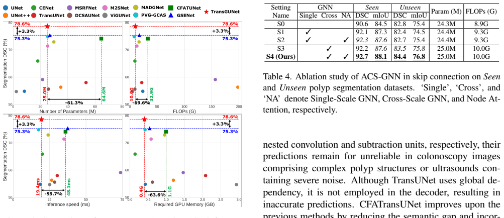
- If the demonstrated gains hold, skip connections in future segmentation networks can be built from graph convolutions instead of nested dense blocks or transformer stacks, matching or beating those designs with roughly 25M parameters and 10G FLOPs.
- Domain transfer across imaging modalities becomes more feasible: the reported 47.2% DSC on AMOS-MRI after Synapse CT-only training is more than 11 percentage points over the next-best model, suggesting that graph-structured cross-scale features carry modality-invariant anatomical cues.
- The entropy filter implies that spatial attention maps improve not by adding more channels but by discarding high-entropy ones, so spatial attention modules in other architectures could adopt the same selection rule.
- The method is backbone-agnostic in the reported ablations: swapping in CNN and transformer encoders retains gains, so the skip-connection design can be retrofitted to existing encoders.
Reading between the lines
- A natural extension the authors do not pursue is to learn the channel-selection rule instead of fixing it to Shannon entropy; if a learned selector matched or beat entropy on new modalities, it would show the benefit is filtering noise rather than entropy per se.
- Because the graph is built at $\frac{H}{8}\times\frac{W}{8}$, the same construction should transfer to 3D volumes by treating voxels as nodes; the reported memory footprint suggests 3D use is plausible but untested.
- The entropy heuristic could be compared against attention-confidence or predictive-uncertainty measures; a testable prediction is that any criterion that removes uniformly activated channels yields similar gains.
Editorial analysis
A structured set of objections, weighed in public.
Referee Report
Summary. The paper proposes TransGUNet, a 2D medical image segmentation architecture that modifies U-Net skip connections with an attentional cross-scale graph neural network (ACS-GNN) and an entropy-driven feature selection (EFS) mechanism for spatial attention. The method uses a P2T encoder-decoder, reports results on six in-distribution ('seen') and eight external ('unseen') datasets spanning multi-organ, skin, COVID-19, breast ultrasound, and polyp segmentation, and compares against twelve baselines in terms of DSC, mIoU, parameters, FLOPs, inference time, and memory. The headline claims are that TransGUNet achieves the best average performance on both seen and unseen settings, that the gains are statistically significant, and that the improvements are particularly large in cross-modality generalization (e.g., Synapse to AMOS-MRI).
Significance. If the empirical claims are sustained, this is a worthwhile contribution to skip-connection design: it introduces a lightweight GNN-based cross-scale fusion alternative to transformer-based skip connections and a channel-selection mechanism for spatial attention, supported by a broad evaluation across modalities. The independent unseen datasets that were not used in hyperparameter selection (AMOS, PH2, COVID19-2, STU) provide genuinely useful generalization evidence, and the paper's use of three repeats with standard deviations is a strength. The efficiency comparison (25.0M parameters, 10.0G FLOPs) is also a useful data point. However, the statistical support for the word 'significantly' is invalid as reported, and the unseen polyp datasets were used to select several hyperparameters, which weakens the claimed generalization margins on those datasets. The entropy-selection mechanism is also not tested against alternative selection criteria, so its specific contribution is underdetermined.
major comments (3)
- [Tables 2 and 3] The reported Wilcoxon signed-rank p-values are not achievable under the experimental design as described. With the six seen datasets as paired observations, the smallest possible one-sided exact p-value is 2^-6 = 0.0156, and with eight unseen datasets it is 2^-8 = 0.0039; yet Table 2 reports p-values as small as 9.3E-06 and Table 3 reports 1.7E-08. Pooling DSC and mIoU into 12 or 16 paired observations still gives minimum one-sided p-values of about 2.4E-04 and 1.5E-05, which remain far above many printed entries. The paper never states the sample unit for the test; if per-image scores were used, the test would need to account for clustering by dataset, and no such analysis is provided. The Section 4.3 claim that TransGUNet 'significantly outperforms' the baselines is therefore unverifiable as reported. Please state the exact test procedure, sample size, and effect definition, and recompute or verify all p-values.
- [Section 4.4, Fig. 7, Appendix Tables 5, 10, 11] The hyperparameters M, the target resolution (Ht,Wt), the ECA kernel size k, and the repetition count G are selected by maximizing performance on the same aggregated 'Seen' and 'Unseen' polyp splits that later appear in Table 3. The 'Unseen' columns in these ablation studies include CVC-300, CVC-ColonDB, and ETIS, which are exactly the datasets reported as unseen generalization results. This means that the claimed gains on those three polyp test sets are partially the result of test-set selection rather than independent generalization. The non-polyp unseen datasets (AMOS, PH2, COVID19-2, STU) are not affected by this leakage, but the overall 'eight unseen datasets' average and the Section 4.3 generalization claim are. Please either fix all hyperparameters before evaluating any test set, or hold out a disjoint validation split from the polyp training data, and re-report the affected numbers.
- [Eq. (4), Eq. (5), Section 4.4] The paper attributes the EFS improvement to the use of low Shannon-entropy channels, but it never tests the entropy criterion against alternative channel-selection rules. The ablation in Fig. 7 compares M=256 with M<256, which only shows that filtering some channels helps; it does not show that sorting by low entropy is the reason. A control selecting M random channels, M highest-entropy channels, or M channels by another statistic (e.g., variance or mean activation) is needed to establish that the entropy score itself is load-bearing. Without such a control, the gains attributed to 'entropy-driven' selection could equally come from dimensionality reduction or from any fixed channel subset. Please add this control or soften the mechanistic claim.
minor comments (3)
- [Table 1 vs Appendix Table 8] The efficiency numbers are inconsistent between the main table and the appendix: for example, UNet inference time is 18.6 ms in Table 1 but 10.1 ms in Table 8, UNet++ is 26.5 vs 22.9 ms, and M2SNet is 34.9 vs 32.1 ms, while ViGUNet has numerical entries in Table 1 but dashes in Table 8. Please harmonize the tables and state the measurement conditions (batch size, input resolution, GPU, warm-up).
- [Section 4.2] The sentence 'In the Appendix, we provide the experiment results on various hyperparameter settings (Tab. 9, 5, 10, 11)' is inaccurate because Appendix Table 9 is a backbone ablation, not a hyperparameter study; please correct the table pointers.
- [Section 4.3] The sentence 'Although MADGNet achieved the state-of-the-art performance in seen clinical settings' is ambiguous, since Table 2 shows TransGUNet with the best seen-average DSC; if the intended meaning is 'best among the previously published baselines,' please rephrase to avoid an apparent internal contradiction.
Circularity Check
Partial circularity: the 'unseen' polyp test sets were used to select hyperparameters, so three of the eight domain-generalization results are test-set-tuned predictions.
-
fitted input called prediction
[Section 4.2 (Key hyperparameters), Section 4.4 (Ablation Study on EFS-based Spatial Attention, Fig. 7), and Appendix Tables 5, 9, 10, 11, feeding Table 3's CVC-300, CVC-ColonDB, and ETIS rows.]
"Key hyperparameters for TransGUNet on all datasets were set to Cr = 64 for efficiency and (Ht, Wt) = ( H 8 , W 8 ), K = 11, k= 3 in ACS-GNN and M = 64in EFS-based spatial attention. In the Appendix, we provide the experiment results on various hyperparameter settings (Tab. 9, 5, 10, 11). ... In Fig. 7, (·, ·) denotes DSC on seen and unseen clinical setting of polyp segmentation. Fig. 7 shows that M = 256results in worse performance than the cases utilizing the EFS-based spatial attention (M = {8, 16, 32, 64, 128})."
The hyperparameters M=64, target resolution (H/8,W/8), ECA kernel k=3, and repetition G=1 are selected by comparing DSC on the 'seen and unseen clinical setting of polyp segmentation'. The 'unseen' polyp datasets are CVC-300, CVC-ColonDB, and ETIS, which are the same rows later presented as unseen-generalization evidence in Table 3. Thus the reported 90.0, 82.0, and 81.3 DSC values on those three datasets are outcomes of test-set-guided model selection rather than independent predictions: the configuration was chosen because it performed well on those specific test sets. The independent unseen datasets (AMOS-CT/MRI, PH2, COVID19-2, STU) were not part of this selection, so the circularity is partial but real for the polyp 'unseen' claim.
full rationale
The architecture derivation itself is self-contained: ACS-GNN is specified by its own equations (KNN graph construction, MRConv, ECA-style node attention), EFS is defined by the Shannon-entropy formula in Eq. (4), and the ablations test the components rather than importing a result from a self-citation. The authors' self-citations [44,45] are used for standard loss definitions and as comparison baselines, not as load-bearing evidence, so they do not raise the circularity score. The main circular step is in the evaluation protocol: the paper labels CVC-300, CVC-ColonDB, and ETIS as 'unseen clinical settings' in Table 3, but Section 4.4 and Appendix Tables 5, 9, 10, and 11 show that key hyperparameters and even the backbone were selected using DSC on those same 'seen and unseen' polyp datasets. That makes those three unseen numbers fitted rather than predicted, partially compromising the headline 'significantly outperforms' claim over eight unseen datasets. The five independent unseen datasets provide external grounding, so the circularity is not total. Separately, the Wilcoxon p-values in Tables 2 and 3 are below the attainable exact minimum for n=6 and n=8 dataset-level paired observations (2^-6 = 0.0156 and 2^-8 = 0.0039), so the significance claim is not supported by a valid described test; this is a statistical-support problem, not a circularity, and does not change the circularity score. Likewise, the entropy heuristic in Eq. (4) is an untested assumption, but it is not circular.
Assumptions & free parameters
free parameters (6)
- M (number of lowest-entropy channels kept) =
64
- k (ECA kernel size for node attention) =
3
- G (repetition count of ACS-GNN+EFS) =
1
- target_resolution (Ht, Wt) =
(H/8, W/8)
- Cr (channel reduction) =
64
- K (number of neighbors in dilated KNN graph) =
11
assumptions (5)
- domain assumption Shannon entropy of a feature channel is a valid measure of feature informativeness.
- domain assumption Graph-based cross-scale fusion reduces the semantic gap between encoder and decoder.
- domain assumption P2T encoder provides sufficient representation for medical segmentation across modalities.
- domain assumption Synapse-to-AMOS and other seen-to-unseen transfers are valid tests of domain generalizability.
- standard math Wilcoxon signed-rank test assumptions hold for paired segmentation metrics.
Cite this review
Pith. "Pith review of TransGUNet: Transformer Meets Graph-based Skip Connection for Medical Image Segmentation." pith.science (2026). https://pith.science/paper/OGCUVE6M
@misc{pith2026250209931,
  author       = {Pith},
  title        = {Pith review of: TransGUNet: Transformer Meets Graph-based Skip Connection for Medical Image Segmentation},
  year         = {2026},
  howpublished = {\url{https://pith.science/paper/OGCUVE6M}},
  note         = {Machine review of arXiv:2502.09931}
}
read the original abstract
Skip connection engineering is primarily employed to address the semantic gap between the encoder and decoder, while also integrating global dependencies to understand the relationships among complex anatomical structures in medical image segmentation. Although several models have proposed transformer-based approaches to incorporate global dependencies within skip connections, they often face limitations in capturing detailed local features with high computational complexity. In contrast, graph neural networks (GNNs) exploit graph structures to effectively capture local and global features. Leveraging these properties, we introduce an attentional cross-scale graph neural network (ACS-GNN), which enhances the skip connection framework by converting cross-scale feature maps into a graph structure and capturing complex anatomical structures through node attention. Additionally, we observed that deep learning models often produce uninformative feature maps, which degrades the quality of spatial attention maps. To address this problem, we integrated entropy-driven feature selection (EFS) with spatial attention, calculating an entropy score for each channel and filtering out high-entropy feature maps. Our innovative framework, TransGUNet, comprises ACS-GNN and EFS-based spatial attentio} to effectively enhance domain generalizability across various modalities by leveraging GNNs alongside a reliable spatial attention map, ensuring more robust features within the skip connection. Through comprehensive experiments and analysis, TransGUNet achieved superior segmentation performance on six seen and eight unseen datasets, demonstrating significantly higher efficiency compared to previous methods.
Figures


Figures from the paper (9 more)





Reference graph
Works this paper leans on
-
[1]
https : / / www
Covid19 dataset. https : / / www . kaggle . com / datasets / piyushsamant11 / pidata - new - names. 6
-
[2]
Dataset of breast ultrasound images
Walid Al-Dhabyani, Mohammed Gomaa, Hussien Khaled, and Aly Fahmy. Dataset of breast ultrasound images. Data in brief, 28:104863, 2020. 6, 1, 7, 9
2020
-
[3]
Segmentation out- side the cranial vault challenge
Landman Bennett, Xu Zhoubing, Igelsias Juan, Styner Mar- tin, Langerak Thomas, and Klein Arno. Segmentation out- side the cranial vault challenge. In Medical Image Com- puting and Computer-Assisted Intervention–MICCAI 2015: 18th International Conference, Munich, Germany, October 5-9, 2015, Proceedings, Part III 18. Springer, 2015. 6, 1, 11, 12
2015
-
[4]
Wm-dova maps for accurate polyp highlighting in colonoscopy: Validation vs
Jorge Bernal, F Javier S ´anchez, Gloria Fern ´andez- Esparrach, Debora Gil, Cristina Rodr ´ıguez, and Fernando Vilari˜no. Wm-dova maps for accurate polyp highlighting in colonoscopy: Validation vs. saliency maps from physi- cians. Computerized medical imaging and graphics, 43:99– 111, 2015. 6, 1, 8, 10
2015
-
[5]
Swin-unet: Unet-like pure transformer for medical image segmentation
Hu Cao, Yueyue Wang, Joy Chen, Dongsheng Jiang, Xi- aopeng Zhang, Qi Tian, and Manning Wang. Swin-unet: Unet-like pure transformer for medical image segmentation. In European conference on computer vision, pages 205–218. Springer, 2022. 2
2022
-
[6]
A generalized surface loss for reducing the hausdorff dis- tance in medical imaging segmentation
Adrian Celaya, Beatrice Riviere, and David Fuentes. A generalized surface loss for reducing the hausdorff dis- tance in medical imaging segmentation. arXiv preprint arXiv:2302.03868, 2023. 4
arXiv 2023
-
[7]
Transunet: Transformers make strong encoders for medi- cal image segmentation
Jieneng Chen, Yongyi Lu, Qihang Yu, Xiangde Luo, Ehsan Adeli, Yan Wang, Le Lu, Alan L Yuille, and Yuyin Zhou. Transunet: Transformers make strong encoders for medi- cal image segmentation. arXiv preprint arXiv:2102.04306,
-
[8]
Lesion-inspired denoising network: Connecting medical image denoising and lesion detection
Kecheng Chen, Kun Long, Yazhou Ren, Jiayu Sun, and Xi- aorong Pu. Lesion-inspired denoising network: Connecting medical image denoising and lesion detection. In Proceed- ings of the 29th ACM International Conference on Multime- dia, pages 3283–3292, 2021. 2, 5
2021
Show all 89 references
-
[9]
Tailoring therapies—improving the management of early breast cancer: St gallen international expert consensus on the primary therapy of early breast cancer 2015
Alan S Coates, Eric P Winer, Aron Goldhirsch, Richard D Gelber, Michael Gnant, M Piccart-Gebhart, Beat Th¨urlimann, H-J Senn, Panel Members, Fabrice Andr´e, et al. Tailoring therapies—improving the management of early breast cancer: St gallen international expert consensus on ...
2015
-
[10]
Polyp-pvt: Polyp segmentation with pyramid vision transformers
Bo Dong, Wenhai Wang, Deng-Ping Fan, Jinpeng Li, Huazhu Fu, and Ling Shao. Polyp-pvt: Polyp segmentation with pyramid vision transformers. arxiv 2021.arXiv preprint arXiv:2108.06932. 3
2021 arXiv
-
[11]
An image is worth 16x16 words: Transformers for image recognition at scale
Alexey Dosovitskiy, Lucas Beyer, Alexander Kolesnikov, Dirk Weissenborn, Xiaohua Zhai, Thomas Unterthiner, Mostafa Dehghani, Matthias Minderer, Georg Heigold, Syl- vain Gelly, Jakob Uszkoreit, and Neil Houlsby. An image is worth 16x16 words: Transformers for image recognition ...
2021
-
[12]
3d dental mesh segmentation using semantics-based feature learning with graph-transformer
Fan Duan and Li Chen. 3d dental mesh segmentation using semantics-based feature learning with graph-transformer. In International Conference on Medical Image Computing and Computer-Assisted Intervention , pages 456–465. Springer,
-
[13]
Structure-measure: A new way to evaluate foreground maps
Deng-Ping Fan, Ming-Ming Cheng, Yun Liu, Tao Li, and Ali Borji. Structure-measure: A new way to evaluate foreground maps. In Proceedings of the IEEE international conference on computer vision, pages 4548–4557, 2017. 4
2017
-
[14]
Enhanced-alignment mea- sure for binary foreground map evaluation
Deng-Ping Fan, Cheng Gong, Yang Cao, Bo Ren, Ming- Ming Cheng, and Ali Borji. Enhanced-alignment mea- sure for binary foreground map evaluation. arXiv preprint arXiv:1805.10421, 2018. 4
2018 arXiv
-
[15]
Pranet: Parallel reverse attention network for polyp segmentation
Deng-Ping Fan, Ge-Peng Ji, Tao Zhou, Geng Chen, Huazhu Fu, Jianbing Shen, and Ling Shao. Pranet: Parallel reverse attention network for polyp segmentation. In International conference on medical image computing and computer- assisted intervention, pages 263–273. Springer, 2020. 5, 6
2020
-
[16]
Res2net: A new multi-scale backbone architecture
Shang-Hua Gao, Ming-Ming Cheng, Kai Zhao, Xin-Yu Zhang, Ming-Hsuan Yang, and Philip Torr. Res2net: A new multi-scale backbone architecture. IEEE transactions on pattern analysis and machine intelligence , 43(2):652–662,
-
[17]
Utnet: a hybrid transformer architecture for medical image segmen- tation
Yunhe Gao, Mu Zhou, and Dimitris N Metaxas. Utnet: a hybrid transformer architecture for medical image segmen- tation. In Medical Image Computing and Computer Assisted Intervention–MICCAI 2021: 24th International Conference, Strasbourg, France, September 27–October 1, 2021, Pr...
2021
-
[18]
Do as ai say: susceptibility in deployment of clinical decision-aids
Susanne Gaube, Harini Suresh, Martina Raue, Alexander Merritt, Seth J Berkowitz, Eva Lermer, Joseph F Coughlin, 9 John V Guttag, Errol Colak, and Marzyeh Ghassemi. Do as ai say: susceptibility in deployment of clinical decision-aids. NPJ digital medicine, 4(1):31, 2021. 2
2021
-
[19]
Ce-net: Context encoder network for 2d medical image segmentation
Zaiwang Gu, Jun Cheng, Huazhu Fu, Kang Zhou, Huay- ing Hao, Yitian Zhao, Tianyang Zhang, Shenghua Gao, and Jiang Liu. Ce-net: Context encoder network for 2d medical image segmentation. IEEE transactions on medical imaging, 38(10):2281–2292, 2019. 5, 6, 8
2019
-
[20]
David Gutman, Noel CF Codella, Emre Celebi, Brian Helba, Michael Marchetti, Nabin Mishra, and Allan Halpern. Skin lesion analysis toward melanoma detection: A challenge at the international symposium on biomedical imaging (isbi) 2016, hosted by the international skin imaging c...
2016 arXiv
-
[21]
Vision gnn: An image is worth graph of nodes
Kai Han, Yunhe Wang, Jianyuan Guo, Yehui Tang, and En- hua Wu. Vision gnn: An image is worth graph of nodes. Ad- vances in neural information processing systems , 35:8291– 8303, 2022. 2, 3
2022
-
[22]
Vision hgnn: An image is more than a graph of nodes
Yan Han, Peihao Wang, Souvik Kundu, Ying Ding, and Zhangyang Wang. Vision hgnn: An image is more than a graph of nodes. In Proceedings of the IEEE/CVF Interna- tional Conference on Computer Vision, pages 19878–19888,
-
[23]
Image analysis using mathematical morphology
Robert M Haralick, Stanley R Sternberg, and Xinhua Zhuang. Image analysis using mathematical morphology. IEEE transactions on pattern analysis and machine intelli- gence, (4):532–550, 1987. 1
1987
-
[24]
Unetr: Transformers for 3d med- ical image segmentation
Ali Hatamizadeh, Yucheng Tang, Vishwesh Nath, Dong Yang, Andriy Myronenko, Bennett Landman, Holger R Roth, and Daguang Xu. Unetr: Transformers for 3d med- ical image segmentation. In Proceedings of the IEEE/CVF winter conference on applications of computer vision, pages 574–58...
2022
-
[25]
Deep residual learning for image recognition
Kaiming He, Xiangyu Zhang, Shaoqing Ren, and Jian Sun. Deep residual learning for image recognition. In Proceed- ings of the IEEE conference on computer vision and pattern recognition, pages 770–778, 2016. 3
2016
-
[26]
Hiformer: Hierarchical multi-scale representations using transformers for medical image seg- mentation
Moein Heidari, Amirhossein Kazerouni, Milad Soltany, Reza Azad, Ehsan Khodapanah Aghdam, Julien Cohen- Adad, and Dorit Merhof. Hiformer: Hierarchical multi-scale representations using transformers for medical image seg- mentation. In Proceedings of the IEEE/CVF Winter Confer- ...
-
[27]
Kvasir-seg: A segmented polyp dataset
Debesh Jha, Pia H Smedsrud, Michael A Riegler, P ˚al Halvorsen, Thomas de Lange, Dag Johansen, and H ˚avard D Johansen. Kvasir-seg: A segmented polyp dataset. In Mul- tiMedia Modeling: 26th International Conference, MMM 2020, Daejeon, South Korea, January 5–8, 2020, Proceed- i...
2020
-
[28]
Amos: A large-scale abdominal multi-organ benchmark for versatile medical image segmentation
Yuanfeng Ji, Haotian Bai, Chongjian Ge, Jie Yang, Ye Zhu, Ruimao Zhang, Zhen Li, Lingyan Zhanng, Wanling Ma, Xi- ang Wan, et al. Amos: A large-scale abdominal multi-organ benchmark for versatile medical image segmentation. Ad- vances in neural information processing systems, 3...
2022
-
[29]
Vig-unet: vision graph neural networks for medical image segmentation
Juntao Jiang, Xiyu Chen, Guanzhong Tian, and Yong Liu. Vig-unet: vision graph neural networks for medical image segmentation. In 2023 IEEE 20th International Symposium on Biomedical Imaging (ISBI), pages 1–5. IEEE, 2023. 3, 5, 6, 8
2023
-
[30]
COVID-19 CT Lung and Infection Segmenta- tion Dataset
Ma Jun, Ge Cheng, Wang Yixin, An Xingle, Gao Jiantao, Yu Ziqi, Zhang Minqing, Liu Xin, Deng Xueyuan, Cao Shucheng, Wei Hao, Mei Sen, Yang Xiaoyu, Nie Ziwei, Li Chen, Tian Lu, Zhu Yuntao, Zhu Qiongjie, Dong Guoqiang, and He Jian. COVID-19 CT Lung and Infection Segmenta- tion Da...
2020
-
[31]
Design of an image edge detection filter using the sobel operator
Nick Kanopoulos, Nagesh Vasanthavada, and Robert L Baker. Design of an image edge detection filter using the sobel operator. IEEE Journal of solid-state circuits , 23(2): 358–367, 1988. 5
1988
-
[32]
Snakes: Active contour models
Michael Kass, Andrew Witkin, and Demetri Terzopoulos. Snakes: Active contour models. International journal of computer vision, 1(4):321–331, 1988. 2
1988
-
[33]
Semi-supervised classi- fication with graph convolutional networks
Thomas N Kipf and Max Welling. Semi-supervised classi- fication with graph convolutional networks. arXiv preprint arXiv:1609.02907, 2016. 3
2016 arXiv
-
[34]
Deepgcns: Can gcns go as deep as cnns? In Proceedings of the IEEE/CVF international conference on computer vision, pages 9267–9276, 2019
Guohao Li, Matthias Muller, Ali Thabet, and Bernard Ghanem. Deepgcns: Can gcns go as deep as cnns? In Proceedings of the IEEE/CVF international conference on computer vision, pages 9267–9276, 2019. 4
2019
-
[35]
Deeper insights into graph convolutional networks for semi-supervised learn- ing
Qimai Li, Zhichao Han, and Xiao-Ming Wu. Deeper insights into graph convolutional networks for semi-supervised learn- ing. In Proceedings of the AAAI conference on artificial in- telligence, 2018. 4
2018
-
[36]
Gse-nets: Global structure enhancement de- coder for thyroid nodule segmentation
Xiang Li, Chong Fu, Qun Wang, Wenchao Zhang, Chen Ye, and Tao Ma. Gse-nets: Global structure enhancement de- coder for thyroid nodule segmentation. Biomedical Signal Processing and Control, 102:107340, 2025. 3, 6, 8
2025
-
[37]
Cafe-net: Cross- attention and feature exploration network for polyp segmen- tation
Guoqi Liu, Sheng Yao, Dong Liu, Baofang Chang, Zongyu Chen, Jiajia Wang, and Jiangqi Wei. Cafe-net: Cross- attention and feature exploration network for polyp segmen- tation. Expert Systems with Applications, 238:121754, 2024. 3
2024
-
[38]
Covsegnet: A multi encoder– decoder architecture for improved lesion segmentation of covid-19 chest ct scans
Tanvir Mahmud, Md Awsafur Rahman, Shaikh Anowarul Fattah, and Sun-Yuan Kung. Covsegnet: A multi encoder– decoder architecture for improved lesion segmentation of covid-19 chest ct scans. IEEE Transactions on Artificial In- telligence, 2(3):283–297, 2021. 2
2021
-
[39]
Simple learned weighted sums of inferior temporal neuronal firing rates accurately predict human core object recognition performance
Najib J Majaj, Ha Hong, Ethan A Solomon, and James J DiCarlo. Simple learned weighted sums of inferior temporal neuronal firing rates accurately predict human core object recognition performance. Journal of Neuroscience, 35(39): 13402–13418, 2015. 4
2015
-
[40]
How to evaluate foreground maps? In Proceedings of the IEEE con- ference on computer vision and pattern recognition , pages 248–255, 2014
Ran Margolin, Lihi Zelnik-Manor, and Ayellet Tal. How to evaluate foreground maps? In Proceedings of the IEEE con- ference on computer vision and pattern recognition , pages 248–255, 2014. 4
2014
-
[41]
Vision: A computational investigation into the human representation and processing of visual information
David Marr. Vision: A computational investigation into the human representation and processing of visual information. MIT press, 2010. 4
2010
-
[42]
Ph 2-a dermoscopic image database for research and benchmarking
Teresa Mendonc ¸a, Pedro M Ferreira, Jorge S Marques, Andr´e RS Marcal, and Jorge Rozeira. Ph 2-a dermoscopic image database for research and benchmarking. In 2013 35th annual international conference of the IEEE engineer- 10 ing in medicine and biology society (EMBC) , pages 5437–
2013
-
[43]
V-net: Fully convolutional neural networks for volumetric medical image segmentation
Fausto Milletari, Nassir Navab, and Seyed-Ahmad Ahmadi. V-net: Fully convolutional neural networks for volumetric medical image segmentation. In 2016 fourth international conference on 3D vision (3DV), pages 565–571. Ieee, 2016. 4
2016
-
[44]
M3fpolypsegnet: Segmentation network with multi-frequency feature fusion for polyp localization in colonoscopy images
Ju-Hyeon Nam, Seo-Hyeong Park, Nur Suriza Syazwany, Yerim Jung, Yu-Han Im, and Sang-Chul Lee. M3fpolypsegnet: Segmentation network with multi-frequency feature fusion for polyp localization in colonoscopy images. In 2023 IEEE International Confer- ence on Image Processing (ICI...
2023
-
[45]
Modality-agnostic domain generalizable medical image segmentation by multi-frequency in multi- scale attention
Ju-Hyeon Nam, Nur Suriza Syazwany, Su Jung Kim, and Sang-Chul Lee. Modality-agnostic domain generalizable medical image segmentation by multi-frequency in multi- scale attention. In Proceedings of the IEEE/CVF Conference on Computer Vision and Pattern Recognition, pages 11480–...
2024
-
[46]
Graph neural networks expo- nentially lose expressive power for node classification
Kenta Oono and Taiji Suzuki. Graph neural networks expo- nentially lose expressive power for node classification. InIn- ternational Conference on Learning Representations , 2020. 4
2020
-
[47]
A threshold selection method from gray- level histograms
Nobuyuki Otsu. A threshold selection method from gray- level histograms. IEEE transactions on systems, man, and cybernetics, 9(1):62–66, 1979. 2
1979
-
[48]
Vision science: Photons to phenomenol- ogy
Stephen E Palmer. Vision science: Photons to phenomenol- ogy. MIT press, 1999. 4
1999
-
[49]
The attention system of the human brain
Michael I Posner, Steven E Petersen, et al. The attention system of the human brain. Annual review of neuroscience, 13(1):25–42, 1990. 4
1990
-
[50]
G-cascade: Efficient cascaded graph convolutional decoding for 2d med- ical image segmentation
Md Mostafijur Rahman and Radu Marculescu. G-cascade: Efficient cascaded graph convolutional decoding for 2d med- ical image segmentation. In Proceedings of the IEEE/CVF Winter Conference on Applications of Computer Vision , pages 7728–7737, 2024. 3, 5, 6, 8
2024
-
[51]
A new unsupervised approach for segment- ing and counting cells in high-throughput microscopy image sets
Daniel Riccio, Nadia Brancati, Maria Frucci, and Diego Gragnaniello. A new unsupervised approach for segment- ing and counting cells in high-throughput microscopy image sets. IEEE journal of biomedical and health informatics, 23 (1):437–448, 2018. 2
2018
-
[52]
U- net: Convolutional networks for biomedical image segmen- tation
Olaf Ronneberger, Philipp Fischer, and Thomas Brox. U- net: Convolutional networks for biomedical image segmen- tation. In Medical Image Computing and Computer-Assisted Intervention–MICCAI 2015: 18th International Conference, Munich, Germany, October 5-9, 2015, Proceedings, Pa...
2015
-
[53]
A ct image denois- ing method with residual encoder-decoder network
Helena Shawn, Thompson Chyrikov, Jacob Lanet, Lam-chi Chen, Jim Zhao, and Christina Chajo. A ct image denois- ing method with residual encoder-decoder network. arXiv preprint arXiv:2404.01553, 2024. 2, 5
2024 arXiv
-
[54]
Toward embedded detection of polyps in wce images for early diagnosis of colorectal can- cer
Juan Silva, Aymeric Histace, Olivier Romain, Xavier Dray, and Bertrand Granado. Toward embedded detection of polyps in wce images for early diagnosis of colorectal can- cer. International journal of computer assisted radiology and surgery, 9:283–293, 2014. 6, 1, 8, 10
2014
-
[55]
Msrf-net: a multi- scale residual fusion network for biomedical image segmen- tation
Abhishek Srivastava, Debesh Jha, Sukalpa Chanda, Uma- pada Pal, H ˚avard D Johansen, Dag Johansen, Michael A Riegler, Sharib Ali, and P ˚al Halvorsen. Msrf-net: a multi- scale residual fusion network for biomedical image segmen- tation. IEEE Journal of Biomedical and Health In...
2021
-
[56]
Automated polyp detection in colonoscopy videos using shape and context information
Nima Tajbakhsh, Suryakanth R Gurudu, and Jianming Liang. Automated polyp detection in colonoscopy videos using shape and context information. IEEE transactions on medical imaging, 35(2):630–644, 2015. 6, 1, 8, 10
2015
-
[57]
Image thresholding using type ii fuzzy sets
Hamid R Tizhoosh. Image thresholding using type ii fuzzy sets. Pattern recognition, 38(12):2363–2372, 2005. 2
2005
-
[58]
The fully convolutional trans- former for medical image segmentation
Athanasios Tragakis, Chaitanya Kaul, Roderick Murray- Smith, and Dirk Husmeier. The fully convolutional trans- former for medical image segmentation. In Proceedings of the IEEE/CVF Winter Conference on Applications of Com- puter Vision, pages 3660–3669, 2023. 2
2023
-
[59]
Neural correlates of attention in primate visual cortex
Stefan Treue. Neural correlates of attention in primate visual cortex. Trends in neurosciences, 24(5):295–300, 2001. 4
2001
-
[60]
A benchmark for endoluminal scene segmentation of colonoscopy images
David V ´azquez, Jorge Bernal, F Javier S ´anchez, Gloria Fern´andez-Esparrach, Antonio M L ´opez, Adriana Romero, Michal Drozdzal, Aaron Courville, et al. A benchmark for endoluminal scene segmentation of colonoscopy images. Journal of healthcare engineering, 2017, 2017. 6, 1, 8, 10
2017
-
[61]
Advent: Adversarial entropy min- imization for domain adaptation in semantic segmentation
Tuan-Hung Vu, Himalaya Jain, Maxime Bucher, Matthieu Cord, and Patrick P ´erez. Advent: Adversarial entropy min- imization for domain adaptation in semantic segmentation. In Proceedings of the IEEE/CVF conference on computer vi- sion and pattern recognition, pages 2517–2526, 2019. 5
2019
-
[62]
Gazegnn: A gaze- guided graph neural network for chest x-ray classification
Bin Wang, Hongyi Pan, Armstrong Aboah, Zheyuan Zhang, Elif Keles, Drew Torigian, Baris Turkbey, Elizabeth Krupin- ski, Jayaram Udupa, and Ulas Bagci. Gazegnn: A gaze- guided graph neural network for chest x-ray classification. In Proceedings of the IEEE/CVF Winter Conference o...
2024
-
[63]
Cfatran- sunet: Channel-wise cross fusion attention and transformer for 2d medical image segmentation
Cheng Wang, Le Wang, Nuoqi Wang, Xiaoling Wei, Ting Feng, Minfeng Wu, Qi Yao, and Rongjun Zhang. Cfatran- sunet: Channel-wise cross fusion attention and transformer for 2d medical image segmentation. Computers in Biology and Medicine, 168:107803, 2024. 2, 5, 6, 8
2024
-
[64]
Uctransnet: rethinking the skip connections in u-net from a channel-wise perspective with transformer
Haonan Wang, Peng Cao, Jiaqi Wang, and Osmar R Zaiane. Uctransnet: rethinking the skip connections in u-net from a channel-wise perspective with transformer. In Proceed- ings of the AAAI conference on artificial intelligence , pages 2441–2449, 2022. 2
2022
-
[65]
Eca-net: Efficient channel at- tention for deep convolutional neural networks
Qilong Wang, Banggu Wu, Pengfei Zhu, Peihua Li, Wang- meng Zuo, and Qinghua Hu. Eca-net: Efficient channel at- tention for deep convolutional neural networks. In Proceed- ings of the IEEE/CVF conference on computer vision and pattern recognition, pages 11534–11542, 2020. 4
2020
-
[66]
Boundary and entropy-driven ad- versarial learning for fundus image segmentation
Shujun Wang, Lequan Yu, Kang Li, Xin Yang, Chi-Wing Fu, and Pheng-Ann Heng. Boundary and entropy-driven ad- versarial learning for fundus image segmentation. In Medi- cal Image Computing and Computer Assisted Intervention– MICCAI 2019: 22nd International Conference, Shenzhen, ...
2019
-
[67]
Pvt v2: Improved baselines with pyramid vision transformer
Wenhai Wang, Enze Xie, Xiang Li, Deng-Ping Fan, Kaitao Song, Ding Liang, Tong Lu, Ping Luo, and Ling Shao. Pvt v2: Improved baselines with pyramid vision transformer. Computational Visual Media, 8(3):415–424, 2022. 3
2022
-
[68]
Dynamic graph learning with content-guided spatial- frequency relation reasoning for deepfake detection
Yuan Wang, Kun Yu, Chen Chen, Xiyuan Hu, and Silong Peng. Dynamic graph learning with content-guided spatial- frequency relation reasoning for deepfake detection. In Pro- ceedings of the IEEE/CVF Conference on Computer Vision and Pattern Recognition, pages 7278–7287, 2023. 3
2023
-
[69]
Multi-scale group agent attention- based graph convolutional decoding networks for 2d medi- cal image segmentation
Zhichao Wang, Lin Guo, Shuchang Zhao, Shiqing Zhang, Xiaoming Zhao, Jiangxiong Fang, Guoyu Wang, Hongsheng Lu, Jun Yu, and Qi Tian. Multi-scale group agent attention- based graph convolutional decoding networks for 2d medi- cal image segmentation. IEEE Journal of Biomedical an...
2024
-
[70]
P2t: Pyramid pooling transformer for scene understanding
Yu-Huan Wu, Yun Liu, Xin Zhan, and Ming-Ming Cheng. P2t: Pyramid pooling transformer for scene understanding. IEEE transactions on pattern analysis and machine intelli- gence, 2022. 3
2022
-
[71]
Dcsau- net: A deeper and more compact split-attention u-net for medical image segmentation
Qing Xu, Zhicheng Ma, HE Na, and Wenting Duan. Dcsau- net: A deeper and more compact split-attention u-net for medical image segmentation. Computers in Biology and Medicine, 154:106626, 2023. 5, 6, 8
2023
-
[72]
Graph-and transformer-guided boundary aware network for medical im- age segmentation
Shanshan Xu, Lianhong Duan, Yang Zhang, Zhicheng Zhang, Tiansheng Sun, and Lixia Tian. Graph-and transformer-guided boundary aware network for medical im- age segmentation. Computer Methods and Programs in Biomedicine, 242:107849, 2023. 3
2023
-
[73]
Graph convolu- tional neural networks for web-scale recommender systems
Rex Ying, Ruining He, Kaifeng Chen, Pong Eksombatchai, William L Hamilton, and Jure Leskovec. Graph convolu- tional neural networks for web-scale recommender systems. In Proceedings of the 24th ACM SIGKDD international con- ference on knowledge discovery & data mining , pages ...
2018
-
[74]
Resnest: Split-attention networks
Hang Zhang, Chongruo Wu, Zhongyue Zhang, Yi Zhu, Haibin Lin, Zhi Zhang, Yue Sun, Tong He, Jonas Mueller, R Manmatha, et al. Resnest: Split-attention networks. In Proceedings of the IEEE/CVF conference on computer vi- sion and pattern recognition, pages 2736–2746, 2022. 3
2022
-
[75]
Transgraphnet: A novel network for medical im- age segmentation based on transformer and graph convo- lution
Ju Zhang, Zhiyi Ye, Mingyang Chen, Jiahao Yu, and Yun Cheng. Transgraphnet: A novel network for medical im- age segmentation based on transformer and graph convo- lution. Biomedical Signal Processing and Control , 104: 107510, 2025. 3
2025
-
[76]
Hsnet: A hybrid semantic network for polyp segmentation
Wenchao Zhang, Chong Fu, Yu Zheng, Fangyuan Zhang, Yanli Zhao, and Chiu-Wing Sham. Hsnet: A hybrid semantic network for polyp segmentation. Computers in biology and medicine, 150:106173, 2022. 3
2022
-
[77]
Auto- matic polyp segmentation via multi-scale subtraction net- work
Xiaoqi Zhao, Lihe Zhang, and Huchuan Lu. Auto- matic polyp segmentation via multi-scale subtraction net- work. In Medical Image Computing and Computer Assisted Intervention–MICCAI 2021: 24th International Conference, Strasbourg, France, September 27–October 1, 2021, Pro- ceedi...
2021
-
[78]
M 2SNet: Multi-scale in multi-scale subtraction network for medi- cal image segmentation
Xiaoqi Zhao, Hongpeng Jia, Youwei Pang, Long Lv, Feng Tian, Lihe Zhang, Weibing Sun, and Huchuan Lu. M 2SNet: Multi-scale in multi-scale subtraction network for medi- cal image segmentation. arXiv preprint arXiv:2303.10894,
-
[79]
Unet++: A nested u-net ar- chitecture for medical image segmentation
Zongwei Zhou, Md Mahfuzur Rahman Siddiquee, Nima Tajbakhsh, and Jianming Liang. Unet++: A nested u-net ar- chitecture for medical image segmentation. In Deep Learn- ing in Medical Image Analysis and Multimodal Learning for Clinical Decision Support: 4th International Workshop,...
2018
-
[80]
An rdau-net model for lesion segmentation in breast ultrasound images
Zhemin Zhuang, Nan Li, Alex Noel Joseph Raj, Vijayalak- shmi GV Mahesh, and Shunmin Qiu. An rdau-net model for lesion segmentation in breast ultrasound images. PloS one, 14(8):e0221535, 2019. 6, 1, 7, 9 12 TransGUNet: Transformer Meets Graph-based Skip Connection for Medical I...
2019
-
[82]
Details of the medical segmentation seen clinical settings used in our experiments
Dataset Descriptions Segmentation Task Dataset Resolutions Train Test Multi-organ Segmentation Synapse 512 × 512 18 Scans 12 Scans Skin Cancer Segmentation ISIC2018 Variable 1868 261 COVID19 Infection Segmentation COVID19-1 512 × 512 643 383 Breast Cancer Segmentation BUSI Var...
-
[83]
In contrast, the STU dataset [80] includes only 42 breast ultrasound images collected by Shantou University
comprises 780 images from 600 female patients, including 133 normal cases, 437 benign cases, and 210 malignant tumors. In contrast, the STU dataset [80] includes only 42 breast ultrasound images collected by Shantou University. Due to the limited number of images in the STU da...
2018
-
[84]
To address this, we transform cross-scale feature maps into a graph and apply efficient node-level at- tention
Intuitiveness and Design Principle of Trans- GUNet We want to clarify that the design of TransGUNet is care- fully considered rather than an ad-hoc decision, as follows: a) Medical images contain diverse anatomical structures, making it essential to flexibly capture both local...
-
[85]
Like our approach, PVT-GCASCADE utilizes GNN; however, it does not consider cross-scale information, which is limited to medical images with more diverse lesion sizes
Technical Novelty of TransGUNet PVT-GCASCADE (W ACV2024) vs TransGUNet. Like our approach, PVT-GCASCADE utilizes GNN; however, it does not consider cross-scale information, which is limited to medical images with more diverse lesion sizes. Addi- tionally, unlike PVT-GCASCADE, ...
-
[86]
Broader Impact in Artificial Intelligence TransGUNet’s superior performance in medical image seg- mentation has the potential to reliable medical diagnostics and treatment planning. By providing accurate and reli- able segmentation of complex anatomical structures, it en- able...
-
[87]
More Detailed Ablation Study on Trans- GUNet In this section, we perform a more detailed ablation study on TransGUNet. 10.1. Ablation Study on Backbone in TransGUNet Network Backbone Seen Unseen Param (M) FLOPs (G)Type DSC mIoU DSC mIoU CNN ResNet50 85.9 79.1 67.3 60.5 25.1M 1...
-
[88]
For convenience, we denote T P, F P, and F Nas the num- ber of samples of true positive, false positive, and false neg- ative between two binary masks A and B
Metrics Descriptions In this section, we describe the metrics used in this paper. For convenience, we denote T P, F P, and F Nas the num- ber of samples of true positive, false positive, and false neg- ative between two binary masks A and B. • The Mean Dice Similarity Coeffici...
-
[89]
12, 13, 14, 15 for binary segmenta- tion
More Qualtative and Quantitative Results In this section, we provide the quantitative results with var- ious metrics in Tab. 12, 13, 14, 15 for binary segmenta- tion. Additionally, we also provide the quantitative results in Tab. 16, 17, 18 with each organ for multi-organ segm...
-
[5440]
6, 1, 5, 9
IEEE, 2013. 6, 1, 5, 9
2013
Reviewed August 7, 2026 · model on record in the stance chip above.
Discussion (0). Continue with ORCID to comment.