REVIEW 3 major objections 5 minor 1 cited by
Putting the Segment Anything Model to the Test with 3D Knee MRI - A Comparison with State-of-the-Art Performance
T0 review · 3 major / 5 minor · reviewed 2026-08-16 · deepseek-v4-flash
Pith's one-line read Fine-tuning only SAM's decoder is not enough to segment knee menisci; even full end-to-end training that matches 3D U-Net on Dice leaves SAM behind on morphology.
desk verdict Useful benchmark showing decoder-only SAM fine-tuning is not enough for 3D meniscus segmentation, but the morphology claim needs an artifact-controlled check. read the letter →
The pith
A machine-rendered reading of the paper's core claim, the machinery that carries it, and where it could break.
The reading
What carries the argument
The central object is the Segment Anything Model in its base ViT configuration, treated as a 2D slice-wise segmenter: each 160-slice sagittal volume is split, each slice is bilinearly upsampled and padded to 1024×1024 and replicated to three channels, SAM runs without prompts and outputs one mask, and the slices are stacked back into a 3D volume. The comparison machinery is a 3D U-Net with 16 initial feature maps and three convolution blocks per encoder and decoder, trained from random weights. Two fine-tuning regimes isolate what carries the argument: a frozen image encoder with only the mask decoder trained (about 4 million parameters) versus full end-to-end training (about 94 million parameters). The metrics that carry the conclusions are Dice score for overlap, 95th-percentile Hausdorff distance for boundary morphology, and average transverse thickness difference as a biomarker-relevant quantity.
What would settle it
The claim would be settled by rerunning the same experiment on the same test volumes with one change: give SAM access to 3D context, either by volume-based prompting or by removing small disconnected components before scoring. If a 3D-aware SAM variant reaches a 95th-percentile Hausdorff distance at or below the 3D U-Net's 1.8 mm while keeping Dice at or above 0.87, the paper's conclusion would not survive; if the morphology gap persists under every reasonable 3D adaptation, the conclusion would be strengthened.
Extended reading notes
Core claim
The paper's central claim is that SAM, when adapted to 3D knee MRI by processing sagittal slices independently, can reach but not exceed the Dice-score performance of a standard 3D U-Net for meniscus segmentation, and only when it is fine-tuned end-to-end. Decoder-only fine-tuning is insufficient: frozen SAM features do not carry enough task-relevant information for low-contrast menisci, producing 0.81 Dice versus the U-Net's 0.87. End-to-end fine-tuning restores overlap performance (0.87), matching the 0.88 winning score of the 2019 challenge, yet the 95th-percentile Hausdorff distance remains worse (2.4±1.4 mm versus 1.8±0.8 mm), and SAM predictions contain far more disconnected components, indicating inferior preservation of meniscus morphology. The paper concludes that SAM fine-tuning may therefore be unsuitable for segmentation of similar fine anatomical structures and for deriving morphological biomarkers from such segmentations.
Load-bearing premise
The load-bearing premise is that feeding SAM sagittal slices independently and stacking the resulting masks is a fair way to apply a 2D foundation model to a 3D knee MRI; if a 3D-aware adaptation would behave differently, the paper's conclusion about SAM's unsuitability would not follow.
Editorial extensions
If this is right
- A 2D foundation model should not be assumed transferable to 3D medical segmentation of small, low-contrast structures; slice-wise application with a frozen encoder underperforms a basic 3D U-Net.
- End-to-end fine-tuning of SAM can recover Dice-score parity with a 3D U-Net and with challenge-winning performance, making it a viable overlap-based baseline when full training is affordable.
- If morphology matters, SAM's slice-wise masks are worse than U-Net's: the paper reports a mean Hausdorff distance of 2.4 mm versus 1.8 mm and an average of 10.2 versus 2.3 disconnected components, so SAM output would need more post-processing and could misrepresent geometry.
- For meniscal thickness as a biomarker, both fully fine-tuned SAM and U-Net overestimate average thickness by sub-voxel amounts, so overlap-based parity does not automatically mean identical downstream biomarker estimates.
Reading between the lines
- Going beyond the paper: a volume-aware adaptation of SAM, such as joint 3D prompting, multi-slice attention, or connected-component post-processing, might erase the reported Dice and Hausdorff gaps, and the slice-wise protocol is the natural first thing to vary.
- Going beyond the paper: the same comparison could be run on other small low-contrast joint structures, for example the acetabular labrum or spinal discs, to see whether the pattern is general or specific to menisci.
- Going beyond the paper: because the ground truths came from a single expert annotating slice-wise, part of U-Net's Hausdorff advantage may reflect matching staircase-style annotation rather than true anatomy; a multi-rater or smooth ground-truth benchmark would test whether SAM's textured output is actually closer to real tissue shape.
Signed reviews
Editorial analysis
A structured set of objections, weighed in public.
Referee Report
Summary. The paper evaluates two fine-tuning configurations of the Segment Anything Model (SAM) against a 3D U-Net for automatic meniscus segmentation in 3D knee MRI from the IWOAI 2019 dataset. The authors report that decoder-only fine-tuning (SAM 1) yields a Dice score of 0.81±0.03, described as significantly worse than the U-Net's 0.87±0.03, while end-to-end fine-tuning (SAM 2) reaches 0.87±0.03, comparable to the U-Net and to the IWOAI winning score. On the 95th-percentile Hausdorff distance, both SAM variants are inferior to the U-Net, leading the authors to conclude that SAM may not be suitable for similar fine-structure, low-contrast 3D segmentation tasks. The experiments use consistent splits with the challenge, a held-out test set, and multiple metrics; code is provided.
Significance. If the findings hold, the paper makes a useful empirical contribution to the debate on foundation models for medical imaging: it shows that decoder-only fine-tuning is insufficient for low-contrast small structures, and that end-to-end fine-tuning can match a well-trained 3D U-Net on Dice but leaves a residual morphology gap. The study is clearly designed, uses a public dataset and challenge splits, and releases code. The main claims, however, rest on a small test set and on a Hausdorff comparison that may be affected by removable isolated predictions; also, the 'significantly worse' statement is not formally tested.
major comments (3)
- [Section 4 (Conclusion) and Abstract] The claim that SAM 1 'performed significantly worse' than the 3D U-Net is not supported by a statistical test. With n=28 test volumes, the reported Dice difference (0.81±0.03 vs 0.87±0.03) should be accompanied by a paired test, bootstrap confidence interval, or a non-parametric alternative. The same applies to the Hausdorff-distance comparisons in Table 2. Without such an analysis, the reader cannot judge whether the observed differences are beyond sampling variation.
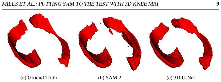
- [Section 3, Table 2, and Figure 5] The Hausdorff-distance evidence for morphological inferiority of SAM is confounded by the isolated prediction components acknowledged in the text (10.2 connected components for SAM 2 versus 2.3 for the U-Net). The 95th-percentile Hausdorff distance is highly sensitive to exactly these spurious islands. Please apply a largest-connected-component filter or a small-component removal step to both models' outputs and re-report Dice and Hausdorff distances. If the gap persists, the morphology claim is strengthened; if it closes, the conclusion that SAM 'is inferior ... in preserving the spatial features of the menisci' would need to be revised or conditioned on the absence of post-processing.
- [Section 4 (Conclusion) and Section 2.4] The generalization that SAM 'may not be suitable for similar 3D medical image segmentation tasks' is too broad for a study that tests only a slice-wise 2D adaptation of SAM. The paper does not compare against a 2D U-Net trained on the same slices, nor does it evaluate a 3D-aware adaptation or volume-based prompting. Adding a 2D U-Net baseline (or a 3D SAM variant) would isolate whether the observed gap is intrinsic to SAM or an artifact of the 2D/3D interface. Without this control, the conclusion should be tempered to the specific slice-wise adaptation evaluated here.
minor comments (5)
- [Table 2] The table contains formatting artifacts, such as '000...888777±±± 000...000333', which should be cleaned to read '0.87±0.03'.
- [Section 3, first paragraph] The winning IWOAI Dice score is reported as '0.88±0.3'; given the other reported standard deviations, this is almost certainly a typo for '0.88±0.03'.
- [Section 3.1] The word 'detatched' should be 'detached' in the sentence describing the separated medial meniscus.
- [Section 2.4] The description 'Three copies of this upsampled image were concatenated' is clear, but it would be helpful to state explicitly that this creates the required 3-channel RGB input for SAM.
- [Section 2.5.3] The average transverse thickness metric would benefit from a precise definition of 'non-zero columns in the transverse plane', including whether medial and lateral menisci are considered jointly or separately.
Circularity Check
No significant circularity: the paper is a held-out empirical benchmark whose claims are supported by direct evaluation against manual ground truth, not by a self-referential derivation.
full rationale
The paper reports a comparative benchmark: SAM fine-tuned in two configurations and a 3D U-Net are trained on fixed train/validation splits and evaluated on a held-out test set (Section 2.1). Dice score, 95th-percentile Hausdorff distance, and average transverse thickness are computed directly from predicted masks against manual ground truths (Sections 2.5 and 2.6, Table 2). No parameter is fitted to the test data, and no claim is derived from an equation that includes the target result as an input. The only self-citation with overlapping authorship is Dube et al. (ref [10]), a meniscal thickness biomarker study cited to motivate why thickness is a clinically relevant measurement; it is not load-bearing for any prediction or comparison. Concerns about slice-wise 2D adaptation or the absence of connected-component post-processing are methodological validity issues, not circularity. The central Dice comparison is also externally anchored to the IWOAI 2019 challenge winner (0.88 ± 0.03), which is independent of the present paper's fitted values. Therefore no specific reduction of a prediction to an input or to a self-citation chain can be exhibited.
Assumptions & free parameters
free parameters (5)
- Learning rate =
SAM 1: 5e-6, SAM 2: 5e-7, 3D U-Net: 1e-3
- Batch size =
SAM 1: 8, SAM 2: 16, 3D U-Net: 4
- Initial feature maps for 3D U-Net =
16
- Intensity windowing threshold =
Clipping window [0, 0.005]
- Crop region =
200x256x160, ~20 voxel margin
assumptions (5)
- domain assumption Manual segmentations of menisci by a single expert from Stryker Imorphics are treated as ground truth.
- domain assumption The OAI-IWOAI 2019 subset is representative of the meniscus segmentation task in knee OA.
- ad hoc to paper Slice-wise 2D processing of SAM is a fair adaptation to 3D MRI.
- ad hoc to paper Windowed intensity range and cropping preserve relevant contrast for meniscus segmentation.
- domain assumption Dice, 95% Hausdorff distance, and average transverse thickness capture clinically meaningful segmentation quality.
Cite this review
Pith. "Pith review of Putting the Segment Anything Model to the Test with 3D Knee MRI - A Comparison with State-of-the-Art Performance." pith.science (2026). https://pith.science/paper/Z7DAFZUQ
@misc{pith2026250413340,
  author       = {Pith},
  title        = {Pith review of: Putting the Segment Anything Model to the Test with 3D Knee MRI - A Comparison with State-of-the-Art Performance},
  year         = {2026},
  howpublished = {\url{https://pith.science/paper/Z7DAFZUQ}},
  note         = {Machine review of arXiv:2504.13340}
}
abstract
Menisci are cartilaginous tissue found within the knee that contribute to joint lubrication and weight dispersal. Damage to menisci can lead to onset and progression of knee osteoarthritis (OA), a condition that is a leading cause of disability, and for which there are few effective therapies. Accurate automated segmentation of menisci would allow for earlier detection and treatment of meniscal abnormalities, as well as shedding more light on the role the menisci play in OA pathogenesis. Focus in this area has mainly used variants of convolutional networks, but there has been no attempt to utilise recent large vision transformer segmentation models. The Segment Anything Model (SAM) is a so-called foundation segmentation model, which has been found useful across a range of different tasks due to the large volume of data used for training the model. In this study, SAM was adapted to perform fully-automated segmentation of menisci from 3D knee magnetic resonance images. A 3D U-Net was also trained as a baseline. It was found that, when fine-tuning only the decoder, SAM was unable to compete with 3D U-Net, achieving a Dice score of $0.81\pm0.03$, compared to $0.87\pm0.03$, on a held-out test set. When fine-tuning SAM end-to-end, a Dice score of $0.87\pm0.03$ was achieved. The performance of both the end-to-end trained SAM configuration and the 3D U-Net were comparable to the winning Dice score ($0.88\pm0.03$) in the IWOAI Knee MRI Segmentation Challenge 2019. Performance in terms of the Hausdorff Distance showed that both configurations of SAM were inferior to 3D U-Net in matching the meniscus morphology. Results demonstrated that, despite its generalisability, SAM was unable to outperform a basic 3D U-Net in meniscus segmentation, and may not be suitable for similar 3D medical image segmentation tasks also involving fine anatomical structures with low contrast and poorly-defined boundaries.
Figures


Figures from the paper (2 more)

Forward citations
Cited by 1 Pith paper
-
A Systematic Benchmark of Intensity Normalisation Methods for 3D Knee MRI Segmentation and Cross-Domain Generalisability
For knee MRI meniscus segmentation, intensity normalisation choice causes ~1% external DSC differences while dataset shift causes ~10%, so normalisation is a minor lever for generalisability.
Reference graph
Works this paper leans on
-
[1]
Medical Image Segmentation Review: The success of U-Net, November 2022
Reza Azad, Ehsan Khodapanah Aghdam, Amelie Rauland, Yiwei Jia, Atlas Had- dadi Avval, Afshin Bozorgpour, Sanaz Karimijafarbigloo, Joseph Paul Cohen, Ehsan Adeli, and Dorit Merhof. Medical Image Segmentation Review: The success of U-Net, November 2022. URL http://arxiv.org/abs/2211.14830. arXiv:2211.14830 [cs, eess]
arXiv 2022
-
[2]
Elon Gale, Sara McLaughlin, Thomas A
Timothy Bhattacharyya, Daniel Gale, Peter Dewire, Saara Totterman, M. Elon Gale, Sara McLaughlin, Thomas A. Einhorn, and David T. Felson. The Clinical Importance of Meniscal Tears Demonstrated by Magnetic Resonance Imaging in Osteoarthritis of the Knee*. JBJS, 85(1):4, January 2003. ISSN 0021-9355. URL https://journals.lww.com/jbjsjournal/Fulltext/2003/01...
work page 2003
-
[3]
Buckwalter, Charles Saltzman, and Thomas Brown
Joseph A. Buckwalter, Charles Saltzman, and Thomas Brown. The Impact of Os- teoarthritis: Implications for Research. Clinical Orthopaedics and Related Re- search (1976-2007), 427:S6, October 2004. doi: 10.1097/01.blo.0000143938.30681. 9d. URL https://journals.lww.com/corr/abstract/2004/10001/ the_impact_of_osteoarthritis__implications_for.4.aspx
-
[4]
A. Chen, C. Gupte, K. Akhtar, P. Smith, and J. Cobb. The Global Economic Cost of Osteoarthritis: How the UK Compares. Arthritis, 2012:698709, 2012. ISSN 2090-
work page 2012
-
[5]
Junlong Cheng, Jin Ye, Zhongying Deng, Jianpin Chen, Tianbin Li, Haoyu Wang, Yanzhou Su, Ziyan Huang, Jilong Chen, Lei Jiang, Hui Sun, Junjun He, Shaot- MILLS ET AL.: PUTTING SAM TO THE TEST WITH 3D KNEE MRI 11 ing Zhang, Min Zhu, and Yu Qiao. SAM-Med2D, August 2023. URL http: //arxiv.org/abs/2308.16184. arXiv:2308.16184 [cs]
arXiv 2023
-
[6]
Lienkamp, Thomas Brox, and Olaf Ron- neberger
Ozgun Cicek, Ahmed Abdulkadir, Soeren S. Lienkamp, Thomas Brox, and Olaf Ron- neberger. 3D U-Net: Learning Dense V olumetric Segmentation from Sparse An- notation. In Sebastien Ourselin, Leo Joskowicz, Mert R. Sabuncu, Gozde Unal, and William Wells, editors, Medical Image Computing and Computer-Assisted In- tervention – MICCAI 2016 , Lecture Notes in Comp...
-
[7]
van Riel, Kaman Chung, Ernst Th
Francesco Ciompi, Bartjan de Hoop, Sarah J. van Riel, Kaman Chung, Ernst Th. Scholten, Matthijs Oudkerk, Pim A. de Jong, Mathias Prokop, and Bram van Gin- neken. Automatic classification of pulmonary peri-fissural nodules in computed to- mography using an ensemble of 2D views and a convolutional neural network out-of- the-box. Medical Image Analysis, 26(1...
-
[8]
Remedios, Shunxing Bao, Bennett A
Ruining Deng, Can Cui, Quan Liu, Tianyuan Yao, Lucas W. Remedios, Shunxing Bao, Bennett A. Landman, Lee E. Wheless, Lori A. Coburn, Keith T. Wilson, Yaohong Wang, Shilin Zhao, Agnes B. Fogo, Haichun Yang, Yucheng Tang, and Yuankai Huo. Segment Anything Model (SAM) for Digital Pathology: Assess Zero-shot Segmenta- tion on Whole Slide Imaging, April 2023. U...
arXiv 2023
Show all 49 references
-
[9]
Arjun D. Desai, Francesco Caliva, Claudia Iriondo, Naji Khosravan, Aliasghar Mortazi, Sachin Jambawalikar, Drew Torigian, Jutta Ellermann, Mehmet Akcakaya, Ulas Bagci, Radhika Tibrewala, Io Flament, Matthew O‘Brien, Sharmila Majumdar, Mathias Per- slev, Akshay Pai, Christian I...
2020 arXiv
-
[10]
B. Dube, M. A. Bowes, S. R. Kingsbury, E. M. A. Hensor, S. Muzumdar, and P. G. Conaghan. Where does meniscal damage progress most rapidly? An anal- ysis using three-dimensional shape models on data from the Osteoarthritis Initia- tive. Osteoarthritis and Cartilage , 26(1):62–7...
2018 doi
-
[11]
Englund, J
M. Englund, J. Niu, A. Guermazi, F. W. Roemer, D. J. Hunter, J. A. Lynch, C. E. Lewis, J. Torner, M. C. Nevitt, Y . Q. Zhang, and D. T. Felson. Effect of meniscal damage on the development of frequent knee pain, aching, or stiffness. Arthritis and Rheumatism, 56(12):4048–4054,...
2007 doi
-
[12]
Fithian, Michael A
Donald C. Fithian, Michael A. Kelly, and Van C. Mow. Material Properties and Structure-Function Relationships in the Menisci. Clinical Orthopaedics 12 MILLS ET AL.: PUTTING SAM TO THE TEST WITH 3D KNEE MRI and Related Research® , 252:19, March 1990. ISSN 0009-921X. URL https:/...
1990
-
[13]
Nicholas Heller, Fabian Isensee, Klaus H. Maier-Hein, Xiaoshuai Hou, Chunmei Xie, Fengyi Li, Yang Nan, Guangrui Mu, Zhiyong Lin, Miofei Han, Guang Yao, Yaozong Gao, Yao Zhang, Yixin Wang, Feng Hou, Jiawei Yang, Guangwei Xiong, Jiang Tian, Cheng Zhong, Jun Ma, Jack Rickman, Jos...
-
[14]
Huttenlocher, G.A
D.P. Huttenlocher, G.A. Klanderman, and W.J. Rucklidge. Comparing images using the Hausdorff distance. IEEE Transactions on Pattern Analysis and Machine Intelli- gence, 15(9):850–863, September 1993. ISSN 1939-3539. doi: 10.1109/34.232073. Conference Name: IEEE Transactions on...
1993 doi
-
[15]
A survey of loss functions for semantic segmentation
Shruti Jadon. A survey of loss functions for semantic segmentation. In 2020 IEEE Conference on Computational Intelligence in Bioinformatics and Computational Biol- ogy (CIBCB), pages 1–7, October 2020. doi: 10.1109/CIBCB48159.2020.9277638. URL http://arxiv.org/abs/2006.14822. ...
2020
-
[16]
Segment Anything Is Not Always Perfect: An Investigation of SAM on Different Real-world Applications, May 2023
Wei Ji, Jingjing Li, Qi Bi, Tingwei Liu, Wenbo Li, and Li Cheng. Segment Anything Is Not Always Perfect: An Investigation of SAM on Different Real-world Applications, May 2023. URL http://arxiv.org/abs/2304.05750. arXiv:2304.05750 [cs]
2023 arXiv
-
[17]
Johnson and David J
Victoria L. Johnson and David J. Hunter. The epidemiology of osteoarthritis. Best Practice & Research Clinical Rheumatology, 28(1):5–15, February 2014. ISSN 1521-
2014
-
[18]
Berg, Wan-Yen Lo, Piotr Dol- lár, and Ross Girshick
Alexander Kirillov, Eric Mintun, Nikhila Ravi, Hanzi Mao, Chloe Rolland, Laura Gustafson, Tete Xiao, Spencer Whitehead, Alexander C. Berg, Wan-Yen Lo, Piotr Dol- lár, and Ross Girshick. Segment Anything, April 2023. URLhttp://arxiv.org/ abs/2304.02643. arXiv:2304.02643 [cs]
2023 arXiv
-
[19]
Kornaat, Johan L
Peter R. Kornaat, Johan L. Bloem, Ruth Y . T. Ceulemans, Naghmeh Riyazi, Frits R. Rosendaal, Rob G. Nelissen, Wayne O. Carter, Marie-Pierre Hellio Le Graverand, and Margreet Kloppenburg. Osteoarthritis of the knee: association between clinical features and MR imaging findings....
2006 doi
-
[20]
ImageNet Classification with Deep Convolutional Neural Networks
Alex Krizhevsky, Ilya Sutskever, and Geoffrey E Hinton. ImageNet Classification with Deep Convolutional Neural Networks. In Advances in Neural Information Processing Systems, volume 25. Curran Associates, Inc., 2012. MILLS ET AL.: PUTTING SAM TO THE TEST WITH 3D KNEE MRI 13
2012
-
[21]
Weaver, Robert D
Leon Lenchik, Laura Heacock, Ashley A. Weaver, Robert D. Boutin, Tessa S. Cook, Ja- son Itri, Christopher G. Filippi, Rao P. Gullapalli, James Lee, Marianna Zagurovskaya, Tara Retson, Kendra Godwin, Joey Nicholson, and Ponnada A. Narayana. Automated Segmentation of Tissues Usi...
2019 doi
-
[22]
Geert Litjens, Thijs Kooi, Babak Ehteshami Bejnordi, Arnaud Arindra Adiyoso Se- tio, Francesco Ciompi, Mohsen Ghafoorian, Jeroen A. W. M. van der Laak, Bram van Ginneken, and Clara I. Sánchez. A survey on deep learning in medical image analysis. Medical Image Analysis , 42:60–...
2017 doi
-
[23]
A Multi-scale CNN and Cur- riculum Learning Strategy for Mammogram Classification
William Lotter, Greg Sorensen, and David Cox. A Multi-scale CNN and Cur- riculum Learning Strategy for Mammogram Classification. In M. Jorge Cardoso, Tal Arbel, Gustavo Carneiro, Tanveer Syeda-Mahmood, João Manuel R.S. Tavares, Mehdi Moradi, Andrew Bradley, Hayit Greenspan, Jo...
2017
-
[24]
Segment Anything in Medical Images, April 2023
Jun Ma and Bo Wang. Segment Anything in Medical Images, April 2023. URLhttp: //arxiv.org/abs/2304.12306. arXiv:2304.12306 [cs, eess]
2023 arXiv
-
[25]
Makris, Pasha Hadidi, and Kyriacos A
Eleftherios A. Makris, Pasha Hadidi, and Kyriacos A. Athanasiou. The knee menis- cus: structure-function, pathophysiology, current repair techniques, and prospects for regeneration. Biomaterials, 32(30):7411–7431, October 2011. ISSN 0142-9612. doi: 10.1016/j.biomaterials.2011....
2011 doi
-
[26]
Johanne Martel-Pelletier, Patrice Paiement, and Jean-Pierre Pelletier. Magnetic res- onance imaging assessments for knee segmentation and their use in combination with machine/deep learning as predictors of early osteoarthritis diagnosis and prog- nosis. Therapeutic Advances i...
2023 doi
-
[27]
Martin Bland and Douglas G
J. Martin Bland and Douglas G. Altman. STATISTICAL METHODS FOR AS- SESSING AGREEMENT BETWEEN TWO METHODS OF CLINICAL MEASURE- MENT. The Lancet , 327(8476):307–310, February 1986. ISSN 0140-6736. doi: 10.1016/S0140-6736(86)90837-8. URL https://www.sciencedirect.com/ science/art...
1986 doi
-
[28]
Man- ual segmentation versus semi-automated segmentation for quantifying vestibu- 14 MILLS ET AL.: PUTTING SAM TO THE TEST WITH 3D KNEE MRI lar schwannoma volume on MRI
Hari McGrath, Peichao Li, Reuben Dorent, Robert Bradford, Shakeel Saeed, Sotirios Bisdas, Sebastien Ourselin, Jonathan Shapey, and Tom Vercauteren. Man- ual segmentation versus semi-automated segmentation for quantifying vestibu- 14 MILLS ET AL.: PUTTING SAM TO THE TEST WITH 3...
2020
-
[29]
Bjoern H. Menze, Andras Jakab, Stefan Bauer, Jayashree Kalpathy-Cramer, Keyvan Farahani, Justin Kirby, Yuliya Burren, Nicole Porz, Johannes Slotboom, Roland Wiest, Levente Lanczi, Elizabeth Gerstner, Marc-André Weber, Tal Arbel, Brian B. Avants, Nicholas Ayache, Patricia Buend...
1993
-
[30]
The osteoarthritis initiative
M Nevitt, D Felson, and Gayle Lester. The osteoarthritis initiative. Protocol for the cohort study , 1, 2006. URL https://nda.nih.gov/static/docs/ StudyDesignProtocolAndAppendices.pdf
2006
-
[31]
C. G. Peterfy, E. Schneider, and M. Nevitt. The osteoarthritis initiative: report on the design rationale for the magnetic resonance imaging protocol for the knee. Os- teoarthritis and Cartilage , 16(12):1433–1441, December 2008. ISSN 1063-4584. doi: 10.1016/j.joca.2008.06.016...
2008 doi
-
[32]
Auto- matic segmentation of knee menisci – A systematic review
Muhammed Masudur Rahman, Lutz Dürselen, and Andreas Martin Seitz. Auto- matic segmentation of knee menisci – A systematic review. Artificial Intelligence in Medicine, 105:101849, May 2020. ISSN 0933-3657. doi: 10.1016/j.artmed.2020. 101849. URL https://www.sciencedirect.com/sc...
2020 doi
-
[33]
U-Net: Convolutional Networks for Biomedical Image Segmentation
Olaf Ronneberger, Philipp Fischer, and Thomas Brox. U-Net: Convolutional Networks for Biomedical Image Segmentation. In Nassir Navab, Joachim Hornegger, William M. Wells, and Alejandro F. Frangi, editors, Medical Image Computing and Computer- Assisted Intervention – MICCAI 201...
2015 doi
-
[34]
D. R. Sarvamangala and Raghavendra V . Kulkarni. Convolutional neural networks in medical image understanding: a survey. Evolutionary Intelligence, 15(1):1–22, March
-
[35]
Elkin, and Vijay Devabhaktuni
Nahian Siddique, Sidike Paheding, Colin P. Elkin, and Vijay Devabhaktuni. U-Net and Its Variants for Medical Image Segmentation: A Review of Theory and Applications. IEEE Access, 9:82031–82057, 2021. ISSN 2169-3536. doi: 10.1109/ACCESS.2021. 3086020. Conference Name: IEEE Access
2021 doi
-
[36]
Swain, A
S. Swain, A. Sarmanova, C. Mallen, C. F. Kuo, C. Coupland, M. Doherty, and W. Zhang. Trends in incidence and prevalence of osteoarthritis in the United Kingdom: findings from the Clinical Practice Research Datalink (CPRD). Osteoarthritis and Cartilage, 28(6):792–801, June 2020...
2020 doi
-
[37]
Metrics for evaluating 3D medical image segmentation: analysis, selection, and tool
Abdel Aziz Taha and Allan Hanbury. Metrics for evaluating 3D medical image segmentation: analysis, selection, and tool. BMC Medical Imaging , 15(1):29, Au- gust 2015. ISSN 1471-2342. doi: 10.1186/s12880-015-0068-x. URL https: //doi.org/10.1186/s12880-015-0068-x
2015 doi
-
[38]
Can SAM Segment Anything? When SAM Meets Camouflaged Object Detection, April 2023
Lv Tang, Haoke Xiao, and Bo Li. Can SAM Segment Anything? When SAM Meets Camouflaged Object Detection, April 2023. URL http://arxiv.org/ abs/2304.04709. arXiv:2304.04709 [cs]
2023 arXiv
-
[39]
Frobell, Richard B
Wolfgang Wirth, Richard B. Frobell, Richard B. Souza, Xiaojuan Li, Bradley T. Wyman, Marie-Pierre Hellio Le Graverand, Thomas M. Link, Sharmila Majumdar, and Felix Eckstein. A three-dimensional quantitative method to measure menis- cus shape, position, and signal intensity usi...
2010 doi
-
[40]
Medical SAM Adapter: Adapting Segment Anything Model for Medical Image Segmentation, May 2023
Junde Wu, Yu Zhang, Rao Fu, Huihui Fang, Yuanpei Liu, Zhaowei Wang, Yanwu Xu, and Yueming Jin. Medical SAM Adapter: Adapting Segment Anything Model for Medical Image Segmentation, May 2023. URL http://arxiv.org/abs/2304. 12620. arXiv:2304.12620 [cs]
2023 arXiv
-
[41]
Yang, Qingchen Zhang, David Armstrong, and M
Hang Yu, Laurence T. Yang, Qingchen Zhang, David Armstrong, and M. Jamal Deen. Convolutional neural networks for medical image analysis: State-of-the-art, comparisons, improvement and perspectives. Neurocomputing, 444:92–110, July
-
[42]
URL https://www.sciencedirect.com/science/article/pii/ S1063458420309183
-
[48]
doi: 10.1016/j.neucom.2020.04.157
ISSN 0925-2312. doi: 10.1016/j.neucom.2020.04.157. URL https://www. sciencedirect.com/science/article/pii/S0925231221001314
2020 doi
-
[49]
Customized Segment Anything Model for Medical Im- age Segmentation, October 2023
Kaidong Zhang and Dong Liu. Customized Segment Anything Model for Medical Im- age Segmentation, October 2023. URL http://arxiv.org/abs/2304.13785. arXiv:2304.13785 [cs]
2023 arXiv
-
[1984]
URL https://www.ncbi.nlm.nih.gov/ pmc/articles/PMC3467755/
doi: 10.1155/2012/698709. URL https://www.ncbi.nlm.nih.gov/ pmc/articles/PMC3467755/
2012 doi
-
[2021]
doi: 10.1016/j.media.2020.101821
ISSN 1361-8415. doi: 10.1016/j.media.2020.101821. URL https://www. sciencedirect.com/science/article/pii/S1361841520301857
2020
-
[2022]
doi: 10.1007/s12065-020-00540-3
ISSN 1864-5917. doi: 10.1007/s12065-020-00540-3. URL https://doi. org/10.1007/s12065-020-00540-3 . MILLS ET AL.: PUTTING SAM TO THE TEST WITH 3D KNEE MRI 15
-
[6429]
URL https://doi.org/10.1007/ s11548-020-02222-y
doi: 10.1007/s11548-020-02222-y. URL https://doi.org/10.1007/ s11548-020-02222-y
-
[6942]
URL https://www.sciencedirect
doi: 10.1016/j.berh.2014.01.004. URL https://www.sciencedirect. com/science/article/pii/S1521694214000059
2014 doi
Reviewed August 16, 2026 · model on record in the stance chip above.
Discussion (0). Continue with ORCID to comment.