REVIEW 5 major objections 5 minor 59 references
IXGS-Intraoperative 3D Reconstruction from Sparse, Arbitrarily Posed Real X-rays
T0 review · 5 major / 5 minor · reviewed 2026-08-16 · deepseek-v4-flash
Pith's one-line read Gaussian splatting reconstructs usable 3D spine volumes from sparse arbitrary X-rays.
desk verdict A plausible feasibility study of splatting-based spine reconstruction from sparse arbitrary X-rays, but the CT-defined volume bounds and style-transfer evaluation mean the 'pure X-ray' claim is sturdier on paper than in practice. read the letter →
The pith
A machine-rendered reading of the paper's core claim, the machinery that carries it, and where it could break.
The reading
What carries the argument
The central object is a collection of learnable 3D Gaussian kernels, each with position, covariance, and central density, optimized by matching their projected 2D radiographs to the input views. Two adaptations carry the argument: a ray-space projection with an integration-bias rectification factor, extended to arbitrary non-circular poses with random volume initialization, and an anatomy-guided standardization step in which a paired style-transfer network converts real X-rays into synthetic radiograph-like images that emphasize bone. The pipeline converts the optimized Gaussians into a voxel density volume via a differentiable voxelizer, then thresholds and crops.
What would settle it
Take a held-out specimen, estimate the volume bounds purely from the calibrated views by triangulating the principal axes, run the full pipeline, and have the same surgeon rate the slices and volume renderings; if ratings fall below 'Acceptable' or the pedicles and endplates become indiscernible, the clinical claim as stated fails.
Extended reading notes
Core claim
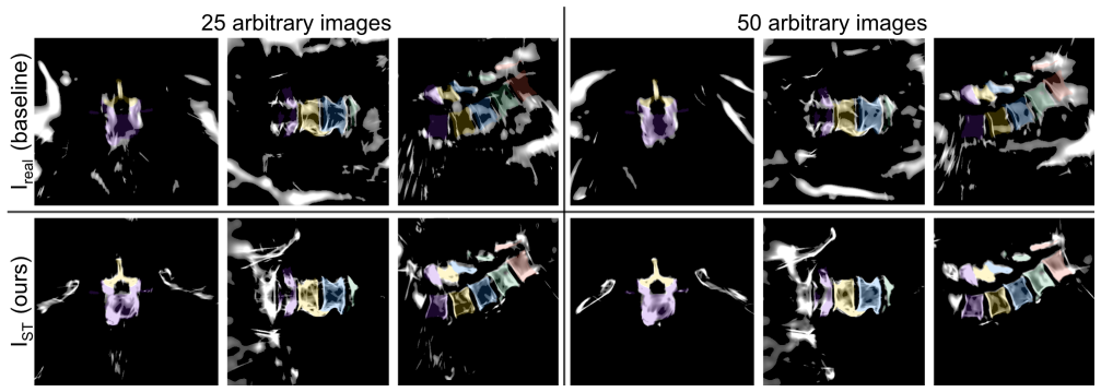
On its own terms, the paper establishes that a pretraining-free Gaussian-splatting pipeline can reconstruct a volumetric lumbar-spine density map from as few as 20-30 arbitrarily posed real intraoperative X-rays, at a level an experienced surgeon rates acceptable to very good for pedicle-screw navigation. The discovery is that the bottleneck is not the splatting optimization itself but the inconsistency of real X-ray appearance: mapping each real radiograph into a standardized, bone-emphasized synthetic-like domain with style transfer before reconstruction is what lifts the volumes from unusable to navigable. With 50 views the standardized inputs reach 25.73 dB PSNR versus 23.22 dB for raw X-rays, still below the 39.0 dB of an idealized circular synthetic baseline, and the reconstructed slices show clearly discernible pedicles, spinous processes, and endplates. The same test set is used across all view-count experiments, so the comparison isolates the effect of view number and input domain.
Load-bearing premise
During evaluation the reconstruction volume's center and size come from the ground-truth CT; the clinical promise depends on estimating them from the calibrated X-ray views alone, which the paper does not demonstrate.
Editorial extensions
If this is right
- If the central claim holds, a C-arm operator could collect 20-30 fluoroscopic shots and obtain a navigational 3D volume without the radiation burden of a cone-beam CT sweep, since the method needs no pretraining and no per-patient training data.
- The style-transfer standardization becomes a reusable front-end for any real-X-ray Gaussian-splatting reconstruction, since raw real X-rays alone produced volumes the authors judged insufficient for expert assessment.
- The trend in surgeon ratings and the plateau in PSNR/SSIM past 20 views suggest a practical acquisition protocol: around 20-30 views are enough, and adding views yields diminishing returns.
- Because the output is a voxel density map with no calibrated Hounsfield units, clinical use would target navigation and visualization, not quantitative CT-like density measurement.
Reading between the lines
- Beyond the paper: if volume bounds can be estimated from calibrated views, for instance by triangulating principal axes, the whole pipeline becomes CT-free and could run on a standard C-arm in the operating room; this is the natural next validation.
- Beyond the paper: the same standardization-plus-splatting recipe may transfer to other bony anatomies such as the femur, pelvis, or cervical spine because there is no anatomy-specific pretraining, but the 20-30-view threshold would need re-measuring per site.
- Beyond the paper: adding a fast initialization, such as a coarse reconstruction or a rough density prior, could cut the reported 13-minute optimization and make intraoperative use practical, since the paper itself notes that convergence can be accelerated.
- Beyond the paper: replacing PSNR/SSIM with a task-based metric, or overlaying ground-truth segmentations automatically, could turn expert 'Acceptable' ratings into a quantitative acceptance criterion for navigation.
Signed reviews
Editorial analysis
A structured set of objections, weighed in public.
Referee Report
Summary. The manuscript presents IXGS, a Gaussian-splatting-based framework for volumetric reconstruction of the lumbar spine from sparse, arbitrarily posed real intraoperative X-ray images. It builds on R2-Gaussian, replaces the FDK-based initialization with random initialization inside a reconstruction volume, and introduces an anatomy-guided radiographic standardization step based on Pix2Pix style transfer that maps real X-rays into a synthetic DRR-like domain. The evaluation uses six ex-vivo specimens: an expert surgeon rates 3D volume renderings and slice views for navigation usability, and PSNR/SSIM on held-out views compare reconstructions from synthetic circular, synthetic arbitrary, raw real, and style-transferred inputs. The authors report that clinically acceptable ratings appear at roughly 20-30 views and that standardization improves quantitative scores.
Significance. If the claims are upheld, the paper makes a useful contribution: it is among the first to apply radiative Gaussian splatting to sparse, arbitrary-pose real intraoperative X-rays, and it provides a practical preprocessing recipe that visibly improves bone clarity. The instance-based nature of the splatting optimization is attractive because it avoids anatomy-specific training of the reconstruction network, and the promised release of code and a small dataset is a strength. However, the reported results are conditioned on CT-derived volume bounds, the evaluation lacks quantitative 3D accuracy and error bars, and the test-domain protocol is not fully consistent across conditions. The expert ratings and the 2D trends are encouraging, but the central 'X-ray-only' feasibility claim is currently stronger than the evidence supports.
major comments (5)
- [Methods, Initialization] The reconstruction volume center and dimensions are defined from ground-truth CT for all reported experiments, as stated in the text: 'For the evaluations presented in this work, where ground truth CT data was available, the center and dimensions of the reconstruction volume were defined based on the CT.' Because kernel positions are randomly sampled within that volume, every expert rating and every PSNR/SSIM result evaluates a pipeline that has been given a geometric prior unavailable in a pure intraoperative X-ray workflow. This does not invalidate the reconstruction optimization itself, but it means the 'from sparse, arbitrarily posed real X-rays' feasibility claim is not yet demonstrated for the full pipeline. Please either implement and evaluate the volume-estimation approach from calibrated views (e.g., the triangulation cited from prior work) or explicitly reframe the claim and add a sensitivity analysis over shifted or scaled reconstruction volumes.
- [Experiments, 3D Evaluation] The paper reports no quantitative 3D accuracy metric, although CT segmentations of L1-L5 are available and are used for overlay visualization. The conclusion that the reconstructions are 'anatomically consistent' rests on expert Likert ratings and 2D PSNR/SSIM, yet the authors themselves argue that such 2D metrics correlate poorly with anatomical plausibility. A voxel-wise or surface-distance comparison between the reconstructed density volume and the CT-derived bone mask is needed to support a volumetric reconstruction claim; the segmentations already in hand make this feasible.
- [Table 1 and Figure 6] The quantitative comparison reports only averages over six specimens with no error bars, per-specimen values, or significance tests. Figure 6 shows that one specimen never received a rating above 'Poor', indicating substantial inter-specimen variability; the 2.51 dB PSNR gain of IST over Ireal and the 20-30-view guideline may not be representative. Please report per-specimen results and variance, and state whether the reported differences are statistically significant.
- [Experiments, 3D Evaluation and Figure 3] The expert evaluation protocol allowed the surgeon to toggle an overlay of ground-truth CT segmentations while rating anatomical accuracy. If ratings were assigned with the overlay visible, high scores may reflect how well the reconstruction aligns with ground truth available to the rater, rather than standalone clinical usability. Please clarify whether the overlay was visible during scoring or only used after scoring, or perform a blinded evaluation without the overlay.
- [Experiments, 2D Quantitative Evaluation and Table 1] The manuscript does not specify whether the held-out test images for the IST condition are the Pix2Pix-transformed versions or the original real X-rays. If the former, the reported IST-vs-Ireal gain partly measures self-consistency of the style-transfer mapping rather than reconstruction fidelity to the original real X-ray content. Please state explicitly which test domain is used for each condition and, ideally, evaluate all reconstructions against the same held-out set, with the remaining domain mismatch discussed.
minor comments (5)
- [Throughout] Several cross-references appear as empty 'Section' placeholders (e.g., in Related Work and Methods), and Figure 6's caption refers to 'Section' without a number; these should be resolved before publication.
- [Methods, Optimization] The sentence beginning 'The optimize the 3D Gaussian kernels' should read 'To optimize the 3D Gaussian kernels'.
- [Results, Table 1] Table 1 reports the Icirc 50-view PSNR as 39.19 dB while the text states 39.0 dB; please make these values consistent.
- [Abstract and Methods, Anatomy-guided Radiographic Standardization] The abstract's claim that 'our framework requires no pretraining' should be qualified, since the Pix2Pix standardization step is trained on paired data; the claim is accurate for the Gaussian optimization but not for the full pipeline as stated.
- [Related Work] The sentence 'Both techniques and synthetic X-ray-based reconstructions32–37.' is grammatically incomplete and should be revised.
Circularity Check
Quantitative IST improvement is evaluated against the same DRR domain used to train the Pix2Pix style-transfer; central 3D reconstruction is otherwise self-contained.
-
fitted input called prediction
[Methods, 'Anatomy-guided Radiographic Standardization', Eqs.]
"L_L1(G) = EIreal,IDRR,z[∥ IDRR−G(Ireal,z)∥1] ... After training, the generator G was used to style-transfer Ireal to the synthetic domain, producing IST = G(Ireal). ... The IDRR images — generated directly from the ground truth CT scans using the exact same arbitrary poses as the corresponding Ireal images — serve as a synthetic benchmark and represent the target appearance domain for our proposed IST radiographic standardization step. ..."
The Pix2Pix generator is fitted by minimizing the L1 distance between G(Ireal) and the synthetic DRR domain IDRR (Eq. 4), so the output domain of the fitted stylization is, by construction, the DRR domain. The quantitative evaluation then identifies IDRR as the target appearance domain for IST and reports the IST-vs-Ireal gain (25.73 vs 23.22 dB) as evidence that the standardization improves reconstruction quality. Because the benchmark rewards closeness to the very domain that Pix2Pix was trained to produce, the measured improvement is partly self-consistency of the fitted stylization rather than an independent measurement of 3D reconstruction fidelity.
full rationale
The central 3D reconstruction pipeline is not circular: the Gaussian splatting is optimized per instance against the input views without anatomy-specific pretraining, rendered from held-out poses, and evaluated both quantitatively and by expert qualitative assessment. The R2-Gaussian backbone is externally published, code-reproduced, and the authors' extension to arbitrary poses with random initialization is a substantive implementation change rather than a renaming of the prior result. The CT-derived volume bounds in the Initialization section are a serious evaluation limitation and are explicitly acknowledged by the authors; however, this is ground-truth conditioning of the test setup, not a derivation that reduces the method to its own inputs, so it does not by itself constitute circularity. The main circularity concern is the quantitative validation of the anatomy-guided standardization: Pix2Pix is trained (Eq. 4) to map Ireal into the IDRR domain, and the benchmark treats IDRR as the target appearance domain for IST, so the reported improvement of IST over Ireal is partly baked into the stylization training objective. This weakens the preprocessing claim but does not undermine the core feasibility conclusion, which also rests on surgeon ratings and held-out novel-view synthesis. Overall score 4 reflects a partial, localized circularity in the standardization evaluation while the central reconstruction claim retains independent content.
Assumptions & free parameters
free parameters (1)
- Volume density threshold for postprocessing =
80th percentile
assumptions (5)
- domain assumption Fiducial-based calibration, using DLT plus RANSAC, gives camera poses accurate enough for the splatting optimization.
- domain assumption Pix2Pix style transfer to the DRR domain preserves anatomy and does not hallucinate or erase structures.
- domain assumption Random Gaussian initialization converges to an acceptable volume from 20 to 50 arbitrary views.
- domain assumption Reconstruction volume bounds are known from CT during evaluation and can be estimated from calibrated X-rays in clinical use.
- domain assumption DRR synthesis with a 0 HU threshold is a valid standardized representation of bone for surgical navigation.
Cite this review
Pith. "Pith review of IXGS-Intraoperative 3D Reconstruction from Sparse, Arbitrarily Posed Real X-rays." pith.science (2026). https://pith.science/paper/WVSHJHYX
@misc{pith2026250414699,
  author       = {Pith},
  title        = {Pith review of: IXGS-Intraoperative 3D Reconstruction from Sparse, Arbitrarily Posed Real X-rays},
  year         = {2026},
  howpublished = {\url{https://pith.science/paper/WVSHJHYX}},
  note         = {Machine review of arXiv:2504.14699}
}
abstract
Spine surgery is a high-risk intervention demanding precise execution, often supported by image-based navigation systems. Recently, supervised learning approaches have gained attention for reconstructing 3D spinal anatomy from sparse fluoroscopic data, significantly reducing reliance on radiation-intensive 3D imaging systems. However, these methods typically require large amounts of annotated training data and may struggle to generalize across varying patient anatomies or imaging conditions. Instance-learning approaches like Gaussian splatting could offer an alternative by avoiding extensive annotation requirements. While Gaussian splatting has shown promise for novel view synthesis, its application to sparse, arbitrarily posed real intraoperative X-rays has remained largely unexplored. This work addresses this limitation by extending the $R^2$-Gaussian splatting framework to reconstruct anatomically consistent 3D volumes under these challenging conditions. We introduce an anatomy-guided radiographic standardization step using style transfer, improving visual consistency across views, and enhancing reconstruction quality. Notably, our framework requires no pretraining, making it inherently adaptable to new patients and anatomies. We evaluated our approach using an ex-vivo dataset. Expert surgical evaluation confirmed the clinical utility of the 3D reconstructions for navigation, especially when using 20 to 30 views, and highlighted the standardization's benefit for anatomical clarity. Benchmarking via quantitative 2D metrics (PSNR/SSIM) confirmed performance trade-offs compared to idealized settings, but also validated the improvement gained from standardization over raw inputs. This work demonstrates the feasibility of instance-based volumetric reconstruction from arbitrary sparse-view X-rays, advancing intraoperative 3D imaging for surgical navigation.
Figures


Figures from the paper (4 more)


Reference graph
Works this paper leans on
-
[1]
Tonetti, J., Boudissa, M., Kerschbaumer, G. & Seurat, O. Role of 3D intraoperative imaging in orthopedic and trauma surgery. Orthop. & Traumatol. Surg. & Res. 106, S19–S25, DOI: 10.1016/j.otsr.2019.05.021 (2020)
-
[2]
Keil, H. et al. Intraoperative revision rates due to three-dimensional imaging in orthopedic trauma surgery: results of a case series of 4721 patients. Eur. J. Trauma Emerg. Surgery: Off. Publ. Eur. Trauma Soc. 49, 373–381, DOI: 10.1007/s00068-022-02083-x (2023)
-
[3]
Hong, J. & Hashizume, M. An effective point-based registration tool for surgical navigation. Surg. Endosc. 24, 944–948, DOI: 10.1007/s00464-009-0568-2 (2010)
-
[4]
Ma, Q. et al. Autonomous Surgical Robot With Camera-Based Markerless Navigation for Oral and Maxillofacial Surgery. IEEE/ASME Transactions on Mechatronics 25, 1084–1094, DOI: 10.1109/TMECH.2020.2971618 (2020). Conference Name: IEEE/ASME Transactions on Mechatronics
arXiv 2020
-
[5]
Suenaga, H. et al. Vision-based markerless registration using stereo vision and an augmented reality surgical navigation system: a pilot study. BMC Med. Imaging 15, 51, DOI: 10.1186/s12880-015-0089-5 (2015). 13/17
-
[6]
Zheng, J., Miao, S., Wang, Z. J. & Liao, R. Pairwise domain adaptation module for CNN-based 2-D/3-D registration. J. Med. Imaging (Bellingham, Wash.) 5, 021204, DOI: 10.1117/1.JMI.5.2.021204 (2018)
-
[7]
Ferrante, E., Oktay, O., Glocker, B. & Milone, D. H. On the Adaptability of Unsupervised CNN-Based Deformable Image Registration to Unseen Image Domains. In Shi, Y ., Suk, H.-I. & Liu, M. (eds.)Machine Learning in Medical Imaging , 294–302, DOI: 10.1007/978-3-030-00919-9_34 (Springer International Publishing, Cham, 2018)
-
[8]
Zhang, J. N., Fan, Y . & Hao, D. J. Risk factors for robot-assisted spinal pedicle screw malposition.Sci. Reports 9, 3025, DOI: 10.1038/s41598-019-40057-z (2019). Publisher: Nature Publishing Group
Show all 59 references
-
[9]
W., Pirris, S
Rahmathulla, G., Nottmeier, E. W., Pirris, S. M., Deen, H. G. & Pichelmann, M. A. Intraoperative image-guided spinal navigation: technical pitfalls and their avoidance. Neurosurg. F ocus.36, E3, DOI: 10.3171/2014.1.FOCUS13516 (2014)
2014 doi
-
[10]
& Elluru, S
Venkatesh, E. & Elluru, S. V . Cone beam computed tomography: basics and applications in dentistry.J. Istanbul Univ. Fac. Dent. 51, S102–S121, DOI: 10.17096/jiufd.00289 (2017)
2017 doi
-
[11]
Costa, F. et al. Spinal Navigation: Standard Preoperative: Versus: Intraoperative Computed Tomography Data Set Acquisition for Computer-Guidance System: Radiological and Clinical Study in 100 Consecutive Patients. Spine 36, 2094–2098, DOI: 10.1097/BRS.0b013e318201129d (2011)
2011 doi
-
[12]
Mendelsohn, D. et al. Patient and surgeon radiation exposure during spinal instrumentation using intraoperative computed tomography-based navigation. The Spine J. 16, 343–354, DOI: 10.1016/j.spinee.2015.11.020 (2016)
2016 doi
-
[13]
Villard, J. et al. Radiation Exposure to the Surgeon and the Patient During Posterior Lumbar Spinal Instrumentation: A Prospective Randomized Comparison of Navigated Versus Non-navigated Freehand Techniques. Spine 39, 1004, DOI: 10.1097/BRS.0000000000000351 (2014)
2014 doi
-
[14]
Dea, N. et al. Economic evaluation comparing intraoperative cone beam CT-based navigation and conventional fluoroscopy for the placement of spinal pedicle screws: a patient-level data cost-effectiveness analysis. The Spine J. 16, 23–31, DOI: 10.1016/j.spinee.2015.09.062 (2016)
2016 doi
-
[15]
& Gradl, G
Beck, M., Mittlmeier, T., Gierer, P., Harms, C. & Gradl, G. Benefit and accuracy of intraoperative 3D-imaging after pedicle screw placement: a prospective study in stabilizing thoracolumbar fractures. Eur. Spine J. 18, 1469–1477, DOI: 10.1007/s00586-009-1050-5 (2009)
2009 doi
-
[16]
& Esfandiari, H
Jecklin, S., Jancik, C., Farshad, M., Fürnstahl, P. & Esfandiari, H. X23D—intraoperative 3D lumbar spine shape reconstruction based on sparse multi-view X-ray data. J. Imaging 8, 271, DOI: 10.3390/jimaging8100271 (2022). Publisher: MDPI
2022 doi
-
[17]
Jecklin, S. et al. Domain adaptation strategies for 3D reconstruction of the lumbar spine using real fluoroscopy data. Med. Image Analysis 98, 103322, DOI: 10.1016/j.media.2024.103322 (2024). Publisher: Elsevier
2024
-
[18]
Zha, R. et al. R$^2$-Gaussian: Rectifying Radiative Gaussian Splatting for Tomographic Reconstruction. Adv. Neural Inf. Process. Syst. 37, 44907–44934 (2024)
2024
-
[19]
Zha, R., Zhang, Y . & Li, H. NAF: Neural Attenuation Fields for Sparse-View CBCT Reconstruction. In Wang, L., Dou, Q., Fletcher, P. T., Speidel, S. & Li, S. (eds.)Medical Image Computing and Computer Assisted Intervention – MICCAI 2022 , 442–452, DOI: 10.1007/978-3-031-16446-0...
2022 doi
-
[20]
& Heidrich, W
Rückert, D., Wang, Y ., Li, R., Idoughi, R. & Heidrich, W. NeAT: neural adaptive tomography.ACM Trans. Graph. 41, 55:1–55:13, DOI: 10.1145/3528223.3530121 (2022)
2022
-
[21]
& Miltchev, R
Rangelov, D., Waanders, S., Waanders, K., van Keulen, M. & Miltchev, R. Impact of Camera Settings on 3D Reconstruction Quality: Insights from NeRF and Gaussian Splatting. Sensors (Basel, Switzerland) 24, 7594, DOI: 10.3390/s24237594 (2024)
2024 doi
-
[22]
& Liu, Y
Ye, S., Dong, Z., Hu, Y ., Wen, Y . & Liu, Y . Gaussian in the Dark: Real-Time View Synthesis From Inconsistent Dark Images Using Gaussian Splatting. Comput. Graph. F orum43, e15213, DOI: 10.1111/cgf.15213 (2024). _eprint: https://onlinelibrary.wiley.com/doi/pdf/10.1111/cgf.15213
2024 doi
- [23]
-
[24]
& Kovler, I
Kasten, Y ., Doktofsky, D. & Kovler, I. End-To-End Convolutional Neural Network for 3D Reconstruction of Knee Bones from Bi-planar X-Ray Images. In Deeba, F., Johnson, P., Würfl, T. & Ye, J. C. (eds.)Machine Learning for Medical Image Reconstruction, 123–133, DOI: 10.1007/978-...
-
[25]
Shiode, R. et al. 2D–3D reconstruction of distal forearm bone from actual X-ray images of the wrist using convolutional neural networks. Sci. Reports 11, 15249, DOI: 10.1038/s41598-021-94634-2 (2021). Publisher: Nature Publishing Group. 14/17
2021 doi
-
[26]
Ge, R. et al. X-CTRSNet: 3D cervical vertebra CT reconstruction and segmentation directly from 2D X-ray images. Knowledge-Based Syst. 236, 107680, DOI: 10.1016/j.knosys.2021.107680 (2022)
2022
-
[27]
& Malik, J
Kar, A., Häne, C. & Malik, J. Learning a Multi-View Stereo Machine. arXiv:1708.05375 [cs] (2017)
2017 arXiv
-
[28]
Luchmann, D. et al. Spinal navigation with AI-driven 3D-reconstruction of fluoroscopy images: an ex-vivo feasibility study. BMC Musculoskelet. Disord. 25, 925, DOI: 10.1186/s12891-024-08052-2 (2024). Publisher: BioMed Central London
2024 doi
-
[29]
Mildenhall, B. et al. NeRF: representing scenes as neural radiance fields for view synthesis. Commun. ACM 65, 99–106, DOI: 10.1145/3503250 (2021)
2021 doi
-
[30]
& Drettakis, G
Kerbl, B., Kopanas, G., Leimkuehler, T. & Drettakis, G. 3D Gaussian Splatting for Real-Time Radiance Field Rendering. ACM Trans. Graph. 42, 139:1–139:14, DOI: 10.1145/3592433 (2023)
2023 doi
-
[31]
Rakotosaona, M.-J. et al. NeRFMeshing: Distilling Neural Radiance Fields into Geometrically-Accurate 3D Meshes. In 2024 International Conference on 3D Vision (3DV) , 1156–1165, DOI: 10.1109/3DV62453.2024.00093 (2024). ISSN: 2475-7888
2024
-
[32]
Corona-Figueroa, A. et al. MedNeRF: Medical Neural Radiance Fields for Reconstructing 3D-aware CT-Projections from a Single X-ray. 2022 44th Annu. Int. Conf. IEEE Eng. Medicine & Biol. Soc. (EMBC) 3843–3848, DOI: 10. 1109/EMBC48229.2022.9871757 (2022). Conference Name: 2022 44...
2022
-
[33]
Radiative Gaussian Splatting for Efficient X-Ray Novel View Synthesis
Cai, Y .et al. Radiative Gaussian Splatting for Efficient X-Ray Novel View Synthesis. In Leonardis, A. et al. (eds.) Computer Vision – ECCV 2024, 283–299, DOI: 10.1007/978-3-031-73232-4_16 (Springer Nature Switzerland, Cham, 2025)
2024 doi
-
[34]
Gao, Z. et al. DDGS-CT: Direction-Disentangled Gaussian Splatting for Realistic V olume Rendering.Adv. Neural Inf. Process. Syst. 37, 39281–39302 (2024)
2024
-
[35]
Wysocki, M. et al. Ultra-NeRF: Neural Radiance Fields for Ultrasound Imaging. In Medical Imaging with Deep Learning , 382–401 (PMLR, 2024). ISSN: 2640-3498
2024
-
[36]
Awojoyogbe, B. O. & Dada, M. O. Neural Radiance Fields (NeRFs) Technique to Render 3D Reconstruction of Magnetic Resonance Images. In Awojoyogbe, B. O. & Dada, M. O. (eds.) Digital Molecular Magnetic Resonance Imaging , 247–258, DOI: 10.1007/978-981-97-6370-2_10 (Springer Natu...
-
[37]
Yang, S. et al. Deform3DGS: Flexible Deformation for Fast Surgical Scene Reconstruction with Gaussian Splatting. In Linguraru, M. G. et al. (eds.) Medical Image Computing and Computer Assisted Intervention – MICCAI 2024 , 132–142, DOI: 10.1007/978-3-031-72089-5_13 (Springer Na...
2024 doi
-
[38]
& Wang, A
Cai, Y ., Wang, J., Yuille, A., Zhou, Z. & Wang, A. Structure-Aware Sparse-View X-ray 3D Reconstruction. 11174–11183 (2024)
2024
-
[39]
X-ray Tomographic Datasets Archives
-
[40]
VertXNet: an ensemble method for vertebral body segmentation and identification from cervical and lumbar spinal X-rays
Chen, Y .et al. VertXNet: an ensemble method for vertebral body segmentation and identification from cervical and lumbar spinal X-rays. Sci. Reports 14, 3341, DOI: 10.1038/s41598-023-49923-3 (2024). Publisher: Nature Publishing Group
2024 doi
-
[41]
C., Cho, H
Kim, K. C., Cho, H. C., Jang, T. J., Choi, J. M. & Seo, J. K. Automatic detection and segmentation of lumbar vertebrae from X-ray images for compression fracture evaluation. Comput. Methods Programs Biomed. 200, 105833, DOI: 10.1016/j.cmpb.2020.105833 (2021)
2021
- [42]
-
[43]
Kirillov, A. et al. Segment Anything. 4015–4026 (2023)
2023
- [44]
-
[45]
Ehab, W., Huang, L. & Li, Y . UNet and Variants for Medical Image Segmentation.Int. J. Netw. Dyn. Intell. 100009–100009, DOI: 10.53941/ijndi.2024.100009 (2024)
2024
-
[46]
& Tallamraju, R
Gupta, S., Lakhotia, S., Rawat, A. & Tallamraju, R. ViTOL: Vision Transformer for Weakly Supervised Object Localization. 4101–4110 (2022). 15/17
2022
-
[47]
Coarse X-ray Lumbar Vertebrae Pose Localization and Registration Using Triangulation Correspon- dence
Yookwan, W.et al. Coarse X-ray Lumbar Vertebrae Pose Localization and Registration Using Triangulation Correspon- dence. Processes 11, 61, DOI: 10.3390/pr11010061 (2023). Number: 1 Publisher: Multidisciplinary Digital Publishing Institute
2023 doi
-
[48]
& Zheng, G
Ye, K., Sun, W., Tao, R. & Zheng, G. A Projective-Geometry-Aware Network for 3D Vertebra Localization in Calibrated Biplanar X-Ray Images. Sensors 25, 1123, DOI: 10.3390/s25041123 (2025). Number: 4 Publisher: Multidisciplinary Digital Publishing Institute
2025 doi
-
[49]
& Liao, H.-Y
Wang, C.-Y ., Bochkovskiy, A. & Liao, H.-Y . M. YOLOv7: Trainable Bag-of-Freebies Sets New State-of-the-Art for Real-Time Object Detectors. 7464–7475 (2023)
2023
-
[50]
& Efros, A
Zhu, J.-Y ., Park, T., Isola, P. & Efros, A. A. Unpaired Image-To-Image Translation Using Cycle-Consistent Adversarial Networks. In 2017 IEEE International Conference on Computer Vision (ICCV) , 2223–2232, DOI: 10.1109/ICCV .2017.244 (IEEE, Venice, 2017)
2017 doi
-
[51]
Duda, R. O. & Hart, P. E. Use of the Hough transformation to detect lines and curves in pictures. Commun. ACM 15, 11–15, DOI: 10.1145/361237.361242 (1972)
1972
-
[52]
I., Karara, H
Abdel-Aziz, Y . I., Karara, H. M. & Hauck, M. Direct Linear Transformation from Comparator Coordinates into Object Space Coordinates in Close-Range Photogrammetry*. Photogramm. Eng. & Remote. Sens. 81, 103–107, DOI: 10.14358/ PERS.81.2.103 (2015)
2015
-
[53]
A., Davis, L
Feldkamp, L. A., Davis, L. C. & Kress, J. W. Practical cone-beam algorithm. JOSA A 1, 612–619, DOI: 10.1364/JOSAA.1. 000612 (1984). Publisher: Optica Publishing Group
1984 doi
-
[54]
& Gross, M
Zwicker, M., Pfister, H., van Baar, J. & Gross, M. EW A splatting.IEEE Transactions on Vis. Comput. Graph. 8, 223–238, DOI: 10.1109/TVCG.2002.1021576 (2002)
2002 arXiv
-
[55]
& Simoncelli, E
Wang, Z., Bovik, A., Sheikh, H. & Simoncelli, E. Image quality assessment: from error visibility to structural similarity. IEEE Transactions on Image Process. 13, 600–612, DOI: 10.1109/TIP.2003.819861 (2004). Conference Name: IEEE Transactions on Image Processing
2004
-
[56]
I., Osher, S
Rudin, L. I., Osher, S. & Fatemi, E. Nonlinear total variation based noise removal algorithms. Phys. D: Nonlinear Phenom. 60, 259–268, DOI: 10.1016/0167-2789(92)90242-F (1992)
1992 doi
- [57]
-
[58]
A., Baltruschat, I
Dohmen, M., Klemens, M. A., Baltruschat, I. M., Truong, T. & Lenga, M. Similarity and quality metrics for MR image- to-image translation. Sci. Reports 15, 3853, DOI: 10.1038/s41598-025-87358-0 (2025). Publisher: Nature Publishing Group
2025 doi
-
[59]
A computer- implemented method, device, system and computer program product for processing anatomic imaging data
Zhang, R., Isola, P., Efros, A. A., Shechtman, E. & Wang, O. The Unreasonable Effectiveness of Deep Features as a Perceptual Metric. 586–595 (2018). Acknowledgements This work has been supported by the OR-X - a Swiss national research infrastructure for translational surgery a...
2018
Reviewed August 16, 2026 · model on record in the stance chip above.
Discussion (0). Continue with ORCID to comment.