REVIEW 3 major objections 8 minor 49 references
Learning via mechanosensitivity and activity in cytoskeletal networks
T0 review · 3 major / 8 minor · reviewed 2026-08-16 · deepseek-v4-flash
Pith's one-line read A disordered Hookean spring network carrying mechanosensitive proteins and motors can learn a desired strain response by remodeling rest lengths, through a contrastive-learning rule powered by strain memory.
desk verdict A useful proof-of-principle for learning in cytoskeletal networks, but the reported simulations run outside the stated temporal-contrastive regime, so the mechanistic claim is not yet demonstrated. read the letter →
The pith
A machine-rendered reading of the paper's core claim, the machinery that carries it, and where it could break.
The reading
What carries the argument
The load-bearing object is the strain memory kernel in Eq. (5): $K(t-t') = \beta_1[\delta(t-t') - (1/\tau_k)e^{-(t-t')/\tau_k}]$, obtained by linearizing fast mechanosensitive-protein kinetics and coupling them to motor binding. It converts local strain history into motor density, hence active force, and makes each edge's learning rule $\dot{L}^0 = \alpha g(f^a)$ respond to the difference between clamped and free strain rather than to instantaneous strain alone. The learning degree of freedom (LDOF) is the edge rest length (or stiffness), whose thresholded, active-force-dependent remodeling carries the contrastive update.
What would settle it
Run the same training protocol with the mechanosensitive protein unbinding rate set comparable to or slower than the driving timescale (for example, $k_{un} \lesssim 1/\tau_f$): if training error still decreases as in the fast-protein case, the protein-relaxation assumption is not load-bearing, and if learning fails, the assumption is confirmed as necessary. A complementary experiment would measure whether adaptation to repeated stretch is impaired when mechanosensitive protein recruitment is blocked in an actomyosin-like system.
Extended reading notes
Core claim
The paper's central claim is that a disordered elastic network whose learning degrees of freedom, the rest length or stiffness of each edge, are updated only when the local active motor force exceeds a threshold can learn a prescribed strain at a target edge in response to a strain applied at a source edge. The mechanism runs through a memory kernel: because mechanosensitive proteins react to strain rate and promote motor binding, motor density obeys $\delta m = \int_{-\infty}^{t} K(t-t')\,\delta\epsilon(t')\,dt'$ with $K(t-t') = \beta_1[\delta(t-t') - \tau_k^{-1}e^{-(t-t')/\tau_k}]$, so the active force at each edge carries a filtered estimate of strain history. When training alternates sharply between a free state and a clamped state, this implicit memory makes the rest-length update $\dot{L}^0_{jk} = \alpha g(f^a_{jk})$ behave as a contrastive learning rule, reducing training error toward zero. The authors show the learning survives nonlinear protein kinetics and nonlinear elasticity, persists under edge turnover when severing is slow enough, extends to classifying strain-gradient signs, and can be driven by self-organized actomyosin-like pulses rather than an external supervisor; in the pulse-driven case the same rule yields adaptation of a low-strain region.
Load-bearing premise
The whole argument rests on the mechanosensitive proteins reacting to each stretch instantly: in Appendix A the protein density is linearized to its instantaneous steady state, so the only memory in the motor dynamics comes from the simple kernel of Eq. (5); if those proteins bind and unbind too slowly, the strain memory the learning needs gets smeared out.
Editorial extensions
If this is right
- Training error at the target edge falls toward zero over repeated free-clamped cycles, so the network stores the source-to-target strain relation in its rest lengths.
- Learning is not tied to one geometry: it works for random source-target pairs, across tested network sizes, and for stiffness remodeling as well as rest-length remodeling.
- Nonlinearities in protein kinetics and nonlinear elastic strain stiffening do not break learning; moderate nonlinearity can even lower training error.
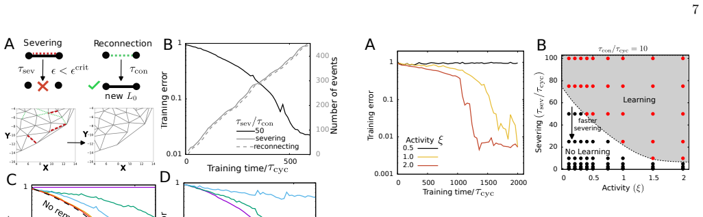
- Network turnover is tolerable: edges can sever and reconnect, and as long as severing is slow relative to reconnection, learning continues; increased contractility can restore learning after turnover-induced loss.
- The same mechanism supports classification of strain-gradient signs and, with length-dependent pulsation amplitude, self-organized learning without an external supervisor; it also drives adaptation to keep a designated region near zero strain.
Reading between the lines
- If this mechanism is general, any cellular machinery with a fast strain-rate-sensitive binding step feeding a slower active process could serve as a physical learning module; the proteins modeled here are one instance.
- The memory-kernel form suggests a quantitative design rule: the contrastive signal works best when the motor turnover timescale $\tau_k = k_u^{-1}$ is short compared with the driving cycle, so tuning motor unbinding rate should tune learning speed, a dependence the simulations use but do not systematically sweep.
- The adaptation result implies that continuous learning and homeostasis may be the same process; a testable extension is to ask whether repeated, changing perturbations preserve the learned low-strain state better than a single static perturbation.
- Because the learning rule uses only local active force, it should transfer to vertex or tissue-scale models, where junction remodeling under myosin pulses could implement the same contrastive update.
Signed reviews
Editorial analysis
A structured set of objections, weighed in public.
Referee Report
Summary. This paper proposes a computational model of a disordered spring network inspired by the actomyosin cytoskeleton. Each edge carries two molecular species: a mechanosensitive protein whose unbinding is strain-rate dependent (Eq. 3) and a motor whose binding is promoted by that protein (Eq. 2). Adiabatic elimination of the protein density gives the motor density an implicit memory of local strain, summarized by the kernel in Eq. (5) with timescale τ_k = 1/k_u and strength β_1 = k_1^b β n_0. The authors argue, building on Ref. [58], that driving the network asymmetrically between free and clamped states (fast clamp, slow release) turns this memory into a temporal-contrastive update of the learning degrees of freedom, which in this model are edge rest lengths (Eq. 6) or stiffnesses (Eq. 9). Simulations show decreasing training error for rest-length and stiffness learning, robustness to nonlinearities and edge turnover, classification of strain-gradient inputs, self-organized learning under actomyosin pulsation with length-dependent feedback, and adaptive maintenance of a low-strain 'perinuclear' region. The paper closes with experimentally testable predictions.
Significance. The analytic core is a strength: the memory kernel in Eq. (5) is derived from the model's own equations (Appendices A-B), the identification of τ_k and β_1 with k_u^{-1} and k_1^b β n_0 is checked by direct differentiation, and the learning outcomes are established by independent simulations of the dynamical system rather than assumed. The paper also tests robustness across several axes (activity, turnover, nonlinearities, network size) and offers falsifiable predictions, which is commendable. If the mechanism is as claimed, the work would be a significant minimal biophysical realization of temporal contrastive learning outside the neural context, extending Ref. [58] with thresholded remodeling, active contractility, turnover, and a homeostasis application. The significance is conditional, however, because the reported simulations appear to lie outside the regime in which the temporal-contrastive interpretation is valid; the mechanistic attribution is therefore not yet established by the evidence presented.
major comments (3)
- [Appendix A / Fig. 2] Appendix A states the contrastive requirement as τ_k < τ_f < τ_s and asserts that τ_cyc = 100 s is much larger than the motor turnover timescale, with τ_f/τ_s = 1/4. The reported simulations use rescaled motor unbinding rates k_u = 0.4–0.5 (Figs. 2–9, e.g., Fig. 2D), which with τ_cyc = 100 s gives τ_k = 1/k_u = 200–250 s, i.e., 2–2.5 cycles, about ten times τ_f = 20 s. This violates the stated condition and places the runs in the slow-memory regime, in which the kernel of Eq. (5) gives δm ≈ β_1 δϵ(t) rather than the strain-rate estimate δm ≈ β_1 τ_k δϵ̇(t) required for the temporal-contrastive argument of Ref. [58]. The observed learning could therefore be a slow-memory averaging effect rather than contrastive learning, and the substantial 'unlearning' reported in Fig. S5 (rest-length updates reversing sign during the slow release phase) is consistent with a strain-tracking, not a strain-rate-estimating, motor density. Note that if k_u = 0.5 were a real-time rate (0.5 s^{-1}), the rescaled value would be k̃_u = k_u τ_cyc = 50 and the stated regime would hold, so the discrepancy could be a units error in the captions; as written, the text and the simulations are inconsistent. Please clarify the units of k_u, rerun training in the regime τ_k < τ_f < τ_s (e.g., rescaled k_u ≳ 5), or provide per-cycle evidence (δm(t) records and a decomposition of the L0 update into clamp and release contributions) showing that the contrastive component dominates in the regime actually simulated. The Sections III–V attribution to temporal contrastive learning should be restricted to the regime in which it is demonstrated.
- [Appendix A, below Eq. (A4)] The memory kernel is derived by adiabatic elimination of the mechanosensitive protein density, δn = n_0 β δϵ̇, justified by the statement 'as the protein dynamics is fast.' No numerical value of the protein unbinding rate k_un^0 is reported for any simulation, so the validity of this elimination on the driving timescales used (τ_f = 20 s for the Fig. 2 parameters, and 2 s for the classification task with τ_cyc = 10 s) is never checked. If the simulations integrate the reduced equations (A7) directly, the fast-protein assumption is imposed rather than tested, and the manuscript should say so explicitly and justify the elimination on timescale grounds; if the full protein dynamics of Eq. (3) is integrated, k_un^0 and k_bn must be given. Please report these values and, ideally, repeat one training run with the full protein dynamics to confirm that Eq. (5) remains the correct memory kernel in the regime of interest.
- [Section III and Eq. (6)] The paper asserts, rather than derives, that the implicit memory of Eqs. (4)-(5) turns the asymmetric drive into a per-edge learning update proportional to the difference between clamped and free strain. This step is imported from Ref. [58] without adaptation to the present model, which differs materially by the threshold nonlinearity g(x) in the learning rule (Eq. 6) and by the mechanical response of the network to the drive. Because edges with |f_a| < g_c receive no update, the cycle-integrated update cannot be exactly proportional to the free-clamped strain difference even in the correct memory regime. The reduction of the training error is established empirically, but the paper's central mechanistic claim requires either an analytic estimate of the per-cycle update (for example, for a single edge under the sawtooth drive) or a numerical decomposition of the update into clamp-driven and release-driven parts, showing that the contrastive part dominates in the simulated regime.
minor comments (8)
- [Appendix A (Supervised learning)] Equation (A8) writes the supervised driving force as f = λ(t)∇(λ/2 |ϵ_T − ϵ*_T|²), which differs from Eq. (7) in sign and in the λ factor inside the gradient; this appears to be a typo and should be corrected for consistency.
- [Section X (Discussion)] The in-text citation '[52?]' is unresolved; please complete or remove it.
- [References] The reference list appears twice with two numbering schemes (Refs. [1]–[46] and Refs. [47]–[92] contain the same entries); please ensure that the manuscript contains a single bibliography whose numbering matches the in-text citations.
- [Abstract] The abstract and introduction contain typos ('occuring phenomenolgy', 'homeostatis', 'mechnosensitive', 'analouges'); please copyedit the manuscript.
- [Fig. 8] The phase boundaries in Fig. 8 are described as determined by visual inspection, with boundaries 'drawn as guides to the eyes'; please provide a quantitative criterion (e.g., a threshold on normalized rest-length change or on connectivity change) so that the conserved-geometry versus altered-geometry classification is reproducible.
- [Fig. S4] The claim that network size has no significant effect on learning is based on a single trajectory per system size; please add several realizations with error bars, or present the statement as a qualitative observation.
- [Model, Eq. (6)] The threshold g_c is reported only as a range (10^{-6}–10^{-5}); please state the specific values used in each figure, since the learning dynamics depend on this hyperparameter.
- [Section V] The claim of 'no significant qualitative or quantitative changes' under nonlinear mechanosensitive-protein dynamics is supported by Fig. S2A, which shows only two nonlinearity strengths; reporting final training-error values would make the claim quantitative.
Circularity Check
No significant circularity: memory kernel is derived from the model's own equations and learning is verified by independent simulations; the only self-citation (Ref. [58]) is explicit and not the sole support.
full rationale
The paper's core derivation is self-contained: Eqs. (2)-(3) are linearized and adiabatically reduced to the motor-density memory kernel, Eqs. (4)-(5), with parameters identified by consistency with Eq. (2) in Appendix B. The learning rule, Eq. (6), is a posited remodeling dynamics, not a fitted surrogate for the reported training error. The decrease of training error, the test-time response in Figs. 2C and 3C, and the classification results in Fig. 4 are produced by direct simulation of the full model, so the central claim does not reduce to the input equations by construction. The main self-citation is Ref. [58], co-authored by M. J. Falk, which supplies the temporal-contrastive-learning framework; the present paper explicitly states that it adapts that framework ('adapting Ref [58]', 'Recent work by Falk et al [58] shows...'). This citation is load-bearing for the interpretive label, but the simulations provide independent evidence that the model learns, so the argument does not reduce to an unverified self-citation. There is a non-circular correctness concern: the reported rescaled k_u=0.5 gives a memory timescale τ_k=2τ_cyc, while the text requires τ_k<τ_f<τ_s for the derivative-estimate argument; if correct, this would mean the simulations operate outside the stated contrastive regime, but that is a parameter-validity issue, not a circularity.
Assumptions & free parameters
free parameters (8)
- g_c =
10^-6 to 10^-5 (rescaled)
- alpha =
1, 10, or 10^3 in different figures
- xi =
0.2 to 5
- beta =
0.1
- k_u =
0.4 to 0.5
- tau_f/tau_s =
1/4 or 1/10
- lambda_max =
0.2 to 10
- epsilon_crit =
0.01
assumptions (7)
- domain assumption The actomyosin cytoskeleton can be coarse-grained as a disordered network of Hookean springs with edge rest length and stiffness as the learning degrees of freedom.
- domain assumption Mechanosensitive proteins recruit motors with rate proportional to bound protein density (Eq. 2), and strain rate reduces protein unbinding (Eq. 3), giving motors implicit strain memory.
- ad hoc to paper Mechanosensitive protein dynamics is fast and reaches steady state instantaneously on the strain-rate timescale (Appendix A).
- ad hoc to paper Asymmetric driving (fast clamp, slow release, tau_f < tau_s) causes the implicit memory to yield a contrastive update (per Ref [58]).
- ad hoc to paper The rest-length update is gated by a threshold: g(x)=x only for |x|>=g_c (Eq. 6).
- domain assumption Temporal contrastive learning with non-equilibrium memory, as proven in Ref [58], applies to this network.
- domain assumption Edge turnover respects catch-bond-like stability: edges with strain > epsilon_crit do not sever (Section VII).
Cite this review
Pith. "Pith review of Learning via mechanosensitivity and activity in cytoskeletal networks." pith.science (2026). https://pith.science/paper/Q2FO2XFJ
@misc{pith2026250415107,
  author       = {Pith},
  title        = {Pith review of: Learning via mechanosensitivity and activity in cytoskeletal networks},
  year         = {2026},
  howpublished = {\url{https://pith.science/paper/Q2FO2XFJ}},
  note         = {Machine review of arXiv:2504.15107}
}
read the original abstract
In this work we show how a network inspired by a coarse-grained description of actomyosin cytoskeleton can learn - in a contrastive learning framework - from environmental perturbations if it is endowed with mechanosensitive proteins and motors. Our work is a proof of principle for how force-sensitive proteins and molecular motors can form the basis of a general strategy to learn in biological systems. Our work identifies a minimal biologically plausible learning mechanism and also explores its implications for commonly occuring phenomenolgy such as adaptation and homeostatis.
Figures


Figures from the paper (5 more)



Reference graph
Works this paper leans on
-
[58]
Temporal contrastive learning through implicit non-equilibrium memory
Martin J Falk, Adam T Strupp, Benjamin Scellier, and Arvind Murugan. Temporal contrastive learning through implicit non-equilibrium memory. Nature Communications, 16(1):2163, 2025
work page 2025
-
[13]
Disordered actomyosin networks are sufficient to produce cooperative and telescopic contractility
Ian Linsmeier, Shiladitya Banerjee, Patrick W Oakes, Wonyeong Jung, Taeyoon Kim, and Michael P Murrell. Disordered actomyosin networks are sufficient to produce cooperative and telescopic contractility. Nature commu- nications, 7(1):12615, 2016
work page 2016
-
[30]
Directional cell migration guided by a strain gradient
Feiyu Yang, Pengcheng Chen, Han Jiang, Tianfa Xie, Yue Shao, Deok-Ho Kim, Bo Li, and Yubing Sun. Directional cell migration guided by a strain gradient. Small, 16 20(4):2302404, 2024
work page 2024
-
[43]
Xian Wang, Haijiao Liu, Min Zhu, Changhong Cao, Zhensong Xu, Yonit Tsatskis, Kimberly Lau, Chikin Kuok, Tobin Filleter, Helen McNeill, et al. Mechanical stability of the cell nucleus–roles played by the cytoskeleton in nuclear deformation and strain recovery. Journal of cell science, 131(13):jcs209627, 2018
work page 2018
-
[47]
Susan J Gunst, Dale D Tang, and Anabelle Opazo Saez. Cytoskeletal remodeling of the airway smooth muscle cell: a mechanism for adaptation to mechanical forces in the lung. Respiratory physiology & neurobiology, 137(2-3):151– 168, 2003
work page 2003
-
[48]
Alexander Nestor-Bergmann, Georgina A Stooke- Vaughan, Georgina K Goddard, Tobias Starborg, Oliver E Jensen, and Sarah Woolner. Decoupling the roles of cell shape and mechanical stress in orienting and cueing epithe- lial mitosis. Cell reports, 26(8):2088–2100, 2019
work page 2019
-
[49]
Mechanics of epithelial tissue homeostasis and morphogenesis
Charl` ene Guillot and Thomas Lecuit. Mechanics of epithelial tissue homeostasis and morphogenesis. Science, 340(6137):1185–1189, 2013
2013
-
[50]
Rigidity of epithelial tissues as a double optimization problem
Sadjad Arzash, Indrajit Tah, Andrea J Liu, and M Lisa Manning. Rigidity of epithelial tissues as a double optimization problem. Physical Review Research, 7(1):013157, 2025
work page 2025
Show all 49 references
-
[51]
A minimal vertex model explains how the amnioserosa avoids fluidization during drosophila dorsal closure
Indrajit Tah, Daniel Haertter, Janice M Crawford, Daniel P Kiehart, Christoph F Schmidt, and Andrea J Liu. A minimal vertex model explains how the amnioserosa avoids fluidization during drosophila dorsal closure. Proceedings of the National Academy of Sciences , 122(1):e232273...
2025
-
[52]
The actin cortex acts as a mechanical memory of morphology in confined migrating cells
Yohalie Kalukula, Marine Luciano, Guillaume Charras, David B Br¨ uckner, and Sylvain Gabriele. The actin cortex acts as a mechanical memory of morphology in confined migrating cells. bioRxiv, pages 2024–08, 2024
2024
-
[53]
Active shape programming drives drosophila wing disc eversion
Jana F Fuhrmann, Abhijeet Krishna, Joris Paijmans, Charlie Duclut, Greta Cwikla, Suzanne Eaton, Marko Popovi´ c, Frank J¨ ulicher, Carl D Modes, and Natalie A Dye. Active shape programming drives drosophila wing disc eversion. Science Advances, 10(32):eadp0860, 2024
2024
-
[54]
Learning without neurons in physical systems
Menachem Stern and Arvind Murugan. Learning without neurons in physical systems. Annual Review of Condensed Matter Physics, 14(1):417–441, 2023
2023
-
[55]
Designing allostery-inspired response in mechanical networks
Jason W Rocks, Nidhi Pashine, Irmgard Bischofberger, Carl P Goodrich, Andrea J Liu, and Sidney R Nagel. Designing allostery-inspired response in mechanical networks. Proceedings of the National Academy of Sciences, 114(10):2520–2525, 2017
2017
-
[56]
Directed aging, memory, and nature’s greed
Nidhi Pashine, Daniel Hexner, Andrea J Liu, and Sidney R Nagel. Directed aging, memory, and nature’s greed. Science advances, 5(12):eaax4215, 2019
2019
-
[57]
Supervised learning in physical networks: From machine learning to learning machines
Menachem Stern, Daniel Hexner, Jason W Rocks, and Andrea J Liu. Supervised learning in physical networks: From machine learning to learning machines. Physical Review X, 11(2):021045, 2021
2021
-
[59]
Disordered actomyosin networks are sufficient to produce 17 cooperative and telescopic contractility
Ian Linsmeier, Shiladitya Banerjee, Patrick W Oakes, Wonyeong Jung, Taeyoon Kim, and Michael P Murrell. Disordered actomyosin networks are sufficient to produce 17 cooperative and telescopic contractility. Nature commu- nications, 7(1):12615, 2016
2016
-
[60]
Molecular motors stiffen non-affine semiflexible polymer networks
CP Broedersz and FC MacKintosh. Molecular motors stiffen non-affine semiflexible polymer networks. Soft Matter, 7(7):3186–3191, 2011
2011
-
[61]
Modeling semiflexible polymer networks
Chase P Broedersz and Fred C MacKintosh. Modeling semiflexible polymer networks. Reviews of Modern Physics, 86(3):995–1036, 2014
2014
-
[62]
Dynamic reorientation of cultured cells and stress fibers under mechanical stress from periodic stretching
Kimihide Hayakawa, Naruki Sato, and Takashi Obinata. Dynamic reorientation of cultured cells and stress fibers under mechanical stress from periodic stretching. Experimental cell research, 268(1):104–114, 2001
2001
-
[63]
Active tension network model suggests an exotic mechanical state realized in epithelial tissues
Nicholas Noll, Madhav Mani, Idse Heemskerk, Sebastian J Streichan, and Boris I Shraiman. Active tension network model suggests an exotic mechanical state realized in epithelial tissues. Nature physics, 13(12):1221–1226, 2017
2017
-
[64]
Adaptive response of actin bundles under mechanical stress
Florian R¨ uckerl, Martin Lenz, Timo Betz, John Manzi, Jean-Louis Martiel, Mahassine Safouane, Rajaa Paterski- Boujemaa, Laurent Blanchoin, and C´ ecile Sykes. Adaptive response of actin bundles under mechanical stress. Biophysical journal, 113(5):1072–1079, 2017
2017
-
[65]
Rhoa medi- ates epithelial cell shape changes via mechanosensitive endocytosis
Kate E Cavanaugh, Michael F Staddon, Edwin Munro, Shiladitya Banerjee, and Margaret L Gardel. Rhoa medi- ates epithelial cell shape changes via mechanosensitive endocytosis. Developmental cell, 52(2):152–166, 2020
2020
-
[66]
Mechanosen- sitive junction remodeling promotes robust epithelial morphogenesis
Michael F Staddon, Kate E Cavanaugh, Edwin M Munro, Margaret L Gardel, and Shiladitya Banerjee. Mechanosen- sitive junction remodeling promotes robust epithelial morphogenesis. Biophysical Journal, 117(9):1739–1750, 2019
2019
-
[67]
Lim domain proteins in cell mechanobiology
Caitlin A Anderson, David R Kovar, Margaret L Gardel, and Jonathan D Winkelman. Lim domain proteins in cell mechanobiology. Cytoskeleton, 78(6):303–311, 2021
2021
-
[68]
Quantitative proteomics of the integrin adhesome show a myosin ii-dependent recruitment of lim domain proteins
Herbert B Schiller, Caroline C Friedel, Cyril Boulegue, and Reinhard F¨ assler. Quantitative proteomics of the integrin adhesome show a myosin ii-dependent recruitment of lim domain proteins. EMBO reports, 12(3):259–266, 2011
2011
-
[69]
Mechan- ical forces alter zyxin unbinding kinetics within focal adhesions of living cells
Tanmay P Lele, JAY Pendse, Sanjay Kumar, Matthew Salanga, John Karavitis, and Donald E Ingber. Mechan- ical forces alter zyxin unbinding kinetics within focal adhesions of living cells. Journal of cellular physiology , 207(1):187–194, 2006
2006
-
[70]
Lim proteins in actin cytoskeleton mechanoresponse
Mark A Smith, Laura M Hoffman, and Mary C Beckerle. Lim proteins in actin cytoskeleton mechanoresponse. Trends in cell biology, 24(10):575–583, 2014
2014
-
[71]
Strain hardening of actin filament networks: regulation by the dynamic cross-linking protein α-actinin
Jingyuan Xu, Yiider Tseng, and Denis Wirtz. Strain hardening of actin filament networks: regulation by the dynamic cross-linking protein α-actinin. Journal of Biological Chemistry, 275(46):35886–35892, 2000
2000
-
[72]
The mechanical properties of actin gels
Paul A Janmey, Soren Hvidt, J K¨ as, Dietmar Lerche, Anthony Maggs, Erich Sackmann, Manfred Schliwa, and Thomas P Stossel. The mechanical properties of actin gels. elastic modulus and filament motions. Journal of Biological Chemistry, 269(51):32503–32513, 1994
1994
-
[73]
Strategies for cellular decision-making
Theodore J Perkins and Peter S Swain. Strategies for cellular decision-making. Molecular systems biology, 5(1):326, 2009
2009
-
[74]
Growth factors, matrices, and forces combine and control stem cells
Dennis E Discher, David J Mooney, and Peter W Zandstra. Growth factors, matrices, and forces combine and control stem cells. Science, 324(5935):1673–1677, 2009
2009
-
[75]
A synthetic protein-level neural network in mammalian cells
Zibo Chen, James M Linton, Shiyu Xia, Xinwen Fan, Dingchen Yu, Jinglin Wang, Ronghui Zhu, and Michael B Elowitz. A synthetic protein-level neural network in mammalian cells. Science, 386(6727):1243–1250, 2024
2024
-
[76]
Directional cell migration guided by a strain gradient
Feiyu Yang, Pengcheng Chen, Han Jiang, Tianfa Xie, Yue Shao, Deok-Ho Kim, Bo Li, and Yubing Sun. Directional cell migration guided by a strain gradient. Small, 20(4):2302404, 2024
2024
-
[77]
Extra-embryonic tissue spreading directs early embryo morphogenesis in killifish
Germ´ an Reig, Mauricio Cerda, N´ estor Sep´ ulveda, Daniela Flores, Victor Castaneda, Masazumi Tada, Steffen H¨ artel, and Miguel L Concha. Extra-embryonic tissue spreading directs early embryo morphogenesis in killifish. Nature communications, 8(1):15431, 2017
2017
-
[78]
Actin filaments function as a tension sensor by tension-dependent binding of cofilin to the filament
Kimihide Hayakawa, Hitoshi Tatsumi, and Masahiro Sokabe. Actin filaments function as a tension sensor by tension-dependent binding of cofilin to the filament. Journal of Cell Biology , 195(5):721–727, 2011
2011
-
[79]
Quantitative evaluation of threshold fiber strain that induces reorganization of cytoskeletal actin fiber structure in osteoblastic cells
Katsuya Sato, Taiji Adachi, Mamoru Matsuo, and Yoshihiro Tomita. Quantitative evaluation of threshold fiber strain that induces reorganization of cytoskeletal actin fiber structure in osteoblastic cells. Journal of Biomechanics, 38(9):1895–1901, 2005
1901
-
[80]
Pulsed contractions of an actin–myosin network drive apical constriction
Adam C Martin, Matthias Kaschube, and Eric F Wieschaus. Pulsed contractions of an actin–myosin network drive apical constriction. Nature, 457(7228):495– 499, 2009
2009
-
[81]
A self-organized biomechanical network drives shape changes during tissue morphogenesis
Akankshi Munjal, Jean-Marc Philippe, Edwin Munro, and Thomas Lecuit. A self-organized biomechanical network drives shape changes during tissue morphogenesis. Nature, 524(7565):351–355, 2015
2015
-
[82]
Actomyosin pulsation and flows in an active elastomer with turnover and network remodeling
Deb Sankar Banerjee, Akankshi Munjal, Thomas Lecuit, and Madan Rao. Actomyosin pulsation and flows in an active elastomer with turnover and network remodeling. Nature communications, 8(1):1121, 2017
2017
-
[83]
Waves of change: Dynamic actomyosin networks in embryonic development
Negar Balaghi and Rodrigo Fernandez-Gonzalez. Waves of change: Dynamic actomyosin networks in embryonic development. Current Opinion in Cell Biology , 91:102435, 2024
2024
-
[84]
Learning outside the brain: Inte- grating cognitive science and systems biology
Jeremy Gunawardena. Learning outside the brain: Inte- grating cognitive science and systems biology. Proceedings of the IEEE , 110(5):590–612, 2022
2022
-
[85]
Extracellular forces cause the nucleus to deform in a highly controlled anisotropic manner
Kristina Haase, Joan KL Macadangdang, Claire H Edrington, Charles M Cuerrier, Sebastian Hadjiantoniou, James L Harden, Ilona S Skerjanc, and Andrew E Pelling. Extracellular forces cause the nucleus to deform in a highly controlled anisotropic manner. Scientific reports, 6(1):2...
2016
-
[86]
Nuclear deformation causes dna damage by increasing replication stress
Pragya Shah, Chad M Hobson, Svea Cheng, Marshall J Colville, Matthew J Paszek, Richard Superfine, and Jan Lammerding. Nuclear deformation causes dna damage by increasing replication stress. Current Biology, 31(4):753– 765, 2021
2021
-
[87]
Nuclear envelope rupture and repair during cancer cell migration
Celine M Denais, Rachel M Gilbert, Philipp Isermann, Alexandra L McGregor, Mariska Te Lindert, Bettina Weigelin, Patricia M Davidson, Peter Friedl, Katarina Wolf, and Jan Lammerding. Nuclear envelope rupture and repair during cancer cell migration. Science, 352(6283):353–358, 2016
2016
-
[88]
Mechanical properties of the cell nucleus and the effect of emerin deficiency
AC Rowat, J Lammerding, and John Hjort Ipsen. Mechanical properties of the cell nucleus and the effect of emerin deficiency. Biophysical journal, 91(12):4649–4664, 2006
2006
-
[89]
Mechanical stability of the cell nucleus–roles played by the cytoskeleton in nuclear deformation and strain recovery
Xian Wang, Haijiao Liu, Min Zhu, Changhong Cao, Zhensong Xu, Yonit Tsatskis, Kimberly Lau, Chikin Kuok, Tobin Filleter, Helen McNeill, et al. Mechanical stability of the cell nucleus–roles played by the cytoskeleton in nuclear deformation and strain recovery. Journal of cell 1...
2018
-
[90]
How cells sense their own shape–mechanisms to probe cell geometry and their implications in cellular organization and function
Armin Haupt and Nicolas Minc. How cells sense their own shape–mechanisms to probe cell geometry and their implications in cellular organization and function. Journal of cell science , 131(6):jcs214015, 2018
2018
-
[91]
The influence of cell mechanics, cell-cell interactions, and proliferation on epithelial packing
Reza Farhadifar, Jens-Christian R¨ oper, Benoit Aigouy, Suzanne Eaton, and Frank J¨ ulicher. The influence of cell mechanics, cell-cell interactions, and proliferation on epithelial packing. Current biology, 17(24):2095–2104, 2007
2007
-
[92]
Vertex models: from cell mechanics to tissue morphogen- esis
Silvanus Alt, Poulami Ganguly, and Guillaume Salbreux. Vertex models: from cell mechanics to tissue morphogen- esis. Philosophical Transactions of the Royal Society B: Biological Sciences, 372(1720):20150520, 2017
2017
Reviewed August 16, 2026 · model on record in the stance chip above.
Discussion (0). Continue with ORCID to comment.