REVIEW 3 major objections 6 minor 55 references
Enhanced Patterned Fluorescence from Polystyrene through Focused Electron Beam Irradiation under Various Gases
T0 review · 3 major / 6 minor · reviewed 2026-08-15 · deepseek-v4-flash
Pith's one-line read Focused electrons in a gas tune polystyrene fluorescence from 451 to 544 nm.
desk verdict A useful qualitative dataset, but the gas-pressure enhancement claims are built on an uncalibrated dose axis and need major revision before the numbers can be trusted. read the letter →
The pith
A machine-rendered reading of the paper's core claim, the machinery that carries it, and where it could break.
The reading
What carries the argument
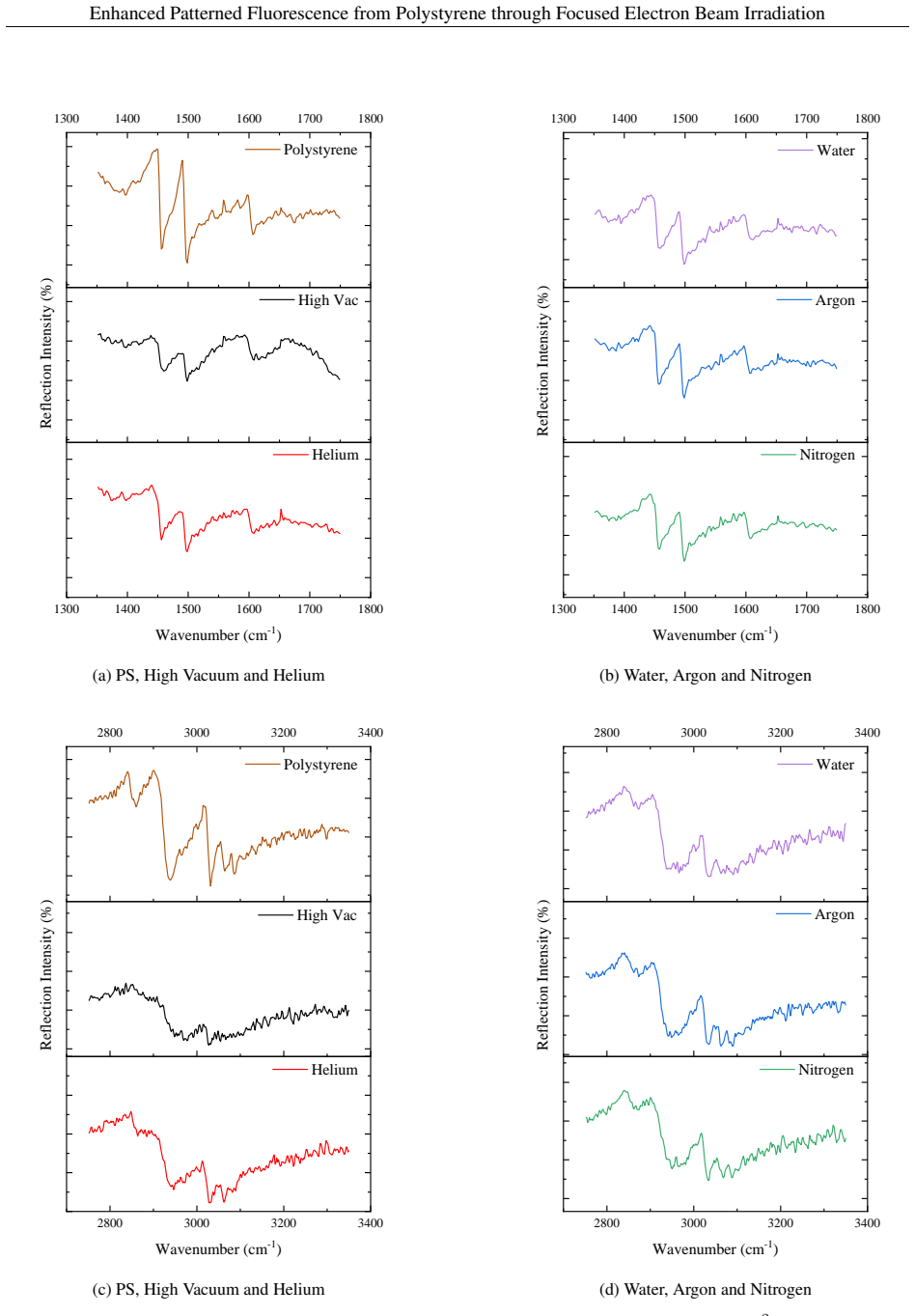
The load-bearing mechanism is electron-beam-induced fluorophore synthesis in polystyrene carried out in a variable-pressure electron microscope, with gas pressure and beam dose as the two controls. The argument separates physical from chemical effects using the beam-skirt formula $R_s = 364 (Z/E)(P/T)^{1/2}L^{3/2}$, which predicts how much a gas spreads the focused beam; because the observed enhancement neither tracks the gases' scattering cross-sections nor weakens with pressure the way scattering would, the paper rules scattering out as the cause. Instead, the mechanism is inferred from substrate comparisons: gas effects are strong on insulating substrates and nearly absent on conductive ones, and yield scales inversely with substrate thermal conductivity, implicating charge dissipation and e-beam heating in fluorophore formation. FTIR supplies the chemical half of the machinery by showing that gas exposure preserves aromatic and aliphatic C-H stretches and phenyl rings relative to high vacuum.
What would settle it
Measure the dose actually deposited in the polystyrene for each gas and pressure, using a Faraday cup or dosimeter at the sample plane or a Monte Carlo transport simulation; if the 18-fold helium enhancement vanishes when gas and vacuum data are compared at equal true absorbed dose, the claimed gas-specific mechanism is an artifact of dose miscalibration.
Extended reading notes
Core claim
On its own terms, the paper's discovery is that gaseous environments change both the chemistry and the physics of electron-beam fluorophore synthesis in polystyrene. Electron irradiation converts non-luminescent PS into a material whose visible photoluminescence is attributed to polycyclic-aromatic or carbon-dot-like fluorophores; the paper adds that performing the exposure in a gas shifts the emission color (blue with pressure, red with dose), sharpens the spectra into at least two peaks, and can raise the photon yield by an order of magnitude or more. FTIR shows the gas suppresses the loss of aromatic and aliphatic C-H bonds and preserves phenyl-ring features, while EDS finds no oxidation, so the new fluorophores are carbon structures rather than carbonyl products. The authors argue that gas scattering cannot explain the enhancement, and instead point to gas-dependent charge dissipation on insulating substrates and to substrate thermal conductivity; the dose for peak yield rises from about 8 mC cm$^{-2}$ at 0.1 mbar of water vapor to 37 mC cm$^{-2}$ at 3 mbar.
Load-bearing premise
The argument assumes that the electron dose, computed from a beam current measured only under high vacuum, is also the dose actually delivered to the film when gas is present; if gas scattering changes the true dose, the gas-versus-vacuum intensity comparisons are not made at matched doses.
Editorial extensions
If this is right
- Fluorescent patterns with locally chosen emission colors can be written directly into polystyrene by varying electron dose and gas pressure during exposure.
- The dose at which photoluminescence peaks rises with gas pressure (about 8 mC cm$^{-2}$ at 0.1 mbar, 15 mC cm$^{-2}$ at 1 mbar, and 37 mC cm$^{-2}$ at 3 mbar of water vapor), so higher pressure allows brighter films before the yield rollover.
- Gas choice should be matched to the substrate: water vapor is most effective on N-BK7 glass, argon on soda lime glass, and helium gives the largest relative boost on sapphire.
- Conductive substrates dissipate the gas effect and lower the photon yield, so insulating supports are preferable when maximum fluorescence is wanted.
- Because gases slow the destruction of C-H bonds and phenyl rings, gas exposure should leave more of the original polymer structure intact at high doses.
Reading between the lines
- A natural extension, not developed in the paper, is multi-color patterning within a single film: since dose and pressure shift color in opposite directions, varying dose or local pressure across one scan could write adjacent regions with different emission colors.
- The inverse correlation between substrate thermal conductivity and yield implies that stage temperature could be a third control knob; the paper discusses only the beam's implicit heating, not deliberate external heating.
- The chemistry is probably not limited to polystyrene: other phenyl-bearing polymers should show similar gas-sensitive fluorophore formation, a prediction the paper does not test.
Signed reviews
Editorial analysis
A structured set of objections, weighed in public.
Referee Report
Summary. The manuscript reports that focused 20 keV electron-beam irradiation of polystyrene (PS) films in an environmental SEM under various gases (water vapor, nitrogen, argon, helium) can tune the photoluminescence (PL) emission wavelength and enhance the PL yield relative to high-vacuum exposure. The authors expose 100 µm square patterns at doses from 1.8 to 45 mC/cm2 and pressures from high vacuum to 3 mbar, then characterize the films with confocal microscopy, FTIR, TEM, and EDS. They report an overall wavelength tuning range of 451–544 nm, PL enhancements up to 10x on N-BK7 under water vapor and up to 18x on sapphire under helium, and the highest absolute yield on soda lime glass under argon. They attribute the effects to reduced C–H bond dissociation under gas, substrate charging, and substrate thermal conductivity, and they explicitly argue that electron scattering in the gas is not the dominant mechanism.
Significance. If the central claims hold, the work offers a simple, maskless route to locally patterned fluorescent regions in a standard polymer, with wavelength tunability controlled by dose and gas pressure, which could be useful for photonics and sensing applications. The manuscript's strengths include a systematic survey across several insulating and conducting substrates, multiple gases, and a range of doses; complementary FTIR, TEM, and EDS characterization; and connection to prior work on gas-modified electron-beam chemistry. The qualitative trends are directly visible in the spectra and integrated-intensity plots. However, the quantitative claims and the mechanistic conclusion about gas scattering rest on a dose calibration that is not established, and the reported enhancement factors are given without uncertainty estimates. These issues need to be resolved before the claims can be accepted.
major comments (3)
- [Section 2.2; Figs. 6, 12, 15, 16] The nominal electron dose is computed from the beam current measured under high vacuum only, but under gas the actual dose delivered to the PS film is lower and gas-dependent. Equation (1) is used only for the skirt radius, not for the transmitted fraction. Using the total scattering cross-sections in Table 1 over a 10 mm gas path at 1 mbar gives approximate transmitted fractions of ~96% for He, ~75% for H2O, ~70% for N2, and ~60% for Ar; at 3 mbar H2O the fraction is roughly 40%. Thus the dose axes in Figs. 6, 8, 12, 15, and 16 are not on a common scale. The observation in Section 3.1 that the PL peak shifts from 8 to 15 to 37 mC/cm2 as water pressure goes from 0.1 to 1 to 3 mbar is the signature one would expect if there is a single actual-dose optimum and the gas simply attenuates the beam by a pressure-dependent factor. The authors should calibrate the delivered dose in gas (e.g., with a Faraday cup in gas or Monte Carlo simulation) and replot the data versus actual dose before claiming gas pressure as a separate tuning axis.
- [Section 3.1; Section 4] The conclusion that 'electron scattering in gas alone is not responsible for the enhanced PL' and the quantitative enhancement magnitudes (10x on N-BK7 under water vapor, 18x on sapphire under helium) are not supported because they rely on the uncalibrated nominal dose. The statement 'This result cannot be explained by simple electron scattering in the gas' (Section 3.1, near Fig. 6) is not justified: a pressure-dependent reduction in actual dose can move the sample along the dose-response curve toward its optimum. The crossover in gas ranking with dose in Fig. 12 is the expected consequence of different attenuation factors sliding each gas along the same dose-response curve. These mechanistic and quantitative conclusions should be restated conditionally or removed until the actual-dose calibration is performed.
- [Sections 3.1–3.3] No error bars, replicate measurements, or statistical analysis are provided for any PL intensity or peak wavelength. The central quantitative claims — the 18x enhancement on sapphire under helium, the 10x enhancement on N-BK7 under water vapor, and the 451–544 nm tuning range — are reported without uncertainty. The qualitative trends are directly visible, but the magnitudes need at least three or more replicate exposures for key conditions (e.g., Fig. 6, Fig. 12, and the sapphire/He comparison) and the results should be reported as mean ± standard deviation.
minor comments (6)
- [Section 3.1.2] The sentence 'It could also be consistent with more efficient indicating that the increase in PL yield with increasing reaction temperature...' is grammatically incomplete and should be rewritten.
- [Section 2.2] The instrument name 'Keathley' should be 'Keithley'.
- [Figure 16 caption] The word 'instulating' should be 'insulating'.
- [Abstract and Section 4] The 18x enhancement on sapphire under helium is stated without identifying the dose and pressure at which it occurs; please specify the exact conditions or point to the relevant panel in Fig. 16.
- [Equation (1)] The units of the constant 364 are not stated; please provide the original formulation from Danilatos or a dimensional check so the reader can verify the skirt-radius values.
- [Section 3.2.1, Figs. 13–14] The FTIR reflection spectra are shown without baseline correction or normalization; the qualitative comparison of peak heights should be supported by a description of how the spectra were processed.
Circularity Check
No significant circularity: the paper is an experimental characterization; dose and pressure are independently controlled inputs, and no fitted parameter is presented as a prediction.
full rationale
The manuscript is an empirical study of the photoluminescence of electron-beam-irradiated polystyrene under various gases. The central variables — electron dose, gas pressure, PL intensity, and emission wavelength — are measured independently: dose is set by beam current and exposure time, pressure is set by the environmental SEM, and PL is measured by confocal microscopy. No model is fitted to a subset of the data and then used to predict a closely related quantity, and no derived result is definitionally equal to an input. The comparison between high-vacuum and gas exposures uses a nominal dose that is not corrected for beam scattering in the gas, since the beam current is measured under vacuum; this is a calibration or correctness risk, not circularity. The paper even acknowledges gas scattering through Eq. (1) and argues against a pure scattering explanation using the nonmonotonic dose response and pressure-dependent shifts. Self-citations to the authors' earlier work on Teflon AF and PMMA appear only as motivation, not as load-bearing inputs to the present analysis. TEM, EDS, and FTIR provide independent characterization supporting the interpretation. No load-bearing step reduces to its own inputs, and no self-citation is load-bearing. Therefore the circularity score is 0.
Assumptions & free parameters
assumptions (2)
- domain assumption PL intensity measured by confocal microscopy is a valid proxy for fluorophore yield and is comparable across different substrates and gas environments.
- domain assumption The nominal electron dose (beam current multiplied by time divided by area) approximates the dose delivered to the PS film under gas, despite gas scattering.
Cite this review
Pith. "Pith review of Enhanced Patterned Fluorescence from Polystyrene through Focused Electron Beam Irradiation under Various Gases." pith.science (2026). https://pith.science/paper/HTZ3KOVG
@misc{pith2026250503167,
  author       = {Pith},
  title        = {Pith review of: Enhanced Patterned Fluorescence from Polystyrene through Focused Electron Beam Irradiation under Various Gases},
  year         = {2026},
  howpublished = {\url{https://pith.science/paper/HTZ3KOVG}},
  note         = {Machine review of arXiv:2505.03167}
}
read the original abstract
We report on a novel method for tuning and enhancing fluorescence from irradiated polystyrene through electron-beam exposure in gaseous environments. We describe the effect of electron dose and ambient gas on the photoluminescence spectra and yield of irradiated PS films on insulating and conductive substrates. PS films were exposed in an environmental scanning electron microscope using a 20 keV electron beam, ambient gas pressures from high vacuum to 3 mbar, and electron doses from 1.8 to 45 mC cm-2. Irradiated PS films were characterized using confocal microscopy, TEM, EDS and FTIR spectroscopy. From emission spectra collected using confocal microscopy we found that the emission wavelength and photon yield of the irradiated film can be tuned by both dose and gas pressure. The emission wavelength blue-shifts with increasing pressure and red-shifts with increasing dose enabling an overall tuning range of 451 - 544 nm. Significant enhancement in the PL intensity, up to 18 times on sapphire substrates under helium when compared to high vacuum, are observed. Overall, the highest PL yield is observed on soda lime glass substrates under argon. Also, the photon-yield on conductive substrates is significantly smaller than that yield from insulating substrates. TEM images revealed e-beam irradiated PS is amorphous in nature and elemental mapping EDS revealed no signs of film oxidation. FTIR spectroscopy revealed that under gaseous environments the decay of aromatic and aliphatic C-H stretches is reduced compared to the high vacuum exposure; in all cases, features associated with the phenyl rings are preserved. Localized e-beam synthesis of fluorophores in PS can be controlled by both dose and by ambient gas pressure. This technique could enable new approaches to photonics where fluorophores with tunable emission properties can be locally introduced by e-beam patterning.
Figures


Figures from the paper (13 more)




Reference graph
Works this paper leans on
-
[1]
Juey H. Lai and Lloyd T. Shepherd. Experimental Observations of Nearly Monodisperse Polystyrene as Negative Electron Resists. Journal of The Electrochemical Society, 126(4):696, April 1979
work page 1979
-
[2]
High Resolution Electron Beam Negative Resist with Very Narrow Molecular Weight Distributions
Kingo Itaya, Kimio Shibayama, and Teruo Fujimoto. High Resolution Electron Beam Negative Resist with Very Narrow Molecular Weight Distributions. Journal of The Electrochemical Society, 129(3):663, March 1982
work page 1982
-
[3]
Shoko Manako, Jun-ichi Fujita, Yukinori Ochiai, Eiichi Nomura Eiichi Nomura, and Shinji Matsui Shinji Matsui. Nanometer-Scale Patterning of Polystyrene Resists in Low-V oltage Electron Beam Lithography.Japanese Journal of Applied Physics, 36(12S):7773, December 1997
work page 1997
-
[4]
Resolution-Limit Study of Chain-Structure Negative Resist by Electron Beam Lithography
Shoko Manako, Jun-ichi Fujita, Yukinori Ochiai, Eiichi Nomura Eiichi Nomura, and Shinji Matsui Shinji Matsui. Resolution-Limit Study of Chain-Structure Negative Resist by Electron Beam Lithography. Japanese Journal of Applied Physics, 36(6A):L724, June 1997
work page 1997
-
[5]
Polystyrene negative resist for high-resolution electron beam lithography
Siqi Ma, Celal Con, Mustafa Yavuz, and Bo Cui. Polystyrene negative resist for high-resolution electron beam lithography. Nanoscale Research Letters, 6(1):446, July 2011
work page 2011
-
[6]
High molecular weight polystyrene as very sensitive electron beam resist
Celal Con, Ripon Dey, Mark Ferguson, Jian Zhang, Raafat Mansour, Mustafa Yavuz, and Bo Cui. High molecular weight polystyrene as very sensitive electron beam resist. Microelectronic Engineering, 98:254–257, October 2012
work page 2012
-
[7]
Dry thermal development of negative electron beam resist polystyrene
Celal Con, Arwa Saud Abbas, Mustafa Yavuz, and Bo Cui. Dry thermal development of negative electron beam resist polystyrene. Advances in Nano Research, 1(2):105–109, June 2013. Number: 2
work page 2013
-
[8]
Effect of molecular weight distribution on e-beam exposure properties of polystyrene
Ripon Kumar Dey and Bo Cui. Effect of molecular weight distribution on e-beam exposure properties of polystyrene. Nanotechnology, 24(24):245302, May 2013
work page 2013
Show all 55 references
-
[9]
Gupta, Amitava Gupta, James Horwitz, and David Kliger
Mool C. Gupta, Amitava Gupta, James Horwitz, and David Kliger. Time-resolved fluorescence and emission depolarization studies on polystyrene: photochemical processes in polymeric systems. 9. Macromolecules, 15(5):1372–1376, September 1982
1982
-
[10]
Photoluminescence of the electron irradiated Polystyrene
Hyeok Moo Lee and Sung Oh Cho. Photoluminescence of the electron irradiated Polystyrene. pages 1CD–ROM, Korea, Republic of, 2006. KNS
2006
-
[11]
Liquid and Solid-State NMR study of the electron irradiated Polystyrene
Hyeok Moo Lee and Sung Oh Cho Cho. Liquid and Solid-State NMR study of the electron irradiated Polystyrene. Korea, Republic of, 2007. KNS
2007
-
[12]
Fabrication of Luminescent Nanoarchitectures by Electron Irradiation of Polystyrene
Hyeok Moo Lee, Yong Nam Kim, Bong Hoon Kim, Sang Ouk Kim, and Sung Oh Cho. Fabrication of Luminescent Nanoarchitectures by Electron Irradiation of Polystyrene. Advanced Materials, 20(11):2094–2098, 2008
2008
-
[13]
Fabrication method of two-photon luminescent organic nano-architectures using electron-beam irradiation
Yoshio Kamura and Kohei Imura. Fabrication method of two-photon luminescent organic nano-architectures using electron-beam irradiation. Applied Physics Letters, 112(24):243104, June 2018
2018
-
[14]
Space-Selective Fabrication of Light-Emitting Carbon Dots in Polymer Films Using Electron-Beam-Induced Chemical Reactions
Yoshio Kamura and Kohei Imura. Space-Selective Fabrication of Light-Emitting Carbon Dots in Polymer Films Using Electron-Beam-Induced Chemical Reactions. Acs Omega, 4(2):3380–3384, February 2019
2019
-
[15]
Alexander, A
P. Alexander, A. Charlesby, and Francis Arthur Freeth. Radiation protection in copolymers of isobutylene and styrene. Proceedings of the Royal Society of London. Series A. Mathematical and Physical Sciences , 230(1180):136–145, January 1997
1997
-
[16]
Charlesby
A. Charlesby. Swelling properties of polystyrene crosslinked by high energy radiation. Journal of Polymer Science, 11(6):521–529, 1953
1953
-
[17]
Radiation Effects on Dimethyl-diphenyl Siloxane Copolymer
Mitsuru Koike and Akibumi Danno. Radiation Effects on Dimethyl-diphenyl Siloxane Copolymer. I. Protective Effect of Phenyl Radical on the Cross-linking. Journal of the Physical Society of Japan, 15(8):1501–1508, August 1960
1960
-
[18]
Burlant, J
W. Burlant, J. Neerman, and V . Serment.γ-radiation of p-substituted polystyrenes. Journal of Polymer Science, 58(166):491–500, 1962. 16 Enhanced Patterned Fluorescence from Polystyrene through Focused Electron Beam Irradiation
1962
-
[19]
C. G. Delides. The protective effect of phenyl group on the crosslinking of irradiated dimethyldiphenylsiloxane. Radiation Physics and Chemistry (1977), 16(5):345–352, January 1980
1977
-
[20]
L. N. Pankratova, L. T. Bugaenko, and A. A. Revina. Effect of aromatic protectors on the radiolysis of poly- organosiloxanes. High Energy Chemistry, 34(1):16–22, January 2000
2000
-
[21]
J. C. Randall, F. J. Zoepfl, and Joseph Silverman. A 13C NMR study of radiation-induced long-chain branching in polyethylene. Die Makromolekulare Chemie, Rapid Communications, 4(3):149–157, 1983
1983
-
[22]
Carbon-13 NMR study of radiation-induced crosslinking of linear polyethylene
Fumitaka Horii, Qingren Zhu, Ryozo Kitamaru, and Hitoshi Yamaoka. Carbon-13 NMR study of radiation-induced crosslinking of linear polyethylene. Macromolecules, 23(4):977–981, February 1990
1990
-
[23]
Number 551 in TECDOC Series
Radiation Damage to Organic Materials in Nuclear Reactors and Radiation Environments (Proceedings of a Final Research Co-ordination Meeting, Takasaki, Japan, 17-20 July 1989). Number 551 in TECDOC Series. INTERNATIONAL ATOMIC ENERGY AGENCY , Vienna, 1990
1989
-
[24]
Effect of x irradiation on optical properties of teflon-af
MS Jahan, DR Ermer, and DW Cooke. Effect of x irradiation on optical properties of teflon-af. Radiation Physics and Chemistry, 41(3):481–486, 1993
1993
-
[25]
The radiation chemistry of the copolymer of tetrafluoroethylene with 2, 2-bis (trifluoromethyl)-4, 5-difluoro-1, 3-dioxole
John S Forsythe, David JT Hill, Anestis L Logothetis, and Andrew K Whittaker. The radiation chemistry of the copolymer of tetrafluoroethylene with 2, 2-bis (trifluoromethyl)-4, 5-difluoro-1, 3-dioxole. Polymer degradation and stability, 63(1):95–101, 1999
1999
-
[26]
Sultan, Sarah K
Mansoor A. Sultan, Sarah K. Lami, Armin Ansary, Douglas R. Strachan, Joseph W. Brill, and J. Todd Hastings. Altering the radiation chemistry of electron-beam lithography with a reactive gas: a study of Teflon AF patterning under water vapor. Nanotechnology, 30(30):305301, May 2019
2019
-
[27]
Brill, Jonathan T
Deepak Kumar, Krishnaroop Chaudhuri, Joseph W. Brill, Jonathan T. Pham, and J. Todd Hastings. Effect of water vapor pressure on positive and negative tone electron-beam patterning of poly(methyl methacrylate). Journal of Vacuum Science & Technology B, 41(1):012604, January 2023
2023
-
[28]
C Pivin, M Sendova-Vassileva, P Colombo, and A Martucci
J. C Pivin, M Sendova-Vassileva, P Colombo, and A Martucci. Photoluminescence of composite ceramics derived from polysiloxanes and polycarbosilanes by ion irradiation. Materials Science and Engineering: B , 69-70:574–577, January 2000
2000
-
[29]
Focused electron beam induced deposition: A perspective
Michael Huth, Fabrizio Porrati, Christian Schwalb, Marcel Winhold, Roland Sachser, Maja Dukic, Jonathan Adams, and Georg Fantner. Focused electron beam induced deposition: A perspective. Beilstein journal of nanotechnology, 3(1):597–619, 2012
2012
-
[30]
Formation mechanism of carbogenic nanoparticles with dual photoluminescence emission
Marta J Krysmann, Antonios Kelarakis, Panagiotis Dallas, and Emmanuel P Giannelis. Formation mechanism of carbogenic nanoparticles with dual photoluminescence emission. Journal of the American Chemical Society, 134(2):747–750, 2012
2012
-
[31]
Investigation from chemical structure to photoluminescent mechanism: a type of carbon dots from the pyrolysis of citric acid and an amine
Yubin Song, Shoujun Zhu, Shitong Zhang, Yu Fu, Li Wang, Xiaohuan Zhao, and Bai Yang. Investigation from chemical structure to photoluminescent mechanism: a type of carbon dots from the pyrolysis of citric acid and an amine. Journal of Materials Chemistry C, 3(23):5976–5984, 2015
2015
-
[32]
Highly photoluminescent carbon dots for multicolor patterning, sensors, and bioimaging
Shoujun Zhu, Qingnan Meng, Lei Wang, Junhu Zhang, Yubin Song, Han Jin, Kai Zhang, Hongchen Sun, Haiyu Wang, and Bai Yang. Highly photoluminescent carbon dots for multicolor patterning, sensors, and bioimaging. Angewandte Chemie International Edition, 52(14):3953–3957, 2013
2013
-
[33]
Hair fiber as a precursor for synthesizing of sulfur-and nitrogen-co-doped carbon dots with tunable luminescence properties
Dong Sun, Rui Ban, Peng-Hui Zhang, Ge-Hui Wu, Jian-Rong Zhang, and Jun-Jie Zhu. Hair fiber as a precursor for synthesizing of sulfur-and nitrogen-co-doped carbon dots with tunable luminescence properties. Carbon, 64:424–434, 2013
2013
-
[34]
Effect of reaction temperature on structure and fluorescence properties of nitrogen-doped carbon dots
Yi Zhang, Yaling Wang, Xiaoting Feng, Feng Zhang, Yongzhen Yang, and Xuguang Liu. Effect of reaction temperature on structure and fluorescence properties of nitrogen-doped carbon dots. Applied Surface Science, 387:1236–1246, 2016
2016
-
[35]
Fast migration of alkali ions in glass irradiated by electrons
Ondrej Gedeon, Karel Jurek, and Václav Hulıénsk`y. Fast migration of alkali ions in glass irradiated by electrons. Journal of non-crystalline solids, 246(1-2):1–8, 1999
1999
-
[36]
Microanalysis of glass containing alkali ions
Ondrej Gedeon, Václav Hulínsk `y, and Karel Jurek. Microanalysis of glass containing alkali ions. Microchimica Acta, 132:505–510, 2000
2000
-
[37]
Chizhik, Narain Karedla, Mariia O
Siddharth Ghosh, Anna M. Chizhik, Narain Karedla, Mariia O. Dekaliuk, Ingo Gregor, Henning Schuhmann, Michael Seibt, Kai Bodensiek, Iwan A. T. Schaap, Olaf Schulz, Alexander P. Demchenko, Jörg Enderlein, and Alexey I. Chizhik. Photoluminescence of Carbon Nanodots: Dipole Emiss...
2014
-
[38]
Carbon-Based Dots Co-doped with Nitrogen and Sulfur for High Quantum Yield and Excitation- Independent Emission
Yongqiang Dong, Hongchang Pang, Hong Bin Yang, Chunxian Guo, Jingwei Shao, Yuwu Chi, Chang Ming Li, and Ting Yu. Carbon-Based Dots Co-doped with Nitrogen and Sulfur for High Quantum Yield and Excitation- Independent Emission. Angewandte Chemie, 125(30):7954–7958, 2013
2013
-
[39]
Carbon Dots with Continuously Tunable Full-Color Emission and Their Application in Ratiometric pH Sensing
Hui Nie, Minjie Li, Quanshun Li, Shaojun Liang, Yingying Tan, Lan Sheng, Wei Shi, and Sean Xiao-An Zhang. Carbon Dots with Continuously Tunable Full-Color Emission and Their Application in Ratiometric pH Sensing. Chemistry of Materials, 26(10):3104–3112, May 2014
2014
-
[40]
Intrinsic and Extrinsic Fluorescence in Carbon Nanodots: Ultrafast Time-Resolved Fluorescence and Carrier Dynamics
Xiaoming Wen, Pyng Yu, Yon-Rui Toh, Xiaotao Hao, and Jau Tang. Intrinsic and Extrinsic Fluorescence in Carbon Nanodots: Ultrafast Time-Resolved Fluorescence and Carrier Dynamics. Advanced Optical Materials, 1(2):173–178, 2013
2013
-
[41]
Temperature-Dependent Fluorescence in Carbon Dots.The Journal of Physical Chemistry C, 116(48):25552–25557, December 2012
Pyng Yu, Xiaoming Wen, Yon-Rui Toh, and Jau Tang. Temperature-Dependent Fluorescence in Carbon Dots.The Journal of Physical Chemistry C, 116(48):25552–25557, December 2012
2012
-
[42]
Das, Yiyang Liu, Sinhea Yeom, Doo Young Kim, and Christopher I
Somes K. Das, Yiyang Liu, Sinhea Yeom, Doo Young Kim, and Christopher I. Richards. Single-Particle Fluorescence Intensity Fluctuations of Carbon Nanodots. Nano Letters, 14(2):620–625, February 2014
2014
-
[43]
White light emission from polystyrene under pulsed ultra violet laser irradiation
Eunkyeom Kim, Jihoon Kyhm, Jung Hyuk Kim, Gi Yong Lee, Doo-Hyun Ko, Il Ki Han, and Hyungduk Ko. White light emission from polystyrene under pulsed ultra violet laser irradiation. Scientific Reports, 3(1):3253, November 2013
2013
-
[44]
G. D. Danilatos. Foundations of Environmental Scanning Electron Microscopy. In Peter W. Hawkes, editor, Advances in Electronics and Electron Physics, volume 71, pages 109–250. Academic Press, January 1988
1988
-
[45]
Toth and M
M. Toth and M. R. Phillips. The effects of space charge on contrast in images obtained using the environmental scanning electron microscope. Scanning, 22(5):319–325, 2000
2000
-
[46]
Charging effects from electron beam lithography
KD Cummings and M Kiersh. Charging effects from electron beam lithography. Journal of Vacuum Science & Technology B: Microelectronics Processing and Phenomena, 7(6):1536–1539, 1989
1989
-
[47]
Jing He and David C. Joy. Measurement of total gas scattering cross-section. Scanning, 25(6):285–290, 2003
2003
-
[48]
Rattenberger, J
J. Rattenberger, J. Wagner, H. Schröttner, S. Mitsche, and A. Zankel. A method to measure the total scattering cross section and effective beam gas path length in a low-vacuum SEM. Scanning, 31(3):107–113, 2009
2009
-
[49]
Nagasawa, R
K. Nagasawa, R. Watanabe, Y . Tanaka, and T. Takada. Charge accumulation in election beam irradiated various polymers. In 2008 International Symposium on Electrical Insulating Materials (ISEIM 2008), pages 147–150, Yokkaichi, September 2008. IEEE
2008
-
[50]
Charge Accumulation in Various Electron-Beam-Irradiated Polymers
Kenichiro Nagasawa, Masato Honjoh, Hiroaki Miyake, Rikio Watanabe, Yasuhiro Tanaka, and Tatsuo Takada. Charge Accumulation in Various Electron-Beam-Irradiated Polymers. IEEJ Transactions on Electrical and Electronic Engineering, 5(4):410–415, 2010
2010
-
[51]
Ito indium tin oxide coated glass /pet substrates, https://www.msesupplies.com/collections/ito-indium-tin-oxide- glass-substrates
-
[52]
Materials database, https://tpsx.arc.nasa.gov/materialsdatabase, 2024
2024
-
[53]
Thermal transport properties of polycrystalline tin-doped indium oxide films
Toru Ashida, Amica Miyamura, Nobuto Oka, Yasushi Sato, Takashi Yagi, Naoyuki Taketoshi, Tetsuya Baba, and Yuzo Shigesato. Thermal transport properties of polycrystalline tin-doped indium oxide films. Journal of applied physics, 105(7), 2009
2009
-
[54]
Schram, F.J
B.L. Schram, F.J. De Heer, M.J. Van Der Wiel, and J. Kistemaker. Ionization cross sections for electrons (0.6–20 keV) in noble and diatomic gases. Physica, 31(1):94–112, January 1965
1965
-
[55]
Schutten, F
J. Schutten, F. J. de Heer, H. R. Moustafa, A. J. H. Boerboom, and J. Kistemaker. Gross- and Partial-Ionization Cross Sections for Electrons on Water Vapor in the Energy Range 0.1–20 keV. The Journal of Chemical Physics, 44(10):3924–3928, May 1966. 18
1966
Reviewed August 15, 2026 · model on record in the stance chip above.
Discussion (0). Continue with ORCID to comment.