REVIEW 4 major objections 6 minor 78 references
SmartTrap: Automated Precision Experiments with Optical Tweezers
T0 review · 4 major / 6 minor · reviewed 2026-08-15 · deepseek-v4-flash
Pith's one-line read An optical-tweezer platform runs complex precision experiments unattended.
desk verdict The platform is genuinely autonomous and the open-source release is valuable; the main fix needed is a real ground-truth check of the CNN's axial localization before the 3D-alignment claims can be trusted. read the letter →
The pith
A machine-rendered reading of the paper's core claim, the machinery that carries it, and where it could break.
The reading
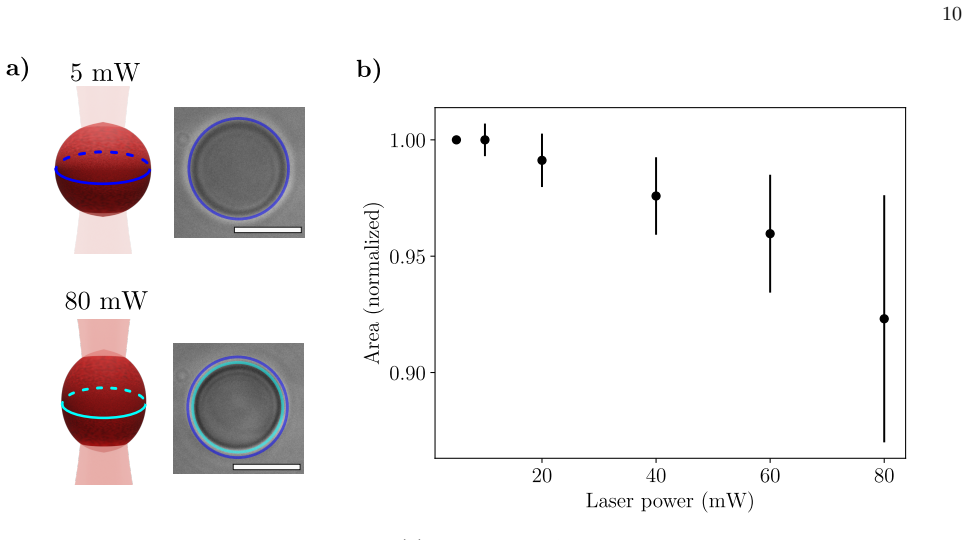
What carries the argument
The load-bearing mechanism is a closed four-step loop—acquire data, process data, decide, execute—running asynchronously so that sensor sampling and camera capture continue while the networks analyze. The central enabling objects are the counter-propagating dual-beam trap, which reports force directly from the change in light momentum and therefore needs no force calibration per particle type; a custom microcontroller that steers the beams with piezoelectric actuators and reads the force sensors; and a microfluidic chamber with a suction micropipette that holds one particle fixed while the trapped particle is moved. Within this loop, the small convolutional network trained on simulated images is the component that supplies 3D awareness: its z-prediction is what lets the system align two particles in the axial direction and also flags when a second particle has entered the trap.
What would settle it
Move a trapped bead of known diameter through focus in measured 1 µm steps while recording the network's z-prediction; if the root-mean-square error exceeds the bead's radius over the range used for alignment, the simulated-only depth network cannot support the claimed autonomous tethering. A complementary check is to count successful DNA attachments over many unattended cycles and compare that rate with what would be expected if the z-alignment were random within a few microns.
Extended reading notes
Core claim
The core discovery is that a sufficiently integrated digital loop can take over procedures that previously required a trained operator at every step. In SmartTrap, a camera feed is analyzed in real time by neural networks: one network detects particles and the micropipette and gives their lateral positions, while a second network estimates the axial (z) position of each particle from a single image, allowing the system to bring a trapped particle and a pipette-held particle into the same focal plane. Custom electronics close force and position feedback loops at roughly 7 kHz, and event-driven logic decides when to trap, align, attach, pull, flush, and repeat. The demonstrations include a 4.5-hour run that measured the hydrodynamic radius of 144 particles, repeated DNA stretching cycles yielding the expected ~65 pN overstretching plateau in the force–extension curve, red blood cell deformation that increases monotonically with laser power, and electrostatic repulsion curves at varying salt concentration that agree with the screened-Coulomb model.
Load-bearing premise
The load-bearing premise is that the convolutional network trained solely on simulated images predicts the axial (z) position of real particles with enough accuracy—within about a micron—for the 2–4 µm particles used here, because the DNA attachment step aligns particles by matching these predictions.
Editorial extensions
If this is right
- A single unattended session can collect hundreds of particle measurements or repeatedly pull dozens of DNA molecules, making statistically rich datasets and parameter sweeps practical.
- Because alignment and force readings are handled by the same code every time, reproducibility improves and operator-dependent bias in how experiments are performed largely disappears.
- The protocol is built from reusable steps—trap, align, attach, measure, flush—so automating a new assay mostly means reconfiguring the state machine rather than rebuilding the instrument.
- Long unattended operation with automatic error recovery opens the door to waiting for rare or transient single-molecule events instead of ending a session when a tether breaks or a sample runs out.
Reading between the lines
- Beyond the paper, a direct extension would be to quantify the depth network's z-error against a calibrated stage and use that number to set the minimum particle size, and hence the shortest tether, for which autonomous alignment is reliable.
- Beyond the paper, the same detect–align–measure logic is not tied to counter-propagating tweezers; retrained on new image data, it could bring automation to other single-object microscopies such as single-beam traps or surface-based force assays.
- Beyond the paper, if the depth estimate were sharpened, the double-trapping check that currently discards bad runs could become a real-time rejection signal that improves data quality before any measurement is recorded.
Signed reviews
Editorial analysis
A structured set of objections, weighed in public.
Referee Report
Summary. The manuscript describes SmartTrap, a counter-propagating optical tweezers platform that integrates custom electronics, microfluidics, and real-time deep-learning image analysis (YOLO for lateral detection and sizing, a convolutional network for axial focus estimation, and a U-Net for tracking close particles) to run experiments without continuous human supervision. Four demonstrations are reported: high-throughput particle size characterization by Stokes drag during a 4.5 h unattended run (144 analyzed particles, measured radius 2.11(4) µm versus the manufacturer's 2.12(5) µm); autonomous DNA pulling of an 11.8 kb λ-DNA fragment with force-extension curves showing an overstretch plateau; optical deformation of red blood cells at increasing laser power; and measurement of electrostatic forces between pairs of colloidal particles at several salt concentrations compared with DLVO theory. The software, electronics schematics, and videos are open source.
Significance. If the claims hold, SmartTrap is a substantial contribution to automated precision optical tweezers: it directly addresses the low-throughput and reproducibility bottleneck of manual operation, spans single-molecule, cellular, and colloidal experiments, and its open-source hardware and software make the platform reproducible by others. The particle characterization result is a strong quantitative check, and the electrostatic experiment is a genuine prediction because the charge parameter Z is fitted to the 0.1 mM data and then used for the other salt concentrations. The DNA overstretch plateau length (~2.8 µm, about 70% of contour length) provides a model-independent physical benchmark. The main weaknesses are the unquantified axial (z) accuracy of the convolutional network, which is load-bearing for the autonomous 3D alignment protocols, and the partly construction-based comparison of the DNA force-extension data to the extensible WLC model. These issues are fixable with additional validation and reporting, so the manuscript merits revision rather than rejection.
major comments (4)
- [Real time image analysis / DNA pulling step 5 / Electrostatic protocol step 5] The load-bearing premise for autonomous 3D alignment is the convolutional network's z-prediction, yet the only real-image validation is the statement that predictions are 'consistent within a range of a few microns.' This is not a quantitative calibration: for the 2-4 µm particles used here, a few microns is comparable to the particle size, and for the electrostatic measurements it is one to two orders of magnitude larger than the surface-to-surface distances of interest. The DNA and electrostatic protocols align the two particles by matching CNN-predicted z-positions, so the safety margin of the alignment loop is unknown. The successful autonomous tethering and DLVO-compatible curves provide indirect evidence that the network is more accurate than the text suggests, but the paper should report a direct real-image validation, for example by translating a trapped particle through focus with the stage while recording the true position, and reporting bias and root-mean-square error of the CNN predictions for the relevant particle sizes.
- [DNA pulling / Fig. 4b / Suppl. Fig. S5 / Eq. (A3)] The claim of 'excellent agreement' between the force-extension data and the extensible WLC model is weakened by the statement that the data were offset to align with the model at 20 pN because the molecular attachment point on the pipette particle is unknown. While this offset is a standard experimental necessity, the current presentation makes part of the agreement with the model a construction rather than a test. The overstretch plateau remains an independent validation because its position and length are not affected by the offset. The authors should report the raw (unoffset) extension data or at least the distribution of offsets across molecules, and should phrase the low-force WLC comparison as a consistency check with one fitted offset rather than as a model-free agreement.
- [DNA pulling experiment / Supplementary Experimental Details] There is an internal inconsistency concerning the expected overstretching force. The main text's DNA protocol uses 60 pN as the attachment-check threshold and states that a force plateau at approximately 65 pN is expected for extensions beyond 4 µm, citing Ref. [30]. However, the Supplementary Experimental Details for the same construct states that 'only one strand at each DNA end is anchored, resulting in a lower overstretching force than would occur if both strands were immobilized' and cites Ref. [52]. If one-strand anchoring lowers the overstretch force, the 60-65 pN threshold may be too high or the text is contradictory. The authors should clarify which expectation applies to their construct and how the autonomous attachment check is robust to this uncertainty.
- [Abstract / Electrostatic interaction between particles / DNA pulling] The abstract's claim that SmartTrap is capable of performing complex experiments 'completely autonomously' is stronger than what the demonstrations support. The DNA pulling protocol begins with a user-configuration routine in which the operator manually locates the pipette and capillary openings, and the electrostatic salt-concentration series requires manual closing of channels and replacement of the solution while retaining the particle pair. The autonomous operation is within a user-prepared configuration and, for the salt series, within a manually executed medium exchange. This is still a valuable and substantial degree of automation, but the wording should be qualified so that the scope of 'completely autonomous' matches the protocol descriptions.
minor comments (6)
- [Throughout] There are several typographical errors: 'streptadavin' should be 'streptavidin' in the DNA pulling protocol, 'inlcuding' should be 'including' in the Real time image analysis section, and 'autonomated' should be 'automated' in the DNA pulling section.
- [Fig. 4b / Suppl. Fig. S5] The figure captions should state explicitly that the extension axes have been offset to align the curves at 20 pN; currently the offset is mentioned only in the main text, which makes the figures appear to show raw data.
- [Electrostatic interaction between particles / Eq. (A4)] The DLVO fit reports Z ≈ 460,000 but no uncertainty or goodness-of-fit measure is given, and the procedure for computing κ at each nominal salt concentration (especially for the nominally salt-free water condition) is not described. Reporting these values would allow readers to assess the strength of the prediction across concentrations.
- [Stretching of red blood cells] The cross-sectional area measurement is described as a Gaussian filter followed by a threshold, but no assessment of the segmentation accuracy or sensitivity to the threshold choice is provided; a brief validation or error estimate is needed for this to be a quantitative measurement.
- [Particle characterization] The text reports that 15 of 159 large-particle measurements failed due to double trapping, but does not state whether these failures correlate with particle size or position and whether they could bias the measured radius distribution; a sentence on this would improve confidence in the 2.11(4) µm result.
- [Real time image analysis / Supplementary video 1] The statement that 'the network consistently predicts the relative focal position' and that predictions on real images are 'consistent within a few microns' should be accompanied by the number of particles and images used in that check, since this is currently the only quantitative-looking claim about the axial network.
Circularity Check
No circularity: SmartTrap's claims are experimental demonstrations, and the model comparisons either fit a stated parameter to a reference condition, use an acknowledged offset, or are validated against external benchmarks.
full rationale
The paper is an experimental platform demonstration rather than a derivation, so the standard circularity patterns do not apply. The central claim that SmartTrap can perform complex experiments autonomously is supported by four independent demonstrations: particle size characterization matching manufacturer specifications, DNA force-extension curves reproducing the known overstretching plateau and consistent with the extensible WLC model, red blood cell stretching comparable to prior optical stretcher results, and electrostatic force measurements compared with DLVO theory. The DLVO comparison is not circular: the charge parameter Z is explicitly fitted only to the 0.1 mM salt-concentration data, and the same Z is then used to predict force curves at other salt concentrations, which is an independent test. The DNA WLC comparison involves an acknowledged offset to align the data at 20 pN because the absolute molecular attachment point is unknown, but the paper does not present this as a parameter-free prediction; the curve shape and the overstretch plateau provide independent content. The deep-learning tracking networks are trained on DeepTrack2 simulations, and the paper cites the authors' own DeepTrack2 package, but this is tool attribution rather than a load-bearing circular inference: the real-image performance is checked against real experiments and supplementary videos, and the successful autonomous protocols provide external behavioral validation. The unquantified sim-to-real accuracy of the z-position CNN is a legitimate robustness or correctness concern, but it is not a case of the paper defining its output in terms of its input or renaming a fitted quantity as a prediction. No load-bearing step reduces to its own inputs, so the appropriate circularity score is 0.
Assumptions & free parameters
free parameters (2)
- Z (number of charge groups per particle) =
460,000
- DNA force-extension offset =
Aligned to model at F = 20 pN
assumptions (5)
- domain assumption Counter-propagating trap force measurement is independent of particle size, shape, and refractive index
- ad hoc to paper Simulated images from DeepTrack2 are sufficiently representative of experimental images for YOLO and CNN training
- standard math Stokes drag law with wall correction [47] applies in the microfluidic chamber
- standard math Extensible WLC model describes DNA elasticity below overstretching
- standard math DLVO theory describes the electrostatic force between the colloidal particles
Cite this review
Pith. "Pith review of SmartTrap: Automated Precision Experiments with Optical Tweezers." pith.science (2026). https://pith.science/paper/NUSL33IR
@misc{pith2026250505290,
  author       = {Pith},
  title        = {Pith review of: SmartTrap: Automated Precision Experiments with Optical Tweezers},
  year         = {2026},
  howpublished = {\url{https://pith.science/paper/NUSL33IR}},
  note         = {Machine review of arXiv:2505.05290}
}
read the original abstract
There is a trend in research towards more automation using smart systems powered by artificial intelligence. While experiments are often challenging to automate, they can greatly benefit from automation by reducing labor and increasing reproducibility. For example, optical tweezers are widely employed in single-molecule biophysics, cell biomechanics, and soft matter physics, but they still require a human operator, resulting in low throughput and limited repeatability. Here, we present a smart optical tweezers platform, which we name SmartTrap, capable of performing complex experiments completely autonomously. SmartTrap integrates real-time 3D particle tracking using deep learning, custom electronics for precise feedback control, and a microfluidic setup for particle handling. We demonstrate the ability of SmartTrap to operate continuously, acquiring high-precision data over extended periods of time, through a series of experiments. By bridging the gap between manual experimentation and autonomous operation, SmartTrap establishes a robust and open source framework for the next generation of optical tweezers research, capable of performing large-scale studies in single-molecule biophysics, cell mechanics, and colloidal science with reduced experimental overhead and operator bias.
Figures


Figures from the paper (3 more)


Reference graph
Works this paper leans on
-
[30]
D. G. Grier, Optical tweezers in colloid and interface sci- ence, Current Opinion in Colloid & Interface Science 2, 264 (1997)
work page 1997
-
[52]
J. Guck, R. Ananthakrishnan, H. Mahmood, T. J. Moon, C. C. Cunningham, and J. K¨ as, The optical stretcher: a novel laser tool to micromanipulate cells, Biophysical Journal 81, 767 (2001)
work page 2001
-
[1]
Check pipette and trap: The algorithm deter- mines whether a particle is present in the optical trap or in the pipette by checking if any particle is located within a prescribed distance of the esti- mated laser position or the pipette tip, respectively. Assuming that it is the start of the procedure, there will be nothing in either the trap or the pipette
-
[2]
Trap streptadavin particle: The program traps 7 FIG. 4: DNA pulling experiment. (a) Illustration of a DNA pulling experiment. The DNA molecule is attached between two particles. One of the particles is kept fixed by a micropipette, while the other is moved back and forth using an optical trap. ( b) Force–extension curves from the experiment (circles) and ...
-
[3]
The tip of the pipette is located using the YOLO algorithm
Suck into pipette: With a SA particle trapped and positioned near the pipette, the next step is to catch it with the pipette. The tip of the pipette is located using the YOLO algorithm. The trapped particle is then approached to the tip, first using the motors for rough alignment, and then the wig- glers to finely adjust the laser position. Once the parti...
-
[4]
The system then returns to step 1 to repeat the process and measure the radius of another par- ticle
Drop the particle: The trapped particle is re- leased by flushing the central chamber with a strong flow. The system then returns to step 1 to repeat the process and measure the radius of another par- ticle. We test SmartTrap with this protocol for a sample and have it run continuously for 4.5 h. During this time 938 particles were trapped, 159 of which w...
-
[5]
Trap DNA particle: Next, the particle with DNA molecules is trapped using the same method as for the first (SA) particle but instead going to the capillary containing the particles with DNA
-
[6]
Attach DNA: Once a SA particle is in the pipette and a DNA particle is trapped near the pipette, it is time to get the DNA to attach to the SA parti- cle. This is done by first positioning the trapped particle above the one in the pipette using the mo- tors and position feedback. Next, the system aligns the focus of the two particles by adjusting the z- p...
Show all 78 references
-
[7]
DNA Pulling Experiment
Perform experiment: Finally, a pulling protocol is loaded onto the micro-controller. The protocol defines how far apart the particles should be moved and at what speed. The micro-controller provides feedback at 7 kHz, much faster than the commu- nication from the host computer...
-
[8]
Flow cells into the chamber: The flow in the central channel is briefly turned on, with the aim of bringing cells into view and removing cells from previous measurements from the optical trap
-
[9]
If there are no visible cells, it reverts back to step 1 to flow more cells
Look for cells: The SmartTrap looks for cells within the field of view. If there are no visible cells, it reverts back to step 1 to flow more cells
-
[10]
The laser power is set to 5 mW so as not to deform the cells during trapping
Trap a cell: With red blood cells in view, the system proceeds to trap the closest one. The laser power is set to 5 mW so as not to deform the cells during trapping
-
[11]
Once the mea- surement is finished, the system will go back to step 1 and the flow will guarantee that the same cell is not measured twice
Measure cross section: The trapping power is changed in steps while the transversal profiles of the cells are recorded in separate videos, one for each power for subsequent analysis. Once the mea- surement is finished, the system will go back to step 1 and the flow will guaran...
-
[12]
Check the pipette and trap: see step 1 in the DNA pulling protocol
-
[13]
Trap a particle: see step 2 in the DNA pulling protocol
-
[14]
Suck into pipette: see step 3 in the DNA pulling protocol
-
[15]
Trap the second particle: A second particle, of the same type as the first is trapped and brought next to the particle already in the pipette
-
[16]
The alignment is similar to that per- formed in the DNA pulling experiment when at- taching DNA
Align the particles: The trapped particle and the one in the pipette are aligned by ensuring that they are in the same focal plane and have the same x-position. The alignment is similar to that per- formed in the DNA pulling experiment when at- taching DNA
-
[17]
U-Net Model
Measure the repulsion: The particles are pushed together until the force exceeds a certain value, typically 10 pN. This is used as the first endpoint of the protocol. The second endpoint is set to a little less than a micron away from the first where the electrostatic force is...
-
[18]
C. Lu, C. Lu, R. T. Lange, J. Foerster, J. Clune, and D. Ha, The ai scientist: Towards fully auto- mated open-ended scientific discovery, arXiv preprint arXiv:2408.06292 (2024)
2024 arXiv
-
[19]
Jumper, R
J. Jumper, R. Evans, A. Pritzel, T. Green, M. Fig- urnov, O. Ronneberger, K. Tunyasuvunakool, R. Bates, A. ˇZ´ ıdek, A. Potapenko, et al. , Highly accurate pro- tein structure prediction with alphafold, Nature 596, 583 (2021)
2021
-
[20]
Midtvedt, J
B. Midtvedt, J. Pineda, H. Klein Moberg, H. Bachi- manchi, J. B. Pereira, C. Manzo, and G. Volpe, Deep Learning Crash Course (No Starch Press, San Francisco, CA, 2025)
2025
-
[21]
Holland and J
I. Holland and J. A. Davies, Automation in the life sci- ence research laboratory, Frontiers in Bioengineering and Biotechnology 8, 571777 (2020)
2020
-
[22]
Ashkin, Acceleration and trapping of particles by ra- diation pressure, Physical Review Letters 24, 156 (1970)
A. Ashkin, Acceleration and trapping of particles by ra- diation pressure, Physical Review Letters 24, 156 (1970)
1970
-
[23]
Ashkin, J
A. Ashkin, J. M. Dziedzic, J. E. Bjorkholm, and S. Chu, Observation of a single-beam gradient force optical trap for dielectric particles, Optics Letters 11, 288 (1986)
1986
-
[24]
Volpe, O
G. Volpe, O. M. Marag` o, H. Rubinsztein-Dunlop, G. Pesce, A. B. Stilgoe, G. Volpe, G. Tkachenko, V. G. Truong, S. N. Chormaic, F. Kalantarifard, et al. , Roadmap for optical tweezers, Journal of Physics: Pho- tonics 5, 022501 (2023)
2023
-
[25]
Magazz` u and C
A. Magazz` u and C. Marcuello, Investigation of soft mat- ter nanomechanics by atomic force microscopy and op- 13 tical tweezers: A comprehensive review, Nanomaterials 13, 963 (2023)
2023
-
[26]
Turlier, D
H. Turlier, D. A. Fedosov, B. Audoly, T. Auth, N. S. Gov, C. Sykes, J.-F. Joanny, G. Gompper, and T. Betz, Equilibrium physics breakdown reveals the active nature of red blood cell flickering, Nature Physics12, 513 (2016)
2016
-
[27]
C. J. Bustamante, Y. R. Chemla, S. Liu, and M. D. Wang, Optical tweezers in single-molecule biophysics, Nature Reviews Methods Primers 1, 25 (2021)
2021
-
[28]
S. Liu, G. Chistol, and C. Bustamante, Mechanical opera- tion and intersubunit coordination of ring-shaped molec- ular motors: insights from single-molecule studies, Bio- physical Journal 106, 1844 (2014)
2014
-
[29]
Zhang and K.-K
H. Zhang and K.-K. Liu, Optical tweezers for single cells, Journal of the Royal Society Interface 5, 671 (2008)
2008
-
[31]
Hertlein, L
C. Hertlein, L. Helden, A. Gambassi, S. Dietrich, and C. Bechinger, Direct measurement of critical casimir forces, Nature 451, 172 (2008)
2008
-
[32]
J. C. Crocker, J. A. Matteo, A. D. Dinsmore, and A. G. Yodh, Entropic attraction and repulsion in binary col- loids probed with a line optical tweezer, Physical Review Letters 82, 4352 (1999)
1999
-
[33]
Cichos, K
F. Cichos, K. Gustavsson, B. Mehlig, and G. Volpe, Ma- chine learning for active matter, Nature Machine Intelli- gence 2, 94 (2020)
2020
-
[34]
Ciarlo, D
A. Ciarlo, D. B. Ciriza, M. Selin, O. M. Marag` o, A. Sasso, G. Pesce, G. Volpe, and M. Goks¨ or, Deep learning for optical tweezers, Nanophotonics (2024)
2024
-
[35]
J. E. Melzer and E. McLeod, Assembly of multicompo- nent structures from hundreds of micron-scale building blocks using optical tweezers, Microsystems & Nanoengi- neering 7, 45 (2021)
2021
-
[36]
Teixeira, F
J. Teixeira, F. C. Moreira, J. Oliveira, V. Rocha, P. A. Jorge, T. Ferreira, and N. A. Silva, Autonomous and in- telligent optical tweezers for improving the reliability and throughput of single particle analysis, Measurement Sci- ence and Technology 35, 025208 (2023)
2023
-
[37]
S. B. Smith, Y. Cui, and C. Bustamante, [7] optical-trap force transducer that operates by direct measurement of light momentum, in Biophotonics, Part B , Methods in Enzymology, Vol. 361 (Academic Press, 2003) pp. 134– 162
2003
-
[38]
Midtvedt, S
B. Midtvedt, S. Helgadottir, A. Argun, J. Pineda, D. Midtvedt, and G. Volpe, Quantitative digital mi- croscopy with deep learning, Applied Physics Reviews 8 (2021)
2021
-
[39]
Redmon, S
J. Redmon, S. Divvala, R. Girshick, and A. Farhadi, You only look once: Unified, real-time object detection, in Proceedings of the IEEE conference on computer vision and pattern recognition (2016) pp. 779–788
2016
-
[40]
Jocher, Ultralytics yolov5, https://github.com/ ultralytics/yolov5 (2020)
G. Jocher, Ultralytics yolov5, https://github.com/ ultralytics/yolov5 (2020)
2020
-
[41]
Team, Deeptrack 2.0, https://github.com/ deepTrackAI/deeptrack2 (2025), accessed: 2025-01-08
D. Team, Deeptrack 2.0, https://github.com/ deepTrackAI/deeptrack2 (2025), accessed: 2025-01-08
2025
-
[42]
D. L. Woods, J. M. Wyma, E. W. Yund, T. J. Herron, and B. Reed, Factors influencing the latency of simple reaction time, Frontiers in Human Neuroscience 9, 131 (2015)
2015
-
[43]
Heller, T
I. Heller, T. P. Hoekstra, G. A. King, E. J. Peterman, and G. J. Wuite, Optical tweezers analysis of DNA–protein complexes, Chemical Reviews 114, 3087 (2014)
2014
-
[44]
K. C. Neuman and A. Nagy, Single-molecule force spec- troscopy: optical tweezers, magnetic tweezers and atomic force microscopy, Nature Methods 5, 491 (2008)
2008
-
[45]
R. T. Dame, M. C. Noom, and G. J. Wuite, Bacterial chromatin organization by h-ns protein unravelled using dual DNA manipulation, Nature 444, 387 (2006)
2006
-
[46]
Bustamante, Z
C. Bustamante, Z. Bryant, and S. B. Smith, Ten years of tension: single-molecule DNA mechanics, Nature 421, 423 (2003)
2003
-
[47]
S. B. Smith, Y. Cui, and C. Bustamante, Overstretch- ing b-DNA: the elastic response of individual double- stranded and single-stranded DNA molecules, Science 271, 795 (1996)
1996
-
[48]
Bustamante, J
C. Bustamante, J. F. Marko, E. D. Siggia, and S. Smith, Entropic elasticity of λ-phage DNA, Science 265, 1599 (1994)
1994
-
[49]
Omori, T
T. Omori, T. Ishikawa, D. Barth` es-Biesel, A.-V. Salsac, Y. Imai, and T. Yamaguchi, Tension of red blood cell membrane in simple shear flow, Phys. Rev. E 86, 056321 (2012)
2012
-
[50]
Diez-Silva, M
M. Diez-Silva, M. Dao, J. Han, C.-T. Lim, and S. Suresh, Shape and biomechanical characteristics of human red blood cells in health and disease, MRS Bulletin 35, 382 (2010)
2010
-
[51]
Q. Guo, S. P. Duffy, K. Matthews, A. T. Santoso, M. D. Scott, and H. Ma, Microfluidic analysis of red blood cell deformability, Journal of Biomechanics 47, 1767 (2014)
2014
-
[53]
Cosgrove, Colloid science: principles, methods and applications (John Wiley & Sons, 2010)
T. Cosgrove, Colloid science: principles, methods and applications (John Wiley & Sons, 2010)
2010
-
[54]
Adamczyk and P
Z. Adamczyk and P. Warszy´ nski, Role of electrostatic interactions in particle adsorption, Advances in Colloid and Interface Science 63, 41 (1996)
1996
-
[55]
Gutsche, U
C. Gutsche, U. Keyser, K. Kegler, F. Kremer, and P. Linse, Forces between single pairs of charged colloids in aqueous salt solutions, Physical Review E 76, 031403 (2007)
2007
-
[56]
Baumgartl and C
J. Baumgartl and C. Bechinger, On the limits of digital video microscopy, Europhysics Letters 71, 487 (2005)
2005
-
[57]
Rissone, M
P. Rissone, M. Rico-Pasto, S. B. Smith, and F. Ritort, Dna calorimetric force spectroscopy at single base pair resolution, Nature Communications 16, 2688 (2025)
2025
-
[58]
J. M. Huguet, C. V. Bizarro, N. Forns, S. B. Smith, C. Bustamante, and F. Ritort, Single-molecule deriva- tion of salt dependent base-pair free energies in dna, Pro- ceedings of the National Academy of Sciences 107, 15431 (2010)
2010
-
[59]
S. B. Smith and C. Rivetti, Tweezerslab (Accessed 2025)
2025
-
[60]
Selin, Smarttrap repository, https://github.com/ softmatterlab/SmartTrap (2025), accessed: 2025-01-08
M. Selin, Smarttrap repository, https://github.com/ softmatterlab/SmartTrap (2025), accessed: 2025-01-08
2025
-
[61]
C. J. Bustamante and S. B. Smith, Light-force sensor and method for measuring axial optical-trap forces from changes in light momentum along an optic axis (2006), patent Number: US 7133132 B2
2006
-
[62]
C. J. Bustamante and S. B. Smith, Optical beam trans- lation device and method utilizing a pivoting optical fiber, United States Patent (2007), patent Number: US 7274451 B2
2007
-
[63]
G. G. Stokes, On the effect of the internal friction of fluids on the motion of pendulums, (1851)
-
[64]
S. B. Smith, L. Finzi, and C. Bustamante, Direct mechan- 14 ical measurements of the elasticity of single dna molecules by using magnetic beads, Science 258, 1122 (1992)
1992
-
[65]
Linstrom and W
P. Linstrom and W. E. Mallard, NIST Chemistry Web- Book, NIST Standard Reference Database Number 69, https://webbook.nist.gov/ (2025)
2025
-
[66]
Goodfellow, Y
I. Goodfellow, Y. Bengio, and A. Courville, Deep Learn- ing (MIT Press, 2016) http://www.deeplearningbook. org
2016
-
[67]
Paszke, S
A. Paszke, S. Gross, F. Massa, A. Lerer, J. Brad- bury, G. Chanan, T. Killeen, Z. Lin, N. Gimelshein, L. Antiga, A. Desmaison, A. K¨ opf, E. Z. Yang, Z. De- Vito, M. Raison, A. Tejani, S. Chilamkurthy, B. Steiner, L. Fang, J. Bai, and S. Chintala, Pytorch: An impera- tive styl...
2019 arXiv
-
[68]
Ronneberger, P
O. Ronneberger, P. Fischer, and T. Brox, U-net: Con- volutional networks for biomedical image segmenta- tion, in Medical image computing and computer-assisted intervention–MICCAI 2015: 18th international confer- ence, Munich, Germany, October 5-9, 2015, proceedings, part III 1...
2015
-
[69]
van Mameren, P
J. van Mameren, P. Gross, G. Farge, P. Hooijman, M. Modesti, M. Falkenberg, G. J. Wuite, and E. J. Pe- terman, Unraveling the structure of DNA during over- stretching by using multicolor, single-molecule fluores- cence imaging, Proceedings of the National Academy of Sciences 1...
2009
-
[70]
Bosaeus and S
N. Bosaeus and S. B. Smith, How to make 1/3 lambda DNA for the minitweezers (2009)
2009
-
[71]
M. D. Wang, H. Yin, R. Landick, J. Gelles, and S. M. Block, Stretching DNA with optical tweezers, Biophysical Journal 72, 1335 (1997)
1997
-
[72]
Derjaguin and L
B. Derjaguin and L. D. Landau, Theory of the stability of strongly charged lyophobic sol and of the adhesion of strongly charged particles in solutions of electrolytes, Acta Physicochim. URSS 14, 633 (1941)
1941
-
[73]
Laser wigglers
E. J. W. Verwey, Theory of the stability of lyophobic col- loids., The Journal of Physical Chemistry51, 631 (1947). 15 SUPPLEMENTARY MATERIAL Appendix A: Figures Main channel Capillaries Micropipe�e a) b) FIG. S1: Microfluidics chamber. (a) Illustration of the microfluidics ch...
1947
-
[74]
The video shows where the program detects the pipette and the particle
Supplementary video 1 - 3D tracking This video is a screen recording of the camera view in the user interface with real-time tracking visualization turned on. The video shows where the program detects the pipette and the particle. This is done while simulta- neously moving the...
-
[75]
The video starts with the in- strument moving to a capillary to trap a particle
Supplementary video 2 - Autonomous particle characterization The video shows the autonomous particle characteri- zation being performed. The video starts with the in- strument moving to a capillary to trap a particle. The first particle trapped is one of the target particles, ...
-
[76]
First, a recording of a single autonomous pulling is showed starting with an empty chamber, without particles in ei- ther trap or pipette
Supplementary video 3 - Autonomous DNA pulling Video illustrating the autonomous DNA pulling. First, a recording of a single autonomous pulling is showed starting with an empty chamber, without particles in ei- ther trap or pipette. This part of the video is commented and is a...
-
[77]
The video is a screen recording of the GUI and also includes a timer
Supplementary video 4 - Autonomous red blood cells measurements The videos shows how the red blood cell experiments are performed by the instrument. The video is a screen recording of the GUI and also includes a timer. After a cell has been trapped, initially at low power, cel...
-
[78]
When the video starts there is nothing in either trap or pipette
Supplementary video 5 - Autonomous electrostatic repulsion measurements Showcase of how electrostatic repulsions can be mea- sured autonomously. When the video starts there is nothing in either trap or pipette. The program first fo- cuses the pipette and checks its content. Ne...
2019
Reviewed August 15, 2026 · model on record in the stance chip above.
Discussion (0). Continue with ORCID to comment.