REVIEW 4 major objections 7 minor 46 references
JSover: Joint Spectrum Estimation and Multi-Material Decomposition from Single-Energy CT Projections
T0 review · 4 major / 7 minor · reviewed 2026-08-15 · deepseek-v4-flash
Pith's one-line read Jointly estimating the X-ray spectrum and material volume fractions directly from single-energy CT projections outperforms two-step SEMMD methods in accuracy and speed.
desk verdict A clever one-step single-energy MMD framework with strong empirical gains, but the joint spectrum estimator lacks an identifiability analysis and the spectrum validation is partly circular. read the letter →
The pith
A machine-rendered reading of the paper's core claim, the machinery that carries it, and where it could break.
The reading
What carries the argument
The load-bearing object is the polychromatic forward model $\widetilde{H}$ of Eq. (13), which generates a simulated projection $\hat{\rho}(r)$ by integrating over discrete energies the exponential of a sum of basis-material line integrals weighted by a softmax-combined spectrum $\eta(E)=\sum_i \mathrm{SoftMax}(\gamma_i)\eta_i(E)$. This single differentiable operator ties the two unknowns together: an MLP $F_\Phi:\mathbb{R}^3\to\mathbb{R}^M$ maps spatial coordinates to volume fractions $\alpha(x)$, and the spectrum parameters $\gamma$ are optimized jointly by backpropagation through the same loss. The softmax transformation guarantees non-negativity and unit sum without constraints, and the hash-encoded MLP imposes a low-frequency inductive bias that regularizes the ill-posed inverse problem.
What would settle it
A phantom containing a known material outside the predefined basis set (e.g., a two-material mixture plus a trace of a third element) would test the claim: if JSover still fits the projections well but the recovered volume fractions are systematically biased, the ideal-solution assumption is violated and the joint estimate returns a plausible but wrong decomposition.
Extended reading notes
Core claim
The central claim is that single-energy CT projections contain enough information to estimate both the X-ray spectrum and the volume fractions of M known basis materials, provided the two unknowns are solved jointly under a physics-consistent polychromatic forward model. The paper formalizes this as minimizing a data-consistency loss between measured and simulated projections, with the spectrum represented as a softmax-weighted combination of library spectra (making the optimization unconstrained) and the material maps represented by an implicit neural network. On simulated XCAT phantoms the method reaches volume-fraction RMSE of 0.014–0.027, versus roughly 0.1 for two-step baselines TMA and MSC, and it recovers the reference spectrum closely; on real solution phantoms and a clinical human-body phantom it produces anatomically plausible decompositions. The authors state this is the first unsupervised deep-learning approach to joint spectrum estimation and single-energy multi-material decomposition.
Load-bearing premise
The forward model assumes the scanned object is an ideal mixture of M predefined basis materials with known energy-dependent attenuation, so that the mixture's attenuation is a volume-fraction-weighted sum; real tissues are not always ideal mixtures, and the clinical experiment assumes only soft tissue, bone, and air.
Editorial extensions
If this is right
- Material decomposition becomes possible on existing single-energy scanners without pre-measured spectra, removing a major barrier to clinical adoption.
- Because the forward model is polychromatic, the decomposition is performed before any FBP reconstruction artifacts enter the process, eliminating beam-hardening artifacts at the source.
- The spectrum estimate is a free by-product of the same optimization, useful for scanner calibration and dose modeling.
- The method degrades gracefully under undersampled projections (down to 4x fewer views), so it could reduce radiation dose.
- The unsupervised nature means no paired spectral-CT training data is required, so it can transfer to new scanners or body regions without retraining.
Reading between the lines
- If the basis-material set were expanded or made learnable, JSover's framework could become a general material-mapping tool beyond the predefined adipose, muscle, bone, and air set.
- The softmax spectrum representation might be reused for beam-hardening correction in standard CT reconstruction, since it estimates the spectrum without any calibration step.
- A straightforward test extension would be to run JSover on a phantom with a known three-material mixture and check whether the recovered volume fractions are unbiased; the paper's real phantom experiments only used two-component solutions.
- The INR solver's low-frequency bias, while helpful for regularization, may limit spatial resolution in fine structures; this could be tested by comparing against the TV-regularized variant on a resolution phantom.
Signed reviews
Editorial analysis
A structured set of objections, weighed in public.
Referee Report
Summary. The paper proposes JSover, a one-step optimization framework for jointly estimating the X-ray energy spectrum and multi-material volume fraction maps directly from single-energy CT projections. The forward model is the standard polychromatic CT model with an ideal-solution mixture assumption; the spectrum is represented as a SoftMax-weighted combination of a precomputed library of spectra, and the material maps are represented by an MLP with hash encoding. The method is optimized with a data-consistency loss using backpropagation. Experiments on simulated XCAT phantoms and on real cone-beam and clinical CT data compare JSover against two image-domain baselines, TMA and MSC, reporting substantially lower RMSE for the decomposed maps and shorter runtime, together with qualitative spectrum estimates that match reference spectra.
Significance. If the central claims hold, JSover is a practically valuable contribution: it replaces the two-step FBP-then-decompose pipeline with a single projection-domain optimization, avoids beam-hardening artifacts by construction, requires no external training data, and reports large accuracy gains on simulated and small real phantoms (RMSE reductions from roughly 0.1 to 0.014–0.027). The forward model is standard physics, and the implementation is transparent and reproducible, with consistent hyperparameters and PyTorch code. The spectrum-estimation component is the least supported part of the contribution: no identifiability analysis is given, and the reported spectrum accuracy is only qualitative in the main experiments; the clinical human-body experiment lacks quantitative validation. These issues are fixable with additional analysis and experiments, so the work is potentially significant but currently not fully substantiated.
major comments (4)
- [III-B, Eq. (9)-(11)] The objective in Eq. (9) is acknowledged to be highly underdetermined, but no identifiability or uniqueness analysis is provided for the spectrum-estimation component. For weakly attenuating objects, the linearized forward model depends on the spectrum only through the M weighted LAC integrals ∫η(E) μ_i(E) dE, so multiple library spectra with the same weighted-mean LAC vector produce nearly identical projections; the reported spectrum accuracy in Table IV and Fig. 7 may therefore reflect the library prior and initialization rather than information in the data. Please provide a sensitivity analysis (e.g., varying the initialization of γ and the composition of the library) and report spectrum MAE quantitatively for the main simulated experiments, not only in the architecture ablation.
- [Table II, Section IV-D] The text states that 'JSover-TV and JSover-INR achieve around 0.03 and 0.02, respectively', but on Phantom B the RMSE of JSover-TV (0.0223) is lower than that of JSover-INR (0.0272). This is the opposite of the stated ordering and weakens the claim that the INR representation enhances decomposition quality; please correct the description or explain the discrepancy (e.g., noise sensitivity of the INR variant).
- [Section IV-F] The real human-body experiment lacks any quantitative ground truth for either the material fractions or the spectrum, so the statement that it 'demonstrates the reliability of our method' is not supported. Moreover, the ideal-solution assumption of Eqs. (3)-(7) is unlikely to hold for mixtures of soft tissue, bone, and air in vivo; the effect of this model mismatch on the estimated fractions and spectrum should be discussed or tested on a numerical phantom with non-ideal mixtures.
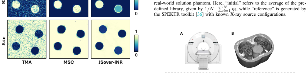
- [Section IV-E, Fig. 10] The reference spectrum for the real solution phantom is a 70 kVP Cu-filtered spectrum, while the library in Fig. 1 consists of 120 kVP and 80 kVP Al-filtered spectra; a convex combination of the library spectra cannot represent the reference exactly. The claimed close match should be quantified (e.g., MAE), and the effect of this representational gap on the spectrum estimate should be discussed.
minor comments (7)
- [III-B1, III-C, II-C] There are several typos: 'Uncostrained Spectra Esimation' and 'uncostrained' should be 'unconstrained', 'spctra' should be 'spectra', and 'Rerepresentation' should be 'Representation'.
- [IV-D] The reference to 'Table I' for the quantitative SEMMD results should be 'Table II'.
- [III-B2, IV-G] The motivation for INR is its low-frequency spectral bias, but the best-performing variant uses hash encoding, which is normally associated with high-frequency detail; please reconcile the stated motivation with the architecture choice.
- [IV-C] Only two image-domain baselines (TMA and MSC) are compared; including a projection-domain or joint-estimation SEMMD baseline would better support the 'state-of-the-art' claim.
- [Eq. (16)] The TV regularizer is defined along X-ray coordinates rather than on the image grid; this is unusual and should be justified or replaced by a standard spatial TV prior.
- [Eq. (9) and Eq. (14)] The notation R denotes the full ray set in Eq. (9) but a random subset in Eq. (14); please use distinct symbols to avoid confusion.
- [Table II vs Tables IV/V] Table II reports RMSE without standard deviations, while Tables IV and V report mean±std; please clarify how many repetitions are performed and whether the RMSE values are averaged over the two phantoms.
Circularity Check
No significant circularity: JSover fits a physics-based forward model to SECT projections and validates against independent material ground truth and SPEKTR reference spectra.
full rationale
The derivation is self-contained. Eq. (8) combines a standard polychromatic CT forward model with an ideal-solution linear-mixture MAC model (Eqs. 3-7); Eq. (9) minimizes projection-domain data consistency. The spectrum is represented as a SoftMax-weighted library combination (Eq. 11), and material fractions are represented by an MLP; both are optimized against the same SECT projections. This common fitting target is not circular because the reported claims are validated against (i) ground-truth volume fractions of simulated XCAT phantoms and prepared solution phantoms, and (ii) SPEKTR reference spectra that are not used as optimization inputs. The library prior constrains the spectrum, but it is an input assumption rather than a restatement of the output; the fitted weights are data-dependent and the ablation (JSover-TV vs JSover-INR) shows the architecture affects the result. Self-citations [23], [29], [30] are related-work context, not load-bearing support for Eqs. (9)-(14). The paper's own statement that Eq. (9) is underdetermined is an honest identifiability caveat, which is a correctness risk, not circularity.
Assumptions & free parameters
free parameters (4)
- Spectrum library weights gamma_i =
Learned per scan; not reported
- INR network parameters Phi =
Learned per scan; not reported
- TV regularization weight lambda =
3e-4
- Hash encoding and optimizer hyperparameters =
L=16, T=2^18, F=8, lr=1e-3, 4000 epochs, 40 rays per step
assumptions (5)
- domain assumption Ideal solution model: mixture MAC is the volume-fraction-weighted sum of basis-material MACs and mixture density is volume-additive.
- domain assumption Basis-material LACs and densities are known from NIST and XCOM tables.
- domain assumption The SPEKTR-generated spectrum library spans the true spectra of the scanners used.
- domain assumption Standard polychromatic CT forward model with a normalized spectrum and no scatter or detector nonlinearity.
- domain assumption INR spectral bias is a valid low-frequency prior for material volume fraction maps.
Cite this review
Pith. "Pith review of JSover: Joint Spectrum Estimation and Multi-Material Decomposition from Single-Energy CT Projections." pith.science (2026). https://pith.science/paper/DC3S7LKI
@misc{pith2026250508123,
  author       = {Pith},
  title        = {Pith review of: JSover: Joint Spectrum Estimation and Multi-Material Decomposition from Single-Energy CT Projections},
  year         = {2026},
  howpublished = {\url{https://pith.science/paper/DC3S7LKI}},
  note         = {Machine review of arXiv:2505.08123}
}
read the original abstract
Multi-material decomposition (MMD) enables quantitative reconstruction of tissue compositions in the human body, supporting a wide range of clinical applications. However, traditional MMD typically requires spectral CT scanners and pre-measured X-ray energy spectra, significantly limiting clinical applicability. To this end, various methods have been developed to perform MMD using conventional (i.e., single-energy, SE) CT systems, commonly referred to as SEMMD. Despite promising progress, most SEMMD methods follow a two-step image decomposition pipeline, which first reconstructs monochromatic CT images using algorithms such as FBP, and then performs decomposition on these images. The initial reconstruction step, however, neglects the energy-dependent attenuation of human tissues, introducing severe nonlinear beam hardening artifacts and noise into the subsequent decomposition. This paper proposes JSover, a fundamentally reformulated one-step SEMMD framework that jointly reconstructs multi-material compositions and estimates the energy spectrum directly from SECT projections. By explicitly incorporating physics-informed spectral priors into the SEMMD process, JSover accurately simulates a virtual spectral CT system from SE acquisitions, thereby improving the reliability and accuracy of decomposition. Furthermore, we introduce implicit neural representation (INR) as an unsupervised deep learning solver for representing the underlying material maps. The inductive bias of INR toward continuous image patterns constrains the solution space and further enhances estimation quality. Extensive experiments on both simulated and real CT datasets show that JSover outperforms state-of-the-art SEMMD methods in accuracy and computational efficiency.
Figures


Figures from the paper (8 more)




Reference graph
Works this paper leans on
-
[1]
Multi-material decomposition of spectral ct images,
P. R. Mendonc ¸a, R. Bhotika, M. Maddah, B. Thomsen, S. Dutta, P. E. Licato, and M. C. Joshi, “Multi-material decomposition of spectral ct images,” inMedical Imaging 2010: Physics of Medical Imaging, vol. 7622, pp. 633–641, SPIE, 2010
work page 2010
-
[2]
Multi-material decomposition using statisti- cal image reconstruction for spectral ct,
Y . Long and J. A. Fessler, “Multi-material decomposition using statisti- cal image reconstruction for spectral ct,”IEEE transactions on medical imaging, vol. 33, no. 8, pp. 1614–1626, 2014
2014
-
[3]
A flexible method for multi-material decomposition of dual-energy ct images,
P. R. Mendonc ¸a, P. Lamb, and D. V . Sahani, “A flexible method for multi-material decomposition of dual-energy ct images,”IEEE transac- tions on medical imaging, vol. 33, no. 1, pp. 99–116, 2013
work page 2013
-
[4]
Accurate multi-material de- composition in dual-energy ct: a phantom study,
Y . Xue, Y . Jiang, C. Yang, Q. Lyu, J. Wang, C. Luo, L. Zhang, C. Desrosiers, K. Feng, X. Sun,et al., “Accurate multi-material de- composition in dual-energy ct: a phantom study,”IEEE Transactions on Computational Imaging, vol. 5, no. 4, pp. 515–529, 2019
work page 2019
-
[5]
Detector- based spectral ct with a novel dual-layer technology: principles and applications,
N. Rassouli, M. Etesami, A. Dhanantwari, and P. Rajiah, “Detector- based spectral ct with a novel dual-layer technology: principles and applications,”Insights into imaging, vol. 8, pp. 589–598, 2017
work page 2017
-
[6]
Photon-counting detector ct: system design and clinical applications of an emerging technology,
S. Leng, M. Bruesewitz, S. Tao, K. Rajendran, A. F. Halaweish, N. G. Campeau, J. G. Fletcher, and C. H. McCollough, “Photon-counting detector ct: system design and clinical applications of an emerging technology,”Radiographics, vol. 39, no. 3, pp. 729–743, 2019
work page 2019
-
[7]
Multi-material decomposition for single energy ct using material sparsity constraint,
Y . Xue, W. Qin, C. Luo, P. Yang, Y . Jiang, T. Tsui, H. He, L. Wang, J. Qin, Y . Xie,et al., “Multi-material decomposition for single energy ct using material sparsity constraint,”IEEE transactions on medical imaging, vol. 40, no. 5, pp. 1303–1318, 2021
work page 2021
-
[8]
Image domain multi-material decomposition using single energy ct,
Y . Xue, C. Luo, Y . Jiang, P. Yang, X. Hu, Q. Zhou, J. Wang, X. Hu, K. Sheng, and T. Niu, “Image domain multi-material decomposition using single energy ct,”Physics in Medicine & Biology, vol. 65, no. 6, p. 065014, 2020
work page 2020
Show all 46 references
-
[9]
A deep learning approach for dual-energy ct imaging using a single-energy ct data,
W. Zhao, T. Lv, P. Gao, L. Shen, X. Dai, K. Cheng, M. Jia, Y . Chen, and L. Xing, “A deep learning approach for dual-energy ct imaging using a single-energy ct data,” in15th International Meeting on Fully Three- Dimensional Image Reconstruction in Radiology and Nuclear Medicin...
2019
-
[10]
A deep learning approach for virtual monochromatic spectral ct imaging with a standard single energy ct scanner,
W. Zhao, T. Lyu, Y . Chen, and L. Xing, “A deep learning approach for virtual monochromatic spectral ct imaging with a standard single energy ct scanner,”arXiv preprint arXiv:2005.09859, 2020
2005 arXiv
-
[11]
Virtual monoenergetic ct imaging via deep learning,
W. Cong, Y . Xi, P. Fitzgerald, B. De Man, and G. Wang, “Virtual monoenergetic ct imaging via deep learning,”Patterns, vol. 1, no. 8, 2020
2020
-
[12]
Pseudo dual- energy ct-derived iodine mapping using single-energy ct data based on a convolution neural network,
Y . Yuasa, T. Shiinoki, K. Fujimoto, and H. Tanaka, “Pseudo dual- energy ct-derived iodine mapping using single-energy ct data based on a convolution neural network,”BJR— Open, vol. 5, no. 1, p. 20220059, 2023
2023
-
[13]
A. C. Kak and M. Slaney,Principles of computerized tomographic imaging. SIAM, 2001
2001
-
[14]
An indirect transmission measurement-based spectrum estimation method for computed tomog- raphy,
W. Zhao, K. Niu, S. Schafer, and K. Royalty, “An indirect transmission measurement-based spectrum estimation method for computed tomog- raphy,”Physics in Medicine & Biology, vol. 60, no. 1, p. 339, 2014
2014
-
[15]
Segmentation-free x-ray energy spectrum estimation for computed tomography,
W. Zhao, Q. Zhang, and T. Niu, “Segmentation-free x-ray energy spectrum estimation for computed tomography,” inMedical Imaging 2016: Physics of Medical Imaging, vol. 9783, pp. 839–845, SPIE, 2016
2016
-
[16]
Segmentation-free x-ray energy spectrum estimation for computed tomography using dual- energy material decomposition,
W. Zhao, L. Xing, Q. Zhang, Q. Xie, and T. Niu, “Segmentation-free x-ray energy spectrum estimation for computed tomography using dual- energy material decomposition,”Journal of Medical Imaging, vol. 4, no. 2, pp. 023506–023506, 2017
2017
-
[17]
Spectrum estimation-guided iterative reconstruction algorithm for dual energy ct,
S. Chang, M. Li, H. Yu, X. Chen, S. Deng, P. Zhang, and X. Mou, “Spectrum estimation-guided iterative reconstruction algorithm for dual energy ct,”IEEE transactions on medical imaging, vol. 39, no. 1, pp. 246–258, 2019
2019
-
[18]
On the spectral bias of neural networks,
N. Rahaman, A. Baratin, D. Arpit, F. Draxler, M. Lin, F. Hamprecht, Y . Bengio, and A. Courville, “On the spectral bias of neural networks,” in International conference on machine learning, pp. 5301–5310, PMLR, 2019
2019
-
[19]
A robust x-ray tube spectra measuring method by attenuation data,
Y . Yang, X. Mou, and X. Chen, “A robust x-ray tube spectra measuring method by attenuation data,” inMedical Imaging 2006: Physics of Medical Imaging, vol. 6142, pp. 1212–1219, SPIE, 2006
2006
-
[20]
A statistical iterative reconstruction framework for dual energy computed tomography without knowing tube spectrum,
S. Chang and X. Mou, “A statistical iterative reconstruction framework for dual energy computed tomography without knowing tube spectrum,” inDevelopments in X-ray Tomography X, vol. 9967, pp. 51–58, SPIE, 2016
2016
-
[21]
A parametric physical model based x-ray spectrum estimation approach for ct imaging,
S. Chang, C. Zhang, X. Mou, Q. Xu, L. He, and X. Chen, “A parametric physical model based x-ray spectrum estimation approach for ct imaging,”IEEE Transactions on Radiation and Plasma Medical Sciences, 2024
2024
-
[22]
Nerp: implicit neural representation learning with prior embedding for sparsely sampled image reconstruc- tion,
L. Shen, J. Pauly, and L. Xing, “Nerp: implicit neural representation learning with prior embedding for sparsely sampled image reconstruc- tion,”IEEE Transactions on Neural Networks and Learning Systems, vol. 35, no. 1, pp. 770–782, 2022
2022
-
[23]
Self-supervised coordinate projection network for sparse-view computed tomography,
Q. Wu, R. Feng, H. Wei, J. Yu, and Y . Zhang, “Self-supervised coordinate projection network for sparse-view computed tomography,” IEEE Transactions on Computational Imaging, vol. 9, pp. 517–529, 2023
2023
-
[24]
Neat: Neural adaptive tomography,
D. R ¨uckert, Y . Wang, R. Li, R. Idoughi, and W. Heidrich, “Neat: Neural adaptive tomography,”ACM Transactions on Graphics (TOG), vol. 41, no. 4, pp. 1–13, 2022
2022
-
[25]
Dper: Diffusion prior driven neural representation for limited angle and sparse view ct reconstruction,
C. Du, X. Lin, Q. Wu, X. Tian, Y . Su, Z. Luo, H. Wei, S. K. Zhou, J. Yu, and Y . Zhang, “Dper: Diffusion prior driven neural representation for limited angle and sparse view ct reconstruction,”arXiv preprint arXiv:2404.17890, 2024
2024 arXiv
-
[26]
Intratomo: self-supervised learning-based tomography via sinogram synthesis and prediction,
G. Zang, R. Idoughi, R. Li, P. Wonka, and W. Heidrich, “Intratomo: self-supervised learning-based tomography via sinogram synthesis and prediction,” inProceedings of the IEEE/CVF International Conference on Computer Vision, pp. 1960–1970, 2021
1960
-
[27]
Dynamic ct reconstruction from limited views with implicit neural representations and parametric motion fields,
A. W. Reed, H. Kim, R. Anirudh, K. A. Mohan, K. Champley, J. Kang, and S. Jayasuriya, “Dynamic ct reconstruction from limited views with implicit neural representations and parametric motion fields,” in Proceedings of the IEEE/CVF International Conference on Computer Vision, p...
2021
-
[28]
Implicit neural representation-based method for metal-induced beam hardening artifact reduction in x-ray ct imaging,
H. S. Park, J. K. Seo, and K. Jeon, “Implicit neural representation-based method for metal-induced beam hardening artifact reduction in x-ray ct imaging,”Medical Physics
-
[29]
Unsupervised polychromatic neural representation for ct metal artifact reduction,
Q. Wu, L. Chen, C. Wang, H. Wei, S. K. Zhou, J. Yu, and Y . Zhang, “Unsupervised polychromatic neural representation for ct metal artifact reduction,”Advances in Neural Information Processing Systems, vol. 36, pp. 69605–69624, 2023
2023
-
[30]
Solving energy-independent density for ct metal artifact reduction via neural representation,
Q. Wu, X. Guo, L. Chen, Y . Liu, D. He, X. Wang, X. Chen, Y . Zhang, S. K. Zhou, J. Yu,et al., “Solving energy-independent density for ct metal artifact reduction via neural representation,”arXiv preprint arXiv:2405.07047, 2024
2024 arXiv
-
[31]
Spectral neural attenuation fields for cone beam ct,
A. Smith, J. Atlas, and N. De Ruiter, “Spectral neural attenuation fields for cone beam ct,” in2024 39th International Conference on Image and Vision Computing New Zealand (IVCNZ), pp. 1–6, IEEE, 2024
2024
-
[32]
Ray-driven spectral ct reconstruction based on neural base-material fields,
L. Shi, C. Liu, P. Yang, J. Qiu, and X. Zhao, “Ray-driven spectral ct reconstruction based on neural base-material fields,”arXiv preprint arXiv:2404.06991, 2024
2024 arXiv
-
[33]
J. K. Seo and E. J. Woo,Nonlinear inverse problems in imaging. John Wiley & Sons, 2012
2012
-
[34]
Icru report 33: Radiation quantities and units,
A. A. Jansen, “Icru report 33: Radiation quantities and units,”Journal of Nuclear Medicine Technology, vol. 8, no. 4, pp. 241–241, 1980
1980
-
[35]
Beutel,Handbook of medical imaging, vol
J. Beutel,Handbook of medical imaging, vol. 3. Spie Press, 2000. 11
2000
-
[36]
Technical note: spektr 3.0—a computational tool for x-ray spectrum,
J. Punnoose, J. Xu, A. Sisniega, W. Zbijewski, and J. Siewerdsen, “Technical note: spektr 3.0—a computational tool for x-ray spectrum,” Medical Physics, vol. 43, no. 8, pp. 4711–4717, 2016
2016
-
[37]
Instant neural graphics primitives with a multiresolution hash encoding,
T. M ¨uller, A. Evans, C. Schied, and A. Keller, “Instant neural graphics primitives with a multiresolution hash encoding,”ACM transactions on graphics (TOG), vol. 41, no. 4, pp. 1–15, 2022
2022
-
[38]
Pytorch: An imperative style, high-performance deep learning library,
A. Paszke, S. Gross, F. Massa, A. Lerer, J. Bradbury, G. Chanan, T. Killeen, Z. Lin, N. Gimelshein, L. Antiga,et al., “Pytorch: An imperative style, high-performance deep learning library,”Advances in neural information processing systems, vol. 32, 2019
2019
-
[39]
Adam: A method for stochastic optimiza- tion,
D. P. Kingma and J. Ba, “Adam: A method for stochastic optimiza- tion,” in3rd International Conference on Learning Representations, ICLR 2015, San Diego, CA, USA, May 7-9, 2015, Conference Track Proceedings(Y . Bengio and Y . LeCun, eds.), 2015
2015
-
[40]
4d xcat phantom for multimodality imaging research,
W. P. Segars, G. Sturgeon, S. Mendonca, J. Grimes, and B. M. Tsui, “4d xcat phantom for multimodality imaging research,”Medical physics, vol. 37, no. 9, pp. 4902–4915, 2010
2010
-
[41]
Tables of x-ray mass attenuation coef- ficients and mass energy-absorption coefficients (version 1.4)
J. H. Hubbell and S. M. Seltzer, “Tables of x-ray mass attenuation coef- ficients and mass energy-absorption coefficients (version 1.4).” Online,
-
[42]
Xcom: Photon cross section database (version 1.5)
M. J. Berger, J. H. Hubbell, S. M. Seltzer, J. Chang, J. S. Coursey, R. Sukumar, D. S. Zucker, and K. Olsen, “Xcom: Photon cross section database (version 1.5).” Online, 2010. Available: http://physics.nist.gov/ xcom [July 24, 2023]
2010
-
[43]
Feasibility study of an improved single-energy material decomposition method for computed tomography,
G. Kim, S. Park, I. Lim, K. Song, and J.-G. Kim, “Feasibility study of an improved single-energy material decomposition method for computed tomography,”IEEE Transactions on Nuclear Science, vol. 69, no. 6, pp. 1366–1374, 2022
2022
-
[44]
Nerf: Representing scenes as neural radiance fields for view synthesis,
B. Mildenhall, P. P. Srinivasan, M. Tancik, J. T. Barron, R. Ramamoorthi, and R. Ng, “Nerf: Representing scenes as neural radiance fields for view synthesis,”Communications of the ACM, vol. 65, no. 1, pp. 99–106, 2021
2021
-
[45]
Fourier features let networks learn high frequency functions in low dimensional domains,
M. Tancik, P. Srinivasan, B. Mildenhall, S. Fridovich-Keil, N. Raghavan, U. Singhal, R. Ramamoorthi, J. Barron, and R. Ng, “Fourier features let networks learn high frequency functions in low dimensional domains,” Advances in neural information processing systems, vol. 33, pp....
2020
-
[2004]
Available: http://physics.nist.gov/xaamdi [2004, July 12]
2004
Reviewed August 15, 2026 · model on record in the stance chip above.
Discussion (0). Continue with ORCID to comment.