REVIEW 3 major objections 5 minor 122 references
Measurements of molecular size and shape on a chip
T0 review · 3 major / 5 minor · reviewed 2026-08-15 · deepseek-v4-flash
Pith's one-line read Escape-time stereometry claims that a molecule's dwell time in nanoscale pockets encodes both its hydrodynamic radius and bounding-sphere diameter, making size, shape, interactions, and conformational change readable from a wide-field…
desk verdict A broadly validated chip-based technique for measuring molecular size and shape in solution, with the non-spherical 'bounding sphere' assumption as the main soft spot. read the letter →
The pith
A machine-rendered reading of the paper's core claim, the machinery that carries it, and where it could break.
The reading
What carries the argument
The entropic fluidic trap: a periodic array of cylindrical indentations of total height $h_2$ in a parallel-plate slit of height $h_1$, where a molecule's residence time is amplified by the ratio $(h_2 - D_s)/(h_1 - D_s)$. The load-bearing identity is Eq. (1), $t_{\mathrm{esc}} = A r_H (h_2 - D_s)/(h_1 - D_s) + t_0$, which converts measured dwell times into the pair ($r_H$, $D_s$). The prefactor $A$ and offset $t_0$ are fixed by Brownian-dynamics simulations of spherical particles, and $h_1$ is calibrated with globular proteins of known $r_H$; the same equation then serves for all molecular species.
What would settle it
Take rigid rod-like molecules of known length, such as 30 to 60 bp dsDNA, measure $t_{\mathrm{esc}}$ in three or more calibrated slit heights, and test whether Eq. (1) with a single fixed $D_s$ (from the helix structure) and an independently measured $r_H$ fits all the data; if the $D_s$ inferred from different height pairs drifts, the effective-sphere assumption fails.
Extended reading notes
Core claim
The central claim is that the escape time of a fluorescently labelled molecule from a cylindrical pocket in a nanoslit obeys $t_{\mathrm{esc}} = A r_H (h_2 - D_s)/(h_1 - D_s) + t_0$, with $A$ a geometry- and viscosity-dependent prefactor fixed by Brownian-dynamics simulation. Because the pocket height $h_2$ and slit height $h_1$ are known, two escape-time measurements in different slit heights give two equations for the two unknowns $r_H$ (the Stokes or hydrodynamic radius) and $D_s$ (the diameter of the smallest sphere enclosing the molecule, which for a rotating non-spherical molecule can be much larger than $2r_H$). The authors take as evidence the agreement of inferred $r_H$ values with structure-based calculations, the recovery of roughly 3.2 Å rise per base pair for B-DNA and 2.3 Å for A-RNA, the resolution of same-mass DNA nanostructures with different shapes, and the ligand-induced compaction of the insulin receptor. They therefore present ETs as a single platform for molecular-weight determination, mixture analysis, affinity and kinetics measurements, and conformational detection in native solution.
Load-bearing premise
The method assumes a non-spherical molecule rotates quickly enough that, while diffusing through the slit, it behaves like an effective sphere of a single diameter $D_s$ (larger than twice its hydrodynamic radius), and that this one $D_s$ controls the entropic factor $(h_2 - D_s)/(h_1 - D_s)$; if real molecules do not sweep out such a sphere under strong confinement, every inferred $D_s$ would be systematically biased.
Editorial extensions
If this is right
- A single minute of imaging can return $r_H$ and $D_s$ for thousands of individual molecules, giving molecular-weight, shape, affinity, and conformational readouts in native buffer without tethering or strong fields.
- Because the readout responds to $D_s$ rather than mass alone, two species with nearly identical diffusion coefficients become distinguishable once the slit height is chosen close to $D_s$.
- Measuring escape times at two different slit heights yields both $r_H$ and $D_s$, and with three or more heights the same data can resolve closely spaced conformational states and map them onto ellipsoidal models.
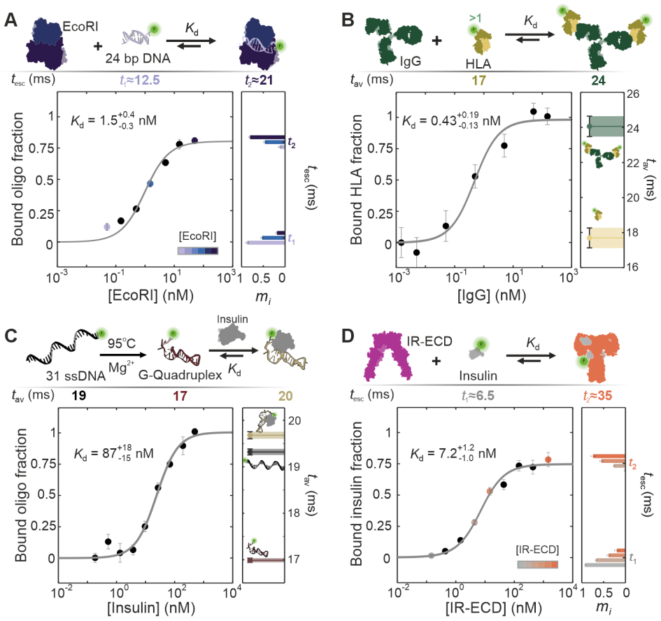
- Binding affinities spanning roughly $10^{-11}$ to $10^{-4}$ M, together with on- and off-rates, follow from the same platform because the bound fraction shifts either a resolved escape-time component or the mean escape time.
- Conformational compaction can dominate the escape-time change on binding, as claimed for the insulin receptor, so the method can report ligand-induced shape changes even when the mass of the complex increases.
Reading between the lines
- If the two-height inference is as robust as claimed, ETs could serve as a routine solution-phase screen between size-exclusion chromatography and small-angle scattering, working on samples too dilute or heterogeneous for either.
- The single-molecule escape-time spectra suggest a general way to count coexisting conformational states and their abundances without fragile multi-exponential fitting; this could be tested on a two-state folding system whose population ratio is controlled externally.
- The claimed sub-1% precision in $t_{\mathrm{esc}}$ implies that longer single-molecule trajectories could resolve mass differences below one carbon atom; a direct test would be a homologous series of small molecules measured in one calibrated chip.
- If $D_s$ values inferred by ETs match $D_{\mathrm{max}}$ values from small-angle scattering on the same constructs, the method could provide a cheap, high-throughput constraint for validating structural models and for machine-learning structure prediction.
Signed reviews
Editorial analysis
A structured set of objections, weighed in public.
Referee Report
Summary. The manuscript reports a microfluidic 'escape-time stereometry' (ETs) method. Singly labeled molecules diffuse through nanoslits with periodic cylindrical pockets; the average time to leave a pocket (t_esc) is argued to obey Eq. (1), t_esc = A rH (h2 - Ds)/(h1 - Ds) + t0, where rH is the hydrodynamic radius and Ds is the diameter of the minimum bounding sphere. The authors calibrate A, t0, and h1 using Brownian Dynamics simulations of spheres and globular proteins of known rH, then use Eq. (1) to infer rH and Ds for dsDNA/dsRNA, DNA nanostructures, a SAM-IV riboswitch, and the insulin receptor ectodomain. The paper also measures binding affinities and kinetics for DNA hybridization, DNA-protein, protein-protein, and aptamer-insulin interactions, and demonstrates a diagnostic readout for insulin in serum based on ligand-induced compaction of IR-ECD.
Significance. If the central mapping holds, this is a broadly applicable, high-throughput solution-phase method for molecular size, shape, conformation, and interaction thermodynamics at single-molecule sensitivity, with clear clinical potential. The paper's strengths are its extensive validation strategy: Eq. (1) is tested by BD simulations for spheres, rH values for globular proteins agree with HYDROPRO and 2f-FCS, DNA rise-per-basepair values match crystallographic expectations, DNA nanostructure Ds values agree with oxDNA, riboswitch inferences are benchmarked against cryo-EM structures, and measured Kd values fall within a factor of 2-3 of literature values. The method is not circular: A and t0 come from simulation, h1 is calibrated with independently known protein radii, and downstream results are checked against multiple independent techniques. However, the central extension from spherical to non-spherical molecules rests on an assumption that is not directly validated, which limits the weight of the shape-related claims.
major comments (3)
- [Main text, after Eq. (1); Section S5.1; Sections S5.4-S5.7] The central mapping Eq. (1) is validated by BD simulations only for spheres (S5.1), yet the paper extends it to non-spherical molecules by asserting that rapid isotropic rotation makes a translating molecule 'sweep out a sphere' of diameter Ds equal to its minimum bounding-sphere diameter. This assumption underlies every shape and conformation inference in the paper, including DNA rise per basepair, nanostructure Ds, riboswitch conformational states, and the IR-ECD compaction claim. The physical situation is not obviously compatible with the assumption: hard-wall confinement in slits with h1 comparable to molecular length restricts isotropic rotation, so the effective excluded height should be an orientation-dependent quantity between the short and long molecular axes. I request a concrete test of the assumption, either by BD simulations of rigid spheroids/cylinders with the aspect ratios and slit heights used here, or by an explicit orientational averaging model. Without such a test, the absolute values of Ds and, more importantly, the inferred differences in Ds between conformational states are not firmly established.
- [Section S5.4, Fig. 3A, Fig. S11] The DNA/RNA rise-per-basepair fits are performed with Ds = b*nbp and with both A and b treated as free parameters (S5.4). For the longest constructs (56-60 bp, L about 19-20 nm) in slits with h1 about 25 nm, the denominator h1 - Ds becomes small. Under the fitted parameters, the escaping time predicted for a rod with Ds equal to its full length is considerably larger than the measured t_esc values for 60 bp DNA reported elsewhere in the manuscript (e.g., Fig. 4B shows about 26 ms for 60 bp in a comparable device). This suggests that either the effective Ds is not the geometric minball diameter, or the fitted parameters absorb this inconsistency. The authors should report the measured and fitted t_esc versus n_bp data with residuals for the specific devices used, and should test the fit's robustness by fixing A from a non-DNA calibration and inferring Ds independently for each DNA length.
- [Section S7.10, Fig. 7E, Eq. (S28)] The Kd determination for IR-ECD binding insulin uses t_av as a proxy for bound fraction with the statement that t_av is 'proportional to the amount of IR-ECD in the liganded state.' However, Section S7.1 derives a nonlinear relation between m2 and t_av (Eq. S28). For the small t_av changes reported (a 7% decrease), the linear and nonlinear mappings may differ by only a small amount, but the quoted Kd values (2-12 nM in PBS, about 100 pM in human serum) are used as quantitative claims. The authors should state explicitly the range of validity of the linear approximation and quantify how much the inferred Kd shifts when Eq. (S28) is used instead of a simple linear interpolation.
minor comments (5)
- [Section S5.2 heading] The heading 'Determination of rH and Ds relation for the SAMI-IV riboswitch' contains a typo: 'SAMI' should be 'SAM-IV.'
- [Eq. (1) and its usage throughout] Please state explicitly the units used in Eq. (1). As written, A has units s/(molecular-length unit), and rH must be expressed in the same unit for the product A*rH to have units of time. The text uses rH in nm in some places and the calibration values in s/micrometer, which can confuse readers.
- [Section S5.1, Eq. (S11)] The reported fit parameters for A have large fractional uncertainties (alpha = 0.16 +/- 0.34 s/nm, beta = -2.35 +/- 0.60, gamma = 0.265 +/- 0.062 s/micrometer). Because A enters every absolute determination of Ds and rH, the paper should include a statement on how these systematic uncertainties propagate into the final Ds and rH values, beyond the statistical uncertainties shown in Figs. 3 and 7.
- [Abstract and Fig. 2B] The abstract claims detection 'down to two carbon atoms,' while the main text reports a demonstrated 25 Da difference (about two carbons) and a 'theoretical ability' to detect about 10 Da. The wording should be adjusted so that the demonstrated resolution is not overstated as a routine capability.
- [Section S7.11] The discussion of concentration inaccuracy is useful but could be more explicit: the statement that Kd is 'relatively insensitive to [A]0' should be accompanied by the parameter ranges over which this holds, particularly for the HLA and serum measurements where [A]0 is around 0.1 nM or lower.
Circularity Check
No significant circularity: Eq. (1) is established by BD simulations and independently calibrated against known protein radii, with downstream inferences cross-validated against HYDROPRO, FCS, oxDNA, SAXS, and cryo-EM structures.
full rationale
The central derivation chain is self-contained. Equation (1) is not obtained by fitting the experimental data it is used to interpret; it is produced by Brownian Dynamics simulations of spherical particles in the trap landscape (Supplementary S5.1), and the prefactor A and offset t0 are simulation outputs. The slit height h1 is then calibrated using globular proteins of known hydrodynamic radius, an external input, and the inferred rH values for a test set agree with HYDROPRO and FCS. DNA rise-per-basepair values are fitted from the escape-time data, but they are presented as measurements to be compared with known crystallographic values, not as predictions derived from the model; the agreement is an external consistency check, not a circular argument. The DNA-nanostructure, riboswitch, and IR-ECD conformational inferences are all cross-validated against oxDNA, cryo-EM, or SAXS-based structural models. The paper's assumption that non-spherical molecules rotate fast enough to sweep out a sphere of diameter Ds is a physical modeling assumption, not a definitional identity with the measured escape time; if invalid it would bias inferences, but it does not make the derivation circular. Self-citations appear for the trap concept and BD method, but the method is re-run and quantified here, so they are not load-bearing. No step was found in which a fitted parameter is renamed as a prediction or in which a result is equivalent to its inputs by construction.
Assumptions & free parameters
free parameters (4)
- A (escape-time prefactor) =
~0.3 s/μm, with A = α(h1/h2)^β + γ; α = 0.16 ± 0.34 s/nm, β = -2.35 ± 0.60, γ = 0.265 ± 0.062 s/μm
- t0 (escape-time offset) =
5.83 ms (≈ exposure time)
- h1 (slit height) =
e.g., 23.8 ± 0.1 nm for one device; calibrated with globular proteins
- b (DNA/RNA rise per basepair) =
3.2 Å (DNA), 2.3 Å (RNA)
assumptions (7)
- standard math Stokes-Einstein relation and Perrin/Tirado hydrodynamic formulas for cylinders and spheroids
- domain assumption At high salt (~160 mM, Debye length ~0.75 nm) electrostatic contributions to the trap are negligible, so the trap is purely entropic
- domain assumption Non-spherical molecules rotate isotropically while translating and sweep out an effective sphere of diameter Ds > 2rH
- domain assumption Globular proteins used for calibration are spherical, i.e., Ds = 2rH
- ad hoc to paper RNA density of 1.6 g/cm3 and fixed spheroid volume of 40 nm3 for the SAM-IV riboswitch
- domain assumption A single ATTO532/Alexa Fluor 532 label does not significantly perturb molecular hydrodynamic properties
- ad hoc to paper For IR-ECD insulin sensing, the change in t_av is attributed solely to conformational compaction and used linearly to infer bound fraction
Cite this review
Pith. "Pith review of Measurements of molecular size and shape on a chip." pith.science (2026). https://pith.science/paper/TTI6RTCY
@misc{pith2026250508452,
  author       = {Pith},
  title        = {Pith review of: Measurements of molecular size and shape on a chip},
  year         = {2026},
  howpublished = {\url{https://pith.science/paper/TTI6RTCY}},
  note         = {Machine review of arXiv:2505.08452}
}
read the original abstract
Size and shape are critical discriminators between molecular species and states. We describe a micro-chip based high-throughput imaging approach offering rapid and precise determination of molecular properties under native solution conditions. Our method detects differences in molecular weight across at least three orders of magnitude, and down to two carbon atoms in small molecules. We quantify the strength of molecular interactions over six orders of magnitude in affinity constant, and track reactions in real-time. Highly parallel measurements on individual molecules serve to characterize sample-state heterogeneity at the highest resolution, offering predictive input to model three-dimensional structure. We further leverage the method's structural sensitivity for diagnostics, exploiting ligand-induced conformational changes in the insulin receptor to sense insulin concentration in serum at the sub-nanoliter and sub-zeptomole scale.
Figures


Figures from the paper (3 more)


Reference graph
Works this paper leans on
-
[2]
E. C. Yusko et al., Real-time shape approximation and fingerprinting of single proteins using a nanopore. Nature Nanotechnology 12, 360–367 (2016). 17
2016
-
[4]
Young et al., Quantitative mass imaging of single biological macromolecules
G. Young et al., Quantitative mass imaging of single biological macromolecules. Science 360, 423-427 (2018)
2018
-
[6]
Schmid, P
S. Schmid, P. Stömmer, H. Dietz, C. Dekker, Nanopore electro-osmotic trap for the label-free study of single proteins and their conformations. Nature Nanotechnology 16, 1244–1250 (2021)
2021
-
[9]
Needham et al., Label-free detection and profiling of individual solution-phase molecules
L.-M. Needham et al., Label-free detection and profiling of individual solution-phase molecules. Nature 629, 1062–1068 (2024)
2024
-
[11]
Friedel, A
M. Friedel, A. Baumketner, J.-E. Shea, Effects of surface tethering on protein folding mechanisms. Proceedings of the National Academy of Sciences 103, 8396-8401 (2006)
2006
-
[13]
Perrin, Mouvement Brownien d'un ellipsoide (II)
F. Perrin, Mouvement Brownien d'un ellipsoide (II). Rotation libre et dépolarisation des fluorescences. Translation et diffusion de molécules ellipsoidales. Journal de Physique et le Radium 7, 1-11 (1936)
1936
-
[15]
P. J. Fleming, K. G. Fleming, HullRad: Fast Calculations of Folded and Disordered Protein and Nucleic Acid Hydrodynamic Properties. Biophysical Journal 114, 856- 869 (2018)
2018
-
[17]
A. G. Kikhney, D. I. Svergun, A practical guide to small angle X-ray scattering (SAXS) of flexible and intrinsically disordered proteins. FEBS Letters 589, 2570- 2577 (2015)
2015
Show all 122 references
-
[19]
Walker-Gibbons, X
R. Walker-Gibbons, X. Zhu, A. Behjatian, T. J. D. Bennett, M. Krishnan, Sensing the structural and conformational properties of single-stranded nucleic acids using electrometry and molecular simulations. Scientific Reports 14, 20582 (2024)
2024
-
[20]
C. B. Müller et al., Precise measurement of diffusion by multi-color dual-focus fluorescence correlation spectroscopy. Europhysics Letters 83, 46001 (2008)
2008
-
[22]
Ruggeri, M
F. Ruggeri, M. Krishnan, Entropic Trapping of a Singly Charged Molecule in Solution. Nano Letters 18, 3773–3779 (2018). 18
2018
-
[23]
Kloes et al., Far-Field Electrostatic Signatures of Macromolecular 3D Conformation
G. Kloes et al., Far-Field Electrostatic Signatures of Macromolecular 3D Conformation. Nano Letters 22, 7834–7840 (2022)
2022
-
[26]
D. K. Wilkins et al., Hydrodynamic Radii of Native and Denatured Proteins Measured by Pulse Field Gradient NMR Techniques. Biochemistry 38, 16424-16431 (1999)
1999
-
[28]
Nojima, K
Y. Nojima, K. Iguchi, Y. Suzuki, A. Sato, The pH-Dependent Formation of PEGylated Bovine Lactoferrin by Branched Polyethylene Glycol (PEG)-N- Hydroxysuccinimide (NHS) Active Esters. Biological and Pharmaceutical Bulletin 32, 523-526 (2009)
2009
-
[30]
R. E. Dickerson et al., The Anatomy of A-, B-, and Z-DNA. Science 216, 475-485 (1982)
1982
-
[33]
Poppleton, R
E. Poppleton, R. Romero, A. Mallya, L. Rovigatti, P. Sulc, OxDNA.org: a public webserver for coarse-grained simulations of DNA and RNA nanostructures. Nucleic Acids Research 49, W491-W498 (2021)
2021
-
[35]
Wei et al., Fluorescent Imaging of Single Nanoparticles and Viruses on a Smart Phone
Q. Wei et al., Fluorescent Imaging of Single Nanoparticles and Viruses on a Smart Phone. ACS Nano 7, 9147–9155 (2013)
2013
-
[36]
R. J. L. Townshend et al., Geometric deep learning of RNA structure. Science 373, 1047-1051 (2021)
2021
-
[37]
Zhang et al., Cryo-EM structure of a 40 kDa SAM-IV riboswitch RNA at 3.7 Å resolution
K. Zhang et al., Cryo-EM structure of a 40 kDa SAM-IV riboswitch RNA at 3.7 Å resolution. Nature Communications 10, 5511 (2019)
2019
-
[39]
C. D. Stoddard et al., Free State Conformational Sampling of the SAM-I Riboswitch Aptamer Domain. Structure 18, 787-797 (2010)
2010
-
[42]
SantaLucia, D
J. SantaLucia, D. Hicks, The thermodynamics of DNA structural motifs. Annu Rev Bioph Biom 33, 415-440 (2004)
2004
-
[43]
Wilson, M
H. Wilson, M. Lee, Q. Wang, Probing DNA-protein interactions using single- molecule diffusivity contrast. Biophysical Reports 1, 100009 (2021)
2021
-
[44]
C. J. Barnstable et al., Production of monoclonal antibodies to group A erythrocytes, HLA and other human cell surface antigens-new tools for genetic analysis. Cell 14, 9- 20 (1978). 19
1978
-
[45]
M. N. Hug et al., HLA antibody affinity determination: From HLA-specific monoclonal antibodies to donor HLA specific antibodies (DSA) in patient serum. HLA 102, 278-300 (2023)
2023
-
[47]
Yoshida et al., Selection of DNA aptamers against insulin and construction of an aptameric enzyme subunit for insulin sensing
W. Yoshida et al., Selection of DNA aptamers against insulin and construction of an aptameric enzyme subunit for insulin sensing. Biosensors and Bioelectronics 24, 1116-1120 (2009)
2009
-
[48]
Hao et al., Real-Time Monitoring of Insulin Using a Graphene Field-Effect Transistor Aptameric Nanosensor
Z. Hao et al., Real-Time Monitoring of Insulin Using a Graphene Field-Effect Transistor Aptameric Nanosensor. ACS Applied Materials & Interfaces 9, 27504– 27511 (2017)
2017
-
[49]
Wu et al., Flow-Cell-Based Technology for Massively Parallel Characterization of Base-Modified DNA Aptamers
D. Wu et al., Flow-Cell-Based Technology for Massively Parallel Characterization of Base-Modified DNA Aptamers. Analytical Chemistry 95, 2645–2652 (2023)
2023
-
[52]
De Meyts, J
P. De Meyts, J. Whittaker, Structural biology of insulin and IGF1 receptors: implications for drug design. Nature Reviews Drug Discovery 1, 769-783 (2002)
2002
-
[54]
Gutmann, K
T. Gutmann, K. H. Kim, M. Grzybek, T. Walz, Ü. Coskun, Visualization of ligand- induced transmembrane signaling in the full-length human insulin receptor. Journal of Cell Biology 217, 1643–1649 (2018)
2018
-
[56]
Xiong et al., Symmetric and asymmetric receptor conformation continuum induced by a new insulin
X. Xiong et al., Symmetric and asymmetric receptor conformation continuum induced by a new insulin. Nature Chemical Biology 18, 511-519 (2022)
2022
-
[58]
Weis et al., The signalling conformation of the insulin receptor ectodomain
F. Weis et al., The signalling conformation of the insulin receptor ectodomain. Nature Communications 9, 4420 (2018)
2018
-
[61]
Ruggeri, M
F. Ruggeri, M. Krishnan, Spectrally resolved single-molecule electrometry. The Journal of Chemical Physics 148, 123307 (2018)
2018
-
[63]
F. L. Sendker et al., Emergence of fractal geometries in the evolution of a metabolic enzyme. Nature 628, 894-900 (2024)
2024
-
[64]
Aguzzi, T
A. Aguzzi, T. O'Connor, Protein aggregation diseases: pathogenicity and therapeutic perspectives. Nature Reviews Drug Discovery 9, 237-248 (2010)
2010
-
[65]
M. T. Marty et al., Bayesian Deconvolution of Mass and Ion Mobility Spectra: From Binary Interactions to Polydisperse Ensembles. Analytical Chemistry 87, 4370–4376 (2015). 20
2015
-
[66]
Femtodrop
S. G. Schultz , A. K. Solomon Determination of the Effective Hydrodynamic Radii of Small Molecules by Viscometry. Journal of General Physiology 44, 1189–1199 (1961). 21 Figures Fig. 1. Principle and overview of escape -time stereometry (ETs) in solution. ( A) Schematic represe...
1961
-
[67]
equivalent spheres
We note that theoretically these should be identical, but in practice we get different uncertainties for 𝐸1 and 𝐸2. We therefore use the larger of the two errors to estimate the overall uncertainty, the final expression given as follows: (𝑚2,e2) = [𝑚2 𝐸2 (1 − 𝑚2)] 2 𝐸2,e 2 + [...
-
[68]
J. Han, H. G. Craighead, Separation of Long DNA Molecules in a Microfabricated Entropic Trap Array. Science 288, 1026-1029 (2000)
2000
-
[69]
E. C. Yusko et al., Real-time shape approximation and fingerprinting of single proteins 5 using a nanopore. Nature Nanotechnology 12, 360–367 (2016)
2016
-
[70]
Ruggeri et al., Single-molecule electrometry
F. Ruggeri et al., Single-molecule electrometry. Nature Nanotechnology 12, 488-495 (2017)
2017
-
[71]
Young et al., Quantitative mass imaging of single biological macromolecules
G. Young et al., Quantitative mass imaging of single biological macromolecules. Science 360, 423-427 (2018). 10
2018
-
[72]
Wilson, Q
H. Wilson, Q. Wang, ABEL-FRET: tether-free single-molecule FRET with hydrodynamic profiling. Nature Methods 18, 816–820 (2021)
2021
-
[73]
Schmid, P
S. Schmid, P. Stömmer, H. Dietz, C. Dekker, Nanopore electro-osmotic trap for the label- free study of single proteins and their conformations. Nature Nanotechnology 16, 1244– 1250 (2021). 15
2021
-
[74]
Špačková et al., Label-free nanofluidic scattering microscopy of size and mass of single diffusing molecules and nanoparticles
B. Špačková et al., Label-free nanofluidic scattering microscopy of size and mass of single diffusing molecules and nanoparticles. Nature Methods 19, 751-758 (2022)
2022
-
[75]
R. P. B. Jacquat et al., Single-Molecule Sizing through Nanocavity Confinement. Nano Letters 23, 1629-1636 (2023)
2023
-
[76]
Needham et al., Label-free detection and profiling of individual solution-phase 20 molecules
L.-M. Needham et al., Label-free detection and profiling of individual solution-phase 20 molecules. Nature 629, 1062–1068 (2024)
2024
-
[77]
Ohayon et al., Full‐Length Single Protein Molecules Tracking and Counting in Thin Silicon Channels
S. Ohayon et al., Full‐Length Single Protein Molecules Tracking and Counting in Thin Silicon Channels. Advanced Materials 36, 2314319 (2024)
2024
-
[78]
Friedel, A
M. Friedel, A. Baumketner, J.-E. Shea, Effects of surface tethering on protein folding mechanisms. Proceedings of the National Academy of Sciences 103, 8396-8401 (2006). 25
2006
-
[79]
Y. Pang, R. Gordon, Optical Trapping of a Single Protein. Nano Letters 12, 402-406 (2012)
2012
-
[80]
Perrin, Mouvement Brownien d'un ellipsoide (II)
F. Perrin, Mouvement Brownien d'un ellipsoide (II). Rotation libre et dépolarisation des fluorescences. Translation et diffusion de molécules ellipsoidales. Journal de Physique et le Radium 7, 1-11 (1936). 30
1936
-
[81]
Ortega, D
A. Ortega, D. Amorós, J. García de la Torre, Prediction of Hydrodynamic and Other Solution Properties of Rigid Proteins from Atomic- and Residue-Level Models. Biophysical Journal 101, 892-898 (2011)
2011
-
[82]
P. J. Fleming, K. G. Fleming, HullRad: Fast Calculations of Folded and Disordered Protein and Nucleic Acid Hydrodynamic Properties. Biophysical Journal 114, 856-869 35 (2018)
2018
-
[83]
Krohn, A
J.-H. Krohn, A. Mamot, N. Kaletta, Y. Qutbuddin, P. Schwille, Fluorescence correlation spectroscopy for particle sizing: A notorious challenge. Biophysical Journal, (2025)
2025
-
[84]
A. G. Kikhney, D. I. Svergun, A practical guide to small angle X-ray scattering (SAXS) of flexible and intrinsically disordered proteins. FEBS Letters 589, 2570-2577 (2015). 40
2015
-
[85]
Bespalova, A
M. Bespalova, A. Behjatian, N. Karedla, R. Walker-Gibbons, M. Krishnan, Opto- Electrostatic Determination of Nucleic Acid Double-Helix Dimensions and the Structure of the Molecule–Solvent Interface. Macromolecules 55, 6200-6210 (2022)
2022
-
[86]
Walker-Gibbons, X
R. Walker-Gibbons, X. Zhu, A. Behjatian, T. J. D. Bennett, M. Krishnan, Sensing the structural and conformational properties of single-stranded nucleic acids using 45 electrometry and molecular simulations. Scientific Reports 14, 20582 (2024)
2024
-
[87]
C. B. Müller et al., Precise measurement of diffusion by multi-color dual-focus fluorescence correlation spectroscopy. Europhysics Letters 83, 46001 (2008). 90
2008
-
[88]
Krainer et al., Single-molecule digital sizing of proteins in solution
G. Krainer et al., Single-molecule digital sizing of proteins in solution. Nature Communications 15, 7740 (2024)
2024
-
[89]
Ruggeri, M
F. Ruggeri, M. Krishnan, Entropic Trapping of a Singly Charged Molecule in Solution. Nano Letters 18, 3773–3779 (2018)
2018
-
[90]
Kloes et al., Far-Field Electrostatic Signatures of Macromolecular 3D Conformation
G. Kloes et al., Far-Field Electrostatic Signatures of Macromolecular 3D Conformation. 5 Nano Letters 22, 7834–7840 (2022)
2022
-
[91]
Krishnan, N
M. Krishnan, N. Mojarad, P. Kukura, V. Sandoghdar, Geometry-induced electrostatic trapping of nanometric objects in a fluid. Nature 467, 692-695 (2010)
2010
-
[92]
P. J. Flory, Principles of Polymer Chemistry. (Cornell University Press, 1953)
1953
-
[93]
D. K. Wilkins et al., Hydrodynamic Radii of Native and Denatured Proteins Measured by 10 Pulse Field Gradient NMR Techniques. Biochemistry 38, 16424-16431 (1999)
1999
-
[94]
J. E. Kohn et al., Random-coil behavior and the dimensions of chemically unfolded proteins. Proceedings of the National Academy of Sciences 101, 12491-12496 (2004)
2004
-
[95]
Nojima, K
Y. Nojima, K. Iguchi, Y. Suzuki, A. Sato, The pH-Dependent Formation of PEGylated Bovine Lactoferrin by Branched Polyethylene Glycol (PEG)-N-Hydroxysuccinimide 15 (NHS) Active Esters. Biological and Pharmaceutical Bulletin 32, 523-526 (2009)
2009
-
[96]
Ruggeri, M
F. Ruggeri, M. Krishnan, Lattice diffusion of a single molecule in solution. Physical Review E 96, 062406 (2017)
2017
-
[97]
R. E. Dickerson et al., The Anatomy of A-, B-, and Z-DNA. Science 216, 475-485 (1982). 20
1982
-
[98]
R. P. Goodman et al., Rapid Chiral Assembly of Rigid DNA Building Blocks for Molecular Nanofabrication. Science 310, 1661-1665 (2005)
2005
-
[99]
Fischer et al., Shape and Interhelical Spacing of DNA Origami Nanostructures Studied by Small-Angle X-ray Scattering
S. Fischer et al., Shape and Interhelical Spacing of DNA Origami Nanostructures Studied by Small-Angle X-ray Scattering. Nano Letters 16, 4282-4287 (2016)
2016
-
[100]
Poppleton, R
E. Poppleton, R. Romero, A. Mallya, L. Rovigatti, P. Sulc, OxDNA.org: a public 25 webserver for coarse-grained simulations of DNA and RNA nanostructures. Nucleic Acids Research 49, W491-W498 (2021)
2021
-
[101]
A. A. Istratov, O. F. Vyvenko, Exponential analysis in physical phenomena. Review of Scientific Instruments 70, 1233–1257 (1999)
1999
-
[102]
Wei et al., Fluorescent Imaging of Single Nanoparticles and Viruses on a Smart 30 Phone
Q. Wei et al., Fluorescent Imaging of Single Nanoparticles and Viruses on a Smart 30 Phone. ACS Nano 7, 9147–9155 (2013)
2013
-
[103]
R. J. L. Townshend et al., Geometric deep learning of RNA structure. Science 373, 1047- 1051 (2021)
2021
-
[104]
Zhang et al., Cryo-EM structure of a 40 kDa SAM-IV riboswitch RNA at 3.7 Å resolution
K. Zhang et al., Cryo-EM structure of a 40 kDa SAM-IV riboswitch RNA at 3.7 Å resolution. Nature Communications 10, 5511 (2019). 35
2019
-
[105]
A. D. Garst, A. Héroux, R. P. Rambo, R. T. Batey, Crystal Structure of the Lysine Riboswitch Regulatory mRNA Element. Journal of Biological Chemistry 283, 22347- 22351 (2008)
2008
-
[106]
C. D. Stoddard et al., Free State Conformational Sampling of the SAM-I Riboswitch Aptamer Domain. Structure 18, 787-797 (2010). 40
2010
-
[107]
Palau, C
W. Palau, C. Di Primo, Single-cycle kinetic analysis of ternary DNA complexes by surface plasmon resonance on a decaying surface. Biochimie 94, 1891-1899 (2012)
2012
-
[108]
Jarmoskaite, I
I. Jarmoskaite, I. AlSadhan, P. P. Vaidyanathan, D. Herschlag, How to measure and evaluate binding affinities. eLife 9, e57264 (2020)
2020
-
[109]
SantaLucia, D
J. SantaLucia, D. Hicks, The thermodynamics of DNA structural motifs. Annu Rev 45 Bioph Biom 33, 415-440 (2004)
2004
-
[110]
Wilson, M
H. Wilson, M. Lee, Q. Wang, Probing DNA-protein interactions using single-molecule diffusivity contrast. Biophysical Reports 1, 100009 (2021). 91
2021
-
[111]
C. J. Barnstable et al., Production of monoclonal antibodies to group A erythrocytes, HLA and other human cell surface antigens-new tools for genetic analysis. Cell 14, 9-20 (1978)
1978
-
[112]
M. N. Hug et al., HLA antibody affinity determination: From HLA-specific monoclonal antibodies to donor HLA specific antibodies (DSA) in patient serum. HLA 102, 278-300 5 (2023)
2023
-
[113]
M. M. Schneider et al., Microfluidic antibody affinity profiling of alloantibody-HLA interactions in human serum. Biosensors and Bioelectronics 228, 115196 (2023)
2023
-
[114]
Yoshida et al., Selection of DNA aptamers against insulin and construction of an aptameric enzyme subunit for insulin sensing
W. Yoshida et al., Selection of DNA aptamers against insulin and construction of an aptameric enzyme subunit for insulin sensing. Biosensors and Bioelectronics 24, 1116-10 1120 (2009)
2009
-
[115]
Hao et al., Real-Time Monitoring of Insulin Using a Graphene Field-Effect Transistor Aptameric Nanosensor
Z. Hao et al., Real-Time Monitoring of Insulin Using a Graphene Field-Effect Transistor Aptameric Nanosensor. ACS Applied Materials & Interfaces 9, 27504–27511 (2017)
2017
-
[116]
Wu et al., Flow-Cell-Based Technology for Massively Parallel Characterization of Base-Modified DNA Aptamers
D. Wu et al., Flow-Cell-Based Technology for Massively Parallel Characterization of Base-Modified DNA Aptamers. Analytical Chemistry 95, 2645–2652 (2023). 15
2023
-
[117]
J. G. Menting et al., How insulin engages its primary binding site on the insulin receptor. Nature 493, 241-245 (2013)
2013
-
[118]
Gutmann et al., Cryo-EM structure of the complete and ligand-saturated insulin receptor ectodomain
T. Gutmann et al., Cryo-EM structure of the complete and ligand-saturated insulin receptor ectodomain. Journal of Cell Biology 219, e201907210 (2020)
2020
-
[119]
De Meyts, J
P. De Meyts, J. Whittaker, Structural biology of insulin and IGF1 receptors: implications 20 for drug design. Nature Reviews Drug Discovery 1, 769-783 (2002)
2002
-
[120]
V. V. Kiselyov, S. Versteyhe, L. Gauguin, P. D. Meyts, Harmonic oscillator model of the insulin and IGF1 receptors’ allosteric binding and activation. Molecular Systems Biology 5, 243 (2009)
2009
-
[121]
Gutmann, K
T. Gutmann, K. H. Kim, M. Grzybek, T. Walz, Ü. Coskun, Visualization of ligand-25 induced transmembrane signaling in the full-length human insulin receptor. Journal of Cell Biology 217, 1643–1649 (2018)
2018
-
[122]
Croll, Tristan et al., Higher-Resolution Structure of the Human Insulin Receptor Ectodomain: Multi-Modal Inclusion of the Insert Domain
I. Croll, Tristan et al., Higher-Resolution Structure of the Human Insulin Receptor Ectodomain: Multi-Modal Inclusion of the Insert Domain. Structure 24, 469-476 (2016)
2016
-
[123]
Xiong et al., Symmetric and asymmetric receptor conformation continuum induced by 30 a new insulin
X. Xiong et al., Symmetric and asymmetric receptor conformation continuum induced by 30 a new insulin. Nature Chemical Biology 18, 511-519 (2022)
2022
-
[124]
P. A. Hoyne et al., High affinity insulin binding by soluble insulin receptor extracellular domain fused to a leucine zipper. FEBS Letters 479, 15-18 (2000)
2000
-
[125]
Weis et al., The signalling conformation of the insulin receptor ectodomain
F. Weis et al., The signalling conformation of the insulin receptor ectodomain. Nature Communications 9, 4420 (2018). 35
2018
-
[126]
Kilár, I
F. Kilár, I. Simon, The effect of iron binding on the conformation of transferrin. A small angle x-ray scattering study. Biophysical Journal 48, 799-802 (1985)
1985
-
[127]
Hwang, S
H. Hwang, S. Myong, Protein induced fluorescence enhancement (PIFE) for probing protein–nucleic acid interactions. Chemical Society Reviews 43, 1221-1229 (2014)
2014
-
[128]
Ruggeri, M
F. Ruggeri, M. Krishnan, Spectrally resolved single-molecule electrometry. The Journal 40 of Chemical Physics 148, 123307 (2018)
2018
-
[129]
Langer et al., A New Spectral Shift-Based Method to Characterize Molecular Interactions
A. Langer et al., A New Spectral Shift-Based Method to Characterize Molecular Interactions. ASSAY and Drug Development Technologies 20, 83-94 (2022)
2022
-
[130]
F. L. Sendker et al., Emergence of fractal geometries in the evolution of a metabolic enzyme. Nature 628, 894-900 (2024). 45
2024
-
[131]
Aguzzi, T
A. Aguzzi, T. O'Connor, Protein aggregation diseases: pathogenicity and therapeutic perspectives. Nature Reviews Drug Discovery 9, 237-248 (2010). 92
2010
-
[132]
M. T. Marty et al., Bayesian Deconvolution of Mass and Ion Mobility Spectra: From Binary Interactions to Polydisperse Ensembles. Analytical Chemistry 87, 4370–4376 (2015)
2015
-
[133]
S. G. Schultz , A. K. Solomon , Determination of the Effective Hydrodynam ic Radii of Small Molecules by Viscometry. Journal of General Physiology 44, 1189–1199 (1961). 5
1961
-
[134]
Emilsson et al
G. Emilsson et al. , Strongly Stretched Protein Resistant Poly(ethylene glycol) Brushes Prepared by Grafting-To. ACS Applied Materials & Interfaces 7, 7505-7515 (2015)
2015
-
[135]
Journal de Mathématiques Pures et Appliquées 13, 377-424 (1868)
Boussinesq, Mémoire sur l'influence des frottements dans les mouvements réguliers des fluides. Journal de Mathématiques Pures et Appliquées 13, 377-424 (1868)
-
[136]
J. R. Taylor, An introduction to error analysis : the study of uncertainties in physical 10 measurements. (University Science Books, Sausalito, Calif, ed. 2nd, 1997)
1997
-
[137]
Dertinger et al., Two‐Focus Fluorescence Correlation Spectroscopy: A New Tool for Accurate and Absolute Diffusion Measurements
T. Dertinger et al., Two‐Focus Fluorescence Correlation Spectroscopy: A New Tool for Accurate and Absolute Diffusion Measurements. ChemPhysChem 8, 433-443 (2007)
2007
-
[138]
Broersma, Viscous Force Constant for a Closed Cylinder
S. Broersma, Viscous Force Constant for a Closed Cylinder. The Journal of Chemical Physics 32, 1632–1635 (1960). 15
1960
-
[139]
M. M. Tirado, J. G. De La Torre, Translational friction coefficients of rigid, symmetric top macromolecules. Application t o circular cylinders. The Journal of Chemical Physics 71, 2581-2587 (1979)
1979
-
[140]
J. G. De La Torre, V. A. Bloomfield, Hydrodynamic properties of complex, rigid, biological macromolecules: theory and applications. Quarterly Reviews of Biophysics 14, 20 81-139 (1981)
1981
-
[141]
H. C. Berg, Random Walks in Biology. (Princeton University Press, 1984)
1984
-
[142]
Ortega, J
A. Ortega, J. Garcı́a de la Torre, Hydrodynamic properties of rodlike and disklike particles in dilute solution. The Journal of Chemical Physics 119, 9914–9919 (2003)
2003
-
[143]
F. E. Arrighi, M. Mandel, J. Bergendahl, T. C. Hsu, Buoyant densities of DNA of mammals. 25 Biochemical Genetics 4, 367-376 (1970)
1970
-
[144]
S. R. De Kloet, B. A. G. Andrean, Buoyant density gradient centrifugation of RNA and DNA in alkali iodide solutions. Biochimica et Biophysica Acta (BBA) - Nucleic Acids and Protein Synthesis 247, 519-527 (1971)
1971
-
[145]
S. H. Koenig, Brownian motion of an ellipsoid. A correction to Perrin's results. 30 Biopolymers 14, 149-160 (1975)
1975
-
[146]
P. V. Ruijgrok, N. R. Verhart, P. Zijlstr a, A. L. Tchebotareva, M. Orrit, Brownian Fluctuations and Heating of an Optically Aligned Gold Nanorod. Physical Review Letters 107, 037401 (2011)
2011
-
[147]
T. E. Ouldridge, A. A. Louis, J. P. K. Doye, Structural, mechanical, and thermodynamic 35 properties of a coarse-grained DNA model. Journal of Chemical Physics 134, 085101 (2011)
2011
-
[148]
Poppleton et al
E. Poppleton et al. , Design, optimization and analysis of large DNA and RNA nanostructures through interactive visualization, editing and molecular simulation. 48, E72-E72 (2020). 40
2020
-
[149]
Zuker, Mfold web server for nucleic acid folding and hybridization prediction
M. Zuker, Mfold web server for nucleic acid folding and hybridization prediction. Nucleic Acids Research 31, 3406-3415 (2003)
2003
-
[150]
J. D. Altman et al., Phenotypic Analysis of Antigen-Specific T Lymphocytes. Science 274, 94-96 (1996)
1996
-
[151]
S. S. Chandran et al., Immunogenicity and therapeutic targeting of a public neoantigen 45 derived from mutated PIK3CA. Nature Medicine 28, 946-957 (2022)
2022
-
[152]
Y. Wu, B. Midinov, R. J. White, Electrochemical Aptamer -Based Sensor for Real -Time Monitoring of Insulin. ACS Sensors 4, 498-503 (2019)
2019
Reviewed August 15, 2026 · model on record in the stance chip above.
Discussion (0). Continue with ORCID to comment.