REVIEW 2 major objections 7 minor 1 cited by
Recent Advances in Medical Imaging Segmentation: A Survey
T0 review · 2 major / 7 minor · reviewed 2026-08-15 · deepseek-v4-flash
Pith's one-line read The paper argues that four method families — generative AI, few-shot learning, foundation models, and universal models — now define the frontier of medical image segmentation, each attacking a different bottleneck.
desk verdict A useful but unoriginal survey of four MIS paradigms; the framing is fine, the background math has sign errors, and the comparative tables carry less weight than they appear to. read the letter →
The pith
A machine-rendered reading of the paper's core claim, the machinery that carries it, and where it could break.
The reading
What carries the argument
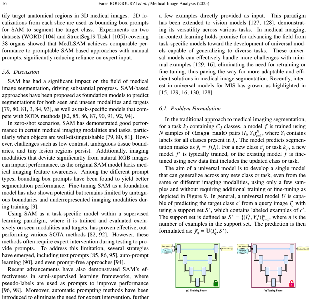
The organizing device is the four-family taxonomy itself, and within each family a specific mechanism carries the explanation. GANs work through an adversarial generator–discriminator game, with CycleGAN adding cycle-consistency to translate between modalities without paired labels; diffusion models segment by denoising a noised mask conditioned on the input image. Few-shot segmentation is built on episodic training with a support set and query image, where prototype networks summarize support features by masked average pooling and classify query pixels by similarity, optionally refined by recurrent mask refinement. Foundation-model work centers on SAM's three-part design — image encoder, prompt encoder, mask decoder — where bounding-box prompts consistently beat point prompts and fine-tuning or adapters transfer it to medical data. Universal models replace per-task training with in-context learning, using interaction blocks (CrossBlock, and SetBlock for stochastic candidates) that fuse query features with a small support set so a single network can segment a new class at inference time.
What would settle it
Run one representative from each family (for example, a CycleGAN-based segmenter, MedSegDiff, a prototype-network FSS method, a fine-tuned SAM, and UniverSeg) on a single common dataset such as BTCV abdominal CT under identical support-set and prompt conditions; if their Dice-score ranking does not reproduce the relative strengths implied by Tables 1 and 2, the survey's cross-method comparisons are not supported.
Extended reading notes
Core claim
The survey's central claim is that generative AI, few-shot segmentation, foundation models, and universal models constitute the current frontier of medical image segmentation because each supplies something the classic fully-supervised, single-task pipeline cannot: synthetic or unlabeled data for GAN and diffusion approaches, episodic support sets for few-shot learning, prompt-based zero-shot generalization for SAM-style foundation models, and in-context support sets that let a universal model segment unseen classes without retraining. It further claims that these families are converging — generative outputs feed segmentation, SAM pseudo-labels feed semi-supervised learning, few-shot prototypes extend into universal models — and that the main obstacle to exploiting this convergence is the absence of shared benchmarks, consistent evaluation settings, and released implementations, which has already produced contrasting results for the same method on the same dataset.
Load-bearing premise
The survey's comparative statements assume that the Dice scores collected across different studies in its tables can be read as comparable evidence, even though no unified evaluation protocol exists — a limitation the paper itself concedes in Section 7.
Editorial extensions
If this is right
- Generative segmentation will keep migrating from data augmentation into semi-supervised and self-supervised training, so unlabeled scans become usable assets instead of discarded data.
- Few-shot segmentation's current 20–40 percentage-point gap to fully supervised performance will only close if the field standardizes episodes and metrics; otherwise results will remain unreadable across papers.
- Promptable foundation models are likely to become the default starting point for new medical segmentation tasks, with automatic prompting replacing expert clicks in annotation workflows.
- A single universal model could segment previously unseen organs or modalities at inference from a few labeled examples, eliminating retraining for new tasks.
- The deciding factor for clinical adoption across all four families will be benchmark and reproducibility infrastructure, not architecture alone.
Reading between the lines
- The survey's own cross-table evidence is weaker than its narrative suggests: Dice scores in Tables 1 and 2 mix datasets, prompt modes, and protocols, so a reader should not rank the four families against each other from this paper alone.
- A natural next experiment the paper gestures at but does not run: combine diffusion-based synthetic data with universal in-context models, using generated volumes as support sets; this could decouple universal models from real annotated data entirely.
- The recurring failure on low-contrast, weak-boundary structures (vessels, tumors) across all families suggests the true common bottleneck is boundary representation, and methods with explicit shape or edge priors may leapfrog purely data-driven ones.
- Defining 'universal model' sharply relative to few-shot and foundation models would let the field build the standardized benchmarks the paper calls for.
Signed reviews
Editorial analysis
A structured set of objections, weighed in public.
Referee Report
Summary. The manuscript is a survey of recent methods for medical image segmentation across four paradigms: generative models (GANs and diffusion models), few-shot segmentation, foundation models (SAM and its adaptations), and universal/in-context learning models. It provides brief theoretical background, descriptions of representative works, summary tables of reported Dice scores, and a discussion of challenges and future directions, with an accompanying GitHub repository for tracking ongoing work.
Significance. If taken as a descriptive catalog, the survey has practical value: it organizes a large literature into four coherent threads, summarizes representative models and datasets, and honestly acknowledges the absence of unified evaluation protocols and benchmark datasets (Sections 3.3, 4.4, and 7). The strengths are the breadth of coverage, the structured tables and figures, and the explicit discussion of reproducibility and evaluation inconsistencies. However, the survey's stated goal of comparing approaches is only partially met: the quantitative tables mix results from heterogeneous evaluation protocols, and the theoretical background in Section 3.1 contains mathematical errors. These issues are fixable but require revision before the survey can be considered a reliable reference.
major comments (2)
- [Section 3.1, Eqs. (2), (4), and (8)] The theoretical background contains mathematical errors that undermine a core part of the survey's pedagogical value. Eq. (8) states x = (1/sqrt(alpha_t)) z_t + (sqrt(1-alpha_t)/sqrt(alpha_t)) epsilon; from the forward process in Eq. (7), z_t = sqrt(alpha_t) x + sqrt(1-alpha_t) epsilon, the correct inversion is x = (z_t - sqrt(1-alpha_t) epsilon)/sqrt(alpha_t). The plus sign implies noise is added during reconstruction, contradicting the text's own description of the reverse process as removing noise. In addition, Eqs. (2) and (4) do not form a standard GAN objective: Eq. (2) defines G's objective as argmax over theta of a min over phi of the discriminator loss, while Eq. (4) then defines the generator loss as sum_j log(1 - sig(...)), which is the quantity G should minimize (or whose negative it should maximize) in the standard saturating formulation. These equations should be reconciled with the usual minimax value V(D,G) = E[log D(x)] + E[log(1 - D(G(z)))], or explicitly flagged as a nonstandard convention.
- [Sections 3.3, 4.4, and Tables 1-2] The survey presents quantitative Dice scores in Tables 1 and 2 as a basis for comparing methods, but the scores are derived from different datasets, prompt settings, and evaluation protocols, as the paper itself concedes in Sections 3.3, 4.4, and 7. For example, Table 1 mixes MedSegDiff on REFUGE-2/BraTS/DDTI with other works on AMOS22/BTCV, and Table 2 mixes St1/St2 Abd-CT and Abd-MRI settings. Consequently, claims such as "diffusion models and hybrid approaches ... pushing the boundaries of segmentation performance to new heights" (Section 3.3) and "the performance gap with supervised scenarios is becoming smaller" (Section 4.4) are not supported by the table evidence. The authors should either restrict comparisons to homogeneous settings (e.g., St1-Abd-CT versus St2-Abd-CT) or explicitly state in the text and table captions that no cross-method performance ordering should be inferred. Table 3 similarly contains qualitative entries such as "competitive with supervised" that are undefined without a stated supervised baseline; a footnote should specify the reference comparison.
minor comments (7)
- [Section 3.1, Eq. (9)] The subscript t on x_t in the loss argument is inconsistent with the notation in Eqs. (5)-(7), where x denotes the clean image and z_t the noised sample; the argument should be sqrt(alpha_t) x + sqrt(1-alpha_t) epsilon (i.e., z_t), not a previously noised x_t.
- [Section 3.2, C-DARL paragraph] The text says "The model employs a total of five losses" but then lists only four (two adversarial, one diffusion, one cycle); please correct the count or add the missing loss term.
- [Section 4.2] The phrase "vused Visceral dataset" should be "used the Visceral dataset."
- [Section 3 heading] The heading "Genrative AI" should be "Generative AI."
- [Table 1] The row for MedSegDiff lists "Brain-Turmor" which should be "Brain-Tumor."
- [Section 5.7] The sentence "Efficient prompt have shown to be crucial" should be "Efficient prompts have been shown to be crucial."
- [Section 6.1] The statement that universal models require "no additional training or fine-tuning" is potentially misleading because models like UnivSeg are trained on large datasets before their inference-time in-context adaptation; please clarify that this refers to no task-specific fine-tuning at inference time.
Circularity Check
Survey is a literature review with no derivation chain, fitted parameters, or load-bearing self-citation; circularity score is 0.
full rationale
This manuscript is a survey of existing medical image segmentation methods. It does not derive new results, fit parameters, or make predictions from a model. Its central claim is to provide an overview of generative AI, few-shot learning, foundation models, and universal models, and to discuss limitations and future directions. The only self-citation is reference [5], the authors' own PDAtt-Unet paper, cited in the introduction as one example of prior deep learning work in the field ('[1, 2, 3, 5]'). This citation is illustrative and not load-bearing: no argument, taxonomy, or conclusion depends on it. The paper's comparative remarks are limited by the acknowledged absence of unified evaluation protocols (Sections 3.3, 4.4, and 7), which is a correctness/evidence limitation, not circularity. No equation in the paper is defined in terms of its own output, no fitted value is renamed as a prediction, and no uniqueness theorem or ansatz is imported from the authors' prior work to force a conclusion. The survey is therefore self-contained as a review and exhibits no significant circularity.
Assumptions & free parameters
assumptions (2)
- domain assumption Reported Dice scores in Tables 1-3 accurately reflect the original papers and are comparable across methods.
- domain assumption The selection of surveyed papers is representative of the state of the art in the four chosen paradigms.
Cite this review
Pith. "Pith review of Recent Advances in Medical Imaging Segmentation: A Survey." pith.science (2026). https://pith.science/paper/O7TV3G3V
@misc{pith2026250509274,
  author       = {Pith},
  title        = {Pith review of: Recent Advances in Medical Imaging Segmentation: A Survey},
  year         = {2026},
  howpublished = {\url{https://pith.science/paper/O7TV3G3V}},
  note         = {Machine review of arXiv:2505.09274}
}
read the original abstract
Medical imaging is a cornerstone of modern healthcare, driving advancements in diagnosis, treatment planning, and patient care. Among its various tasks, segmentation remains one of the most challenging problem due to factors such as data accessibility, annotation complexity, structural variability, variation in medical imaging modalities, and privacy constraints. Despite recent progress, achieving robust generalization and domain adaptation remains a significant hurdle, particularly given the resource-intensive nature of some proposed models and their reliance on domain expertise. This survey explores cutting-edge advancements in medical image segmentation, focusing on methodologies such as Generative AI, Few-Shot Learning, Foundation Models, and Universal Models. These approaches offer promising solutions to longstanding challenges. We provide a comprehensive overview of the theoretical foundations, state-of-the-art techniques, and recent applications of these methods. Finally, we discuss inherent limitations, unresolved issues, and future research directions aimed at enhancing the practicality and accessibility of segmentation models in medical imaging. We are maintaining a \href{https://github.com/faresbougourzi/Awesome-DL-for-Medical-Imaging-Segmentation}{GitHub Repository} to continue tracking and updating innovations in this field.
Figures


Figures from the paper (8 more)



Forward citations
Cited by 1 Pith paper
-
Medical Image Segmentation based on Deep Active Contour and Mean Curvature Loss Function
DACMC loss (active-contour region term plus convolution-approximated mean curvature) yields higher Dice than CE/DC/AC/ACE on small liver CT and spleen MRI test sets with U-Net.
Reference graph
Works this paper leans on
-
[1]
Four challenges in medical image analysis from an industrial perspective,
J. Weese and C. Lorenz, “Four challenges in medical image analysis from an industrial perspective,” Medical image analysis , vol. 33, pp. 44–49, 2016
2016
-
[2]
Anomaly detection-inspired few-shot medical image segmentation through self- supervision with supervoxels,
S. Hansen, S. Gautam, R. Jenssen, and M. Kamp ffmeyer, “Anomaly detection-inspired few-shot medical image segmentation through self- supervision with supervoxels,” Medical Image Analysis , vol. 78, p. 102385, 2022
2022
-
[3]
Segment anything in medical images,
J. Ma, Y . He, F. Li, L. Han, C. You, and B. Wang, “Segment anything in medical images,” Nature Communications, vol. 15, no. 1, p. 654, 2024
2024
-
[4]
Segment anything model for medical image segmentation: Current applications and future directions,
Y . Zhang, Z. Shen, and R. Jiao, “Segment anything model for medical image segmentation: Current applications and future directions,” Com- puters in Biology and Medicine, p. 108238, 2024
2024
-
[5]
PDAtt- Unet: Pyramid dual-decoder attention unet for covid-19 infection seg- mentation from ct-scans,
F. Bougourzi, C. Distante, F. Dornaika, and A. Taleb-Ahmed, “PDAtt- Unet: Pyramid dual-decoder attention unet for covid-19 infection seg- mentation from ct-scans,” Medical Image Analysis, vol. 86, p. 102797, 2023
2023
-
[6]
U-net and its variants for medical image segmentation: A review of theory and applications,
N. Siddique, S. Paheding, C. P. Elkin, and V . Devabhaktuni, “U-net and its variants for medical image segmentation: A review of theory and applications,” IEEE access, vol. 9, pp. 82 031–82 057, 2021
2021
-
[7]
Medical image segmentation review: The success of U-Net,
R. Azad, E. K. Aghdam, A. Rauland et al., “Medical image segmentation review: The success of U-Net,” IEEE Transactions on Pattern Analysis and Machine Intelligence, 2024
2024
-
[8]
Advances in medical image analysis with vision transformers: a comprehensive review,
R. Azad, A. Kazerouni, M. Heidari et al., “Advances in medical image analysis with vision transformers: a comprehensive review,” Medical Image Analysis, vol. 91, p. 103000, 2024
2024
Show all 132 references
-
[9]
Transformers in medical imaging: A survey,
F. Shamshad, S. Khan, S. W. Zamir et al. , “Transformers in medical imaging: A survey,” Medical Image Analysis, vol. 88, p. 102802, 2023
2023
-
[10]
Transforming medical imaging with transformers? a comparative re- view of key properties, current progresses, and future perspectives,
J. Li, J. Chen, Y . Tang, C. Wang, B. A. Landman, and S. K. Zhou, “Transforming medical imaging with transformers? a comparative re- view of key properties, current progresses, and future perspectives,” Medical image analysis, vol. 85, p. 102762, 2023
2023
-
[11]
SynSeg-Net: Synthetic segmentation without target modality ground truth,
Y . Huo, Z. Xu, H. Moon et al., “SynSeg-Net: Synthetic segmentation without target modality ground truth,” IEEE transactions on medical imaging, vol. 38, no. 4, pp. 1016–1025, 2018
2018
-
[12]
Data augmen- tation using generative adversarial networks (CycleGAN) to improve generalizability in CT segmentation tasks,
V . Sandfort, K. Yan, P. J. Pickhardt, and R. M. Summers, “Data augmen- tation using generative adversarial networks (CycleGAN) to improve generalizability in CT segmentation tasks,” Scientific reports , vol. 9, no. 1, p. 16884, 2019. Fares BOUGOURZI et al. / Medical Image Ana...
2025
-
[13]
MedSegDi ff-V2: Diffusion-based medical image segmentation with transformer,
J. Wu, W. Ji, H. Fu, M. Xu, Y . Jin, and Y . Xu, “MedSegDi ff-V2: Diffusion-based medical image segmentation with transformer,” inProc. AAAI, vol. 38, no. 6, 2024, pp. 6030–6038
2024
-
[14]
Prototype correlation matching and class-relation reasoning for few-shot medical image segmentation,
Y . Zhang, H. Li, Y . Gao, H. Duan, Y . Huang, and Y . Zheng, “Prototype correlation matching and class-relation reasoning for few-shot medical image segmentation,” IEEE Transactions on Medical Imaging, 2024
2024
-
[15]
Clip-driven universal model for organ segmentation and tumor detection,
J. Liu, Y . Zhang, J.-N. Chen et al. , “Clip-driven universal model for organ segmentation and tumor detection,” in Proc. ICCV , 2023, pp. 21 152–21 164
2023
-
[16]
Tyche: Stochastic in-context learning for medical image segmentation,
M. Rakic, H. E. Wong, J. J. G. Ortiz et al., “Tyche: Stochastic in-context learning for medical image segmentation,” in Proc. CVPR, 2024, pp. 11 159–11 173
2024
-
[17]
Unsupervised representa- tion learning with deep convolutional generative adversarial networks,
A. Radford, L. Metz, and S. Chintala, “Unsupervised representa- tion learning with deep convolutional generative adversarial networks,” CoRR, vol. abs/1511.06434, 2015
2015 arXiv
-
[18]
Deep unsupervised learning using nonequilibrium thermodynamics,
J. Sohl-Dickstein, E. Weiss, N. Maheswaranathan, and S. Ganguli, “Deep unsupervised learning using nonequilibrium thermodynamics,” in Proc. ICML, vol. 37. PMLR, 2015, pp. 2256–2265
2015
-
[19]
Auto-encoding variational bayes,
D. P. Kingma and M. Welling, “Auto-encoding variational bayes,” CoRR, vol. abs/1312.6114, 2013
2013 arXiv
-
[20]
Variational inference with normalizing flows,
D. Rezende and S. Mohamed, “Variational inference with normalizing flows,” in Proc. MLR, vol. 37. PMLR, 2015, pp. 1530–1538
2015
-
[21]
Deep genera- tive modelling: A comparative review of vaes, gans, normalizing flows, energy-based and autoregressive models,
S. Bond-Taylor, A. Leach, Y . Long, and C. G. Willcocks, “Deep genera- tive modelling: A comparative review of vaes, gans, normalizing flows, energy-based and autoregressive models,”IEEE transactions on pattern analysis and machine intelligence, vol. 44, no. 11, pp. 7327–7347, 2021
2021
-
[22]
Diffusion mod- els in vision: A survey,
F.-A. Croitoru, V . Hondru, R. T. Ionescu, and M. Shah, “Diffusion mod- els in vision: A survey,” IEEE Transactions on Pattern Analysis and Machine Intelligence, vol. 45, no. 9, pp. 10 850–10 869, 2023
2023
-
[23]
S. J. Prince, Understanding deep learning. MIT press, 2023
2023
-
[24]
Generative adversar- ial nets,
I. Goodfellow, J. Pouget-Abadie, M. Mirza et al., “Generative adversar- ial nets,” Proc. NeurIPS, vol. 27, 2014
2014
-
[25]
Precomputed real-time texture synthesis with markovian generative adversarial networks,
C. Li and M. Wand, “Precomputed real-time texture synthesis with markovian generative adversarial networks,” in Proc. MICCAI . Springer, 2016, pp. 702–716
2016
-
[26]
Unpaired image-to-image translation using cycle-consistent adversarial networks,
J.-Y . Zhu, T. Park, P. Isola, and A. A. Efros, “Unpaired image-to-image translation using cycle-consistent adversarial networks,” in Proc. ICCV, 2017, pp. 2223–2232
2017
-
[27]
Conditional generative adversarial nets,
M. Mirza and S. Osindero, “Conditional generative adversarial nets,” 2014
2014
-
[28]
Conditional image synthesis with auxiliary classifier gans,
A. Odena, C. Olah, and J. Shlens, “Conditional image synthesis with auxiliary classifier gans,” in International conference on machine learn- ing. PMLR, 2017, pp. 2642–2651
2017
-
[29]
A style-based generator architecture for generative adversarial networks,
T. Karras, S. Laine, and T. Aila, “A style-based generator architecture for generative adversarial networks,”Proc. CVPR, pp. 4396–4405, 2018
2018
-
[30]
Deep unsupervised learning using nonequilibrium thermodynamics,
J. Sohl-Dickstein, E. Weiss, N. Maheswaranathan, and S. Ganguli, “Deep unsupervised learning using nonequilibrium thermodynamics,” in International conference on machine learning . PMLR, 2015, pp. 2256–2265
2015
-
[31]
Score-based generative modeling through stochastic di fferential equations,
Y . Song, J. Sohl-Dickstein, D. P. Kingmaet al., “Score-based generative modeling through stochastic di fferential equations,” arXiv:2011.13456, 2020
2011 arXiv
-
[32]
Denoising di ffusion probabilistic mod- els,
J. Ho, A. Jain, and P. Abbeel, “Denoising di ffusion probabilistic mod- els,” Proc. NeurIPS, vol. 33, pp. 6840–6851, 2020
2020
-
[33]
Di ffusion models beat gans on image syn- thesis,
P. Dhariwal and A. Nichol, “Di ffusion models beat gans on image syn- thesis,” Proc. NeurIPS, vol. 34, pp. 8780–8794, 2021
2021
-
[34]
Classifier-free di ffusion guidance,
J. Ho and T. Salimans, “Classifier-free di ffusion guidance,” NeurIPS Workshop on Deep Generative Models and Downstream Applications , 2022
2022
-
[35]
Deep adversarial training for multi-organ nuclei segmentation in histopathology images,
F. Mahmood, D. Borders, R. J. Chen et al., “Deep adversarial training for multi-organ nuclei segmentation in histopathology images,” IEEE transactions on medical imaging, vol. 39, no. 11, pp. 3257–3267, 2019
2019
-
[36]
Translating and segmenting mul- timodal medical volumes with cycle-and shape-consistency generative adversarial network,
Z. Zhang, L. Yang, and Y . Zheng, “Translating and segmenting mul- timodal medical volumes with cycle-and shape-consistency generative adversarial network,” in Proc. CVPR, 2018, pp. 9242–9251
2018
-
[37]
MedSegDiff: Medical image segmenta- tion with diffusion probabilistic model,
J. Wu, R. Fu, H. Fang et al., “MedSegDiff: Medical image segmenta- tion with diffusion probabilistic model,” in Medical Imaging with Deep Learning. PMLR, 2024, pp. 1623–1639
2024
-
[38]
Deep adversarial networks for biomedical image segmentation utilizing unannotated images,
Y . Zhang, L. Yang, J. Chen et al. , “Deep adversarial networks for biomedical image segmentation utilizing unannotated images,” in Proc. MICCAI. Springer, 2017, pp. 408–416
2017
-
[39]
Self-supervised vessel segmentation via adversarial learning,
Y . Ma, Y . Hua, H. Denget al., “Self-supervised vessel segmentation via adversarial learning,” in Proc. ICCV, 2021, pp. 7536–7545
2021
-
[40]
Diffusion adversarial representation learn- ing for self-supervised vessel segmentation,
B. Kim, Y . Oh, and J. C. Ye, “Diffusion adversarial representation learn- ing for self-supervised vessel segmentation,” in Proc. ICLR, 2023
2023
-
[41]
C-DARL: Contrastive diffusion adversarial representation learning for label-free blood vessel segmentation,
B. Kim, Y . Oh, B. J. Wood, R. M. Summers, and J. C. Ye, “C-DARL: Contrastive diffusion adversarial representation learning for label-free blood vessel segmentation,”Medical Image Analysis, vol. 91, p. 103022, 2024
2024
-
[42]
Spine-GAN: Seman- tic segmentation of multiple spinal structures,
Z. Han, B. Wei, A. Mercado, S. Leung, and S. Li, “Spine-GAN: Seman- tic segmentation of multiple spinal structures,” Medical image analysis, vol. 50, pp. 23–35, 2018
2018
-
[43]
Automatic segmentation of coronary arteries in x-ray angiograms us- ing multiscale analysis and artificial neural networks,
F. Cervantes-Sanchez, I. Cruz-Aceves, A. Hernandez-Aguirre et al. , “Automatic segmentation of coronary arteries in x-ray angiograms us- ing multiscale analysis and artificial neural networks,”Applied Sciences, vol. 9, no. 24, p. 5507, 2019
2019
-
[44]
Sequential vessel segmentation via deep channel attention network,
D. Hao, S. Ding, L. Qiu et al., “Sequential vessel segmentation via deep channel attention network,” Neural Networks, vol. 128, pp. 172–187, 2020
2020
-
[45]
Locating blood ves- sels in retinal images by piecewise threshold probing of a matched filter response,
A. Hoover, V . Kouznetsova, and M. Goldbaum, “Locating blood ves- sels in retinal images by piecewise threshold probing of a matched filter response,” IEEE Transactions on Medical imaging , vol. 19, no. 3, pp. 203–210, 2000
2000
-
[46]
REFUGE2 challenge: A treasure trove for multi-dimension analysis and evaluation in glaucoma screening,
H. Fang, F. Li, J. Wu et al. , “REFUGE2 challenge: A treasure trove for multi-dimension analysis and evaluation in glaucoma screening,” arXiv:2202.08994, 2022
2022 arXiv
-
[47]
The RSNA-ASNR-MICCAI BraTS 2021 benchmark on brain tumor segmentation and radiogenomic classification,
U. Baid, S. Ghodasara, S. Mohan et al., “The RSNA-ASNR-MICCAI BraTS 2021 benchmark on brain tumor segmentation and radiogenomic classification,” arXiv:2107.02314, 2021
2021 arXiv
-
[48]
An open access thyroid ultra- sound image database,
L. Pedraza, C. Vargas, F. Narv ´aez et al., “An open access thyroid ultra- sound image database,” in Proc. 10th ISMIP, vol. 9287. SPIE, 2015, pp. 188–193
2015
-
[49]
AMOS: A large-scale abdominal multi-organ benchmark for versatile medical image segmentation,
Y . Ji, H. Bai, C. Geet al., “AMOS: A large-scale abdominal multi-organ benchmark for versatile medical image segmentation,” Proc. NeurIPS, vol. 35, pp. 36 722–36 732, 2022
2022
-
[50]
Automatic multi-organ segmentation on abdominal CT with dense v-networks,
E. Gibson, F. Giganti, Y . Huet al., “Automatic multi-organ segmentation on abdominal CT with dense v-networks,”IEEE transactions on medical imaging, vol. 37, no. 8, pp. 1822–1834, 2018
2018
-
[51]
Skin lesion analysis to- ward melanoma detection 2018: A challenge hosted by the international skin imaging collaboration (ISIC),
N. Codella, V . Rotemberg, P. Tschandl et al., “Skin lesion analysis to- ward melanoma detection 2018: A challenge hosted by the international skin imaging collaboration (ISIC),” arXiv:1902.03368, 2019
2018 arXiv
-
[52]
Semantic image syn- thesis with spatially-adaptive normalization,
T. Park, M.-Y . Liu, T.-C. Wang, and J.-Y . Zhu, “Semantic image syn- thesis with spatially-adaptive normalization,” in Proc. CVPR, 2019, pp. 2337–2346
2019
-
[53]
One-shot learn- ing for semantic segmentation,
A. Shaban, S. Bansal, Z. Liu, I. Essa, and B. Boots, “One-shot learn- ing for semantic segmentation,” in British Machine Vision Conference (BMVC), 2017
2017
-
[54]
PANet: Few-shot image seman- tic segmentation with prototype alignment,
K. Wang, J. H. Liew, Y . Zou et al., “PANet: Few-shot image seman- tic segmentation with prototype alignment,” in Proc. ICCV, 2019, pp. 9197–9206
2019
-
[55]
Cloud-based evaluation of anatomical structure segmentation and landmark detection algorithms: VISCERAL anatomy benchmarks,
O. Jimenez-del Toro, H. M ¨uller et al. , “Cloud-based evaluation of anatomical structure segmentation and landmark detection algorithms: VISCERAL anatomy benchmarks,”IEEE transactions on medical imag- ing, vol. 35, no. 11, pp. 2459–2475, 2016
2016
-
[56]
Miccai multi-atlas labeling beyond the cranial vault–workshop and challenge,
B. Landman, Z. Xu, J. Igelsias et al. , “Miccai multi-atlas labeling beyond the cranial vault–workshop and challenge,” in Proc. MIC- CAI Multi-Atlas Labeling Beyond Cranial Vault—Workshop Challenge, vol. 5, 2015, p. 12
2015
-
[57]
CHAOS challenge-combined (CT-MR) healthy abdominal organ segmentation,
A. E. Kavur, N. S. Gezer, M. Barıs ¸et al., “CHAOS challenge-combined (CT-MR) healthy abdominal organ segmentation,”Medical Image Anal- ysis, vol. 69, p. 101950, 2021
2021
-
[58]
Multivariate mixture model for myocardial segmentation combining multi-source images,
X. Zhuang, “Multivariate mixture model for myocardial segmentation combining multi-source images,” IEEE Transactions on Pattern Analy- sis and Machine Intelligence, vol. 41, no. 12, pp. 2933–2946, 2018
2018
-
[59]
Spatial context-aware self-attention model for multi-organ segmentation,
H. Tang, X. Liu, K. Han et al. , “Spatial context-aware self-attention model for multi-organ segmentation,” in Proc. WACV, 2021, pp. 939– 949
2021
-
[60]
Prototypical few-shot segmentation for cross-institution male pelvic structures with spatial registration,
Y . Li, Y . Fu, I. J. Gayo et al., “Prototypical few-shot segmentation for cross-institution male pelvic structures with spatial registration,” Medi- cal Image Analysis, vol. 90, p. 102935, 2023
2023
-
[61]
‘squeeze & excite’guided few-shot segmentation of volumetric images,
A. G. Roy, S. Siddiqui, S. P ¨olsterl et al. , “‘squeeze & excite’guided few-shot segmentation of volumetric images,” Medical image analysis, vol. 59, p. 101587, 2020
2020
-
[62]
Self-supervision with superpixels: 20 Fares BOUGOURZI et al. / Medical Image Analysis (2025) Training few-shot medical image segmentation without annotation,
C. Ouyang, C. Bi ffi, C. Chen et al., “Self-supervision with superpixels: 20 Fares BOUGOURZI et al. / Medical Image Analysis (2025) Training few-shot medical image segmentation without annotation,” in Proc. ECCV. Springer, 2020, pp. 762–780
2025
-
[63]
Recurrent mask refinement for few-shot medical image segmentation,
H. Tang, X. Liu, S. Sun et al., “Recurrent mask refinement for few-shot medical image segmentation,” in Proc. ICCV, 2021, pp. 3918–3928
2021
-
[64]
Few shot medical im- age segmentation with cross attention transformer,
Y . Lin, Y . Chen, K.-T. Cheng, and H. Chen, “Few shot medical im- age segmentation with cross attention transformer,” in Proc. MICCAI. Springer, 2023, pp. 233–243
2023
-
[65]
Rethinking few-shot medical segmenta- tion: a vector quantization view,
S. Huang, T. Xu, N. Shen et al., “Rethinking few-shot medical segmenta- tion: a vector quantization view,” in Proc. CVPR, 2023, pp. 3072–3081
2023
-
[66]
Dual contrastive learning with anatom- ical auxiliary supervision for few-shot medical image segmentation,
H. Wu, F. Xiao, and C. Liang, “Dual contrastive learning with anatom- ical auxiliary supervision for few-shot medical image segmentation,” in Proc. ECCV. Springer, 2022, pp. 417–434
2022
-
[67]
Learning what and where to segment: A new perspective on medical image few-shot segmentation,
Y . Feng, Y . Wang, H. Li et al., “Learning what and where to segment: A new perspective on medical image few-shot segmentation,” Medical Image Analysis, vol. 87, p. 102834, 2023
2023
-
[68]
Language models are few-shot learners,
T. Brown, B. Mann, N. Ryder et al., “Language models are few-shot learners,” in Proc. NeurIPS, vol. 33. Curran Associates, Inc., 2020, pp. 1877–1901
2020
-
[69]
GPT-4 technical report,
J. Achiam, S. Adler, S. Agarwal et al. , “GPT-4 technical report,” arXiv:2303.08774, 2023
2023 arXiv
-
[70]
PaLM: Scaling language modeling with pathways,
A. Chowdhery, S. Narang, J. Devlin et al., “PaLM: Scaling language modeling with pathways,” Journal of Machine Learning Research , vol. 24, no. 240, pp. 1–113, 2023
2023
-
[71]
Llama: Open and e fficient foundation language models,
H. Touvron, T. Lavril, G. Izacard et al. , “Llama: Open and e fficient foundation language models,” arXiv:2302.13971, 2023
2023 arXiv
-
[72]
Learning transferable visual models from natural language supervision,
A. Radford, J. W. Kim, C. Hallacy et al., “Learning transferable visual models from natural language supervision,” in Proc. ICML. PMLR, 2021, pp. 8748–8763
2021
-
[73]
BLIP: Bootstrapping language-image pre-training for unified vision-language understanding and generation,
J. Li, D. Li, C. Xiong, and S. Hoi, “BLIP: Bootstrapping language-image pre-training for unified vision-language understanding and generation,” in Proc. ICML. PMLR, 2022, pp. 12 888–12 900
2022
-
[74]
Segment anything,
A. Kirillov, E. Mintun, N. Ravi et al. , “Segment anything,” in Proc. ICCV, 2023, pp. 4015–4026
2023
-
[75]
Segment everything everywhere all at once,
X. Zou, J. Yang, H. Zhang et al., “Segment everything everywhere all at once,” Proc. NeurIPS, vol. 36, 2024
2024
-
[76]
An image is worth 16x16 words: Transformers for im- age recognition at scale,
A. Dosovitskiy, “An image is worth 16x16 words: Transformers for im- age recognition at scale,” arXiv:2010.11929, 2020
2010 arXiv
-
[77]
Masked autoencoders are scalable vision learners,
K. He, X. Chen, S. Xie et al., “Masked autoencoders are scalable vision learners,” in Proc. CVPR, 2022, pp. 16 000–16 009
2022
-
[78]
Fourier features let networks learn high frequency functions in low dimensional domains,
M. Tancik, P. Srinivasan, B. Mildenhall et al. , “Fourier features let networks learn high frequency functions in low dimensional domains,” Proc. NeurIPS, vol. 33, pp. 7537–7547, 2020
2020
-
[79]
Sam.md: Zero-shot medical image segmentation capabilities of the segment anything model,
T. Wald, S. Roy, G. Koehler et al., “Sam.md: Zero-shot medical image segmentation capabilities of the segment anything model,” inMID, short paper track, 2023
2023
-
[80]
Segment anything model for medical image analysis: an experimental study,
M. A. Mazurowski, H. Dong, H. Gu et al., “Segment anything model for medical image analysis: an experimental study,” Medical Image Analy- sis, vol. 89, p. 102918, 2023
2023
-
[81]
Segment anything model for medical images?
Y . Huang, X. Yang, L. Liu et al., “Segment anything model for medical images?” Medical Image Analysis, vol. 92, p. 103061, 2024
2024
-
[82]
Customized segment anything model for medical image segmentation,
K. Zhang and D. Liu, “Customized segment anything model for medical image segmentation,” arXiv:2304.13785, 2023
2023 arXiv
-
[83]
Transunet: Transformers make strong en- coders for medical image segmentation,
J. Chen, Y . Lu, Yu et al. , “Transunet: Transformers make strong en- coders for medical image segmentation,” arXiv:2102.04306, 2021
2021 arXiv
-
[84]
Sam-med2d,
J. Cheng, J. Ye, Z. Deng, J. Chen, T. Li, H. Wang, Y . Su, Z. Huang, J. Chen, L. Jianget al., “Sam-med2d,”arXiv preprint arXiv:2308.16184, 2023
2023 arXiv
-
[85]
S-SAM: SVD- Based Fine-Tuning of segment anything model for medical image seg- mentation,
J. N. Paranjape, S. Sikder, S. S. Vedula, and V . M. Patel, “S-SAM: SVD- Based Fine-Tuning of segment anything model for medical image seg- mentation,” in Proc MICCAI. Springer, 2024, pp. 720–730
2024
-
[86]
AdaptiveSAM: Towards ef- ficient tuning of SAM for surgical scene segmentation,
J. N. Paranjape, N. G. Nair, S. Sikder et al., “AdaptiveSAM: Towards ef- ficient tuning of SAM for surgical scene segmentation,” in Annual Con- ference on Medical Image Understanding and Analysis. Springer, 2024, pp. 187–201
2024
-
[87]
SAM-Path: A segment anything model for semantic segmentation in digital pathology,
J. Zhang, K. Ma, S. Kapse et al. , “SAM-Path: A segment anything model for semantic segmentation in digital pathology,” in Proc. MIC- CAI. Springer, 2023, pp. 161–170
2023
-
[88]
Structured crowdsourcing en- ables convolutional segmentation of histology images,
M. Amgad, H. Elfandy, H. Hussein et al., “Structured crowdsourcing en- ables convolutional segmentation of histology images,” Bioinformatics, vol. 35, no. 18, pp. 3461–3467, 2019
2019
-
[89]
MILD-Net: Minimal information loss dilated network for gland instance segmentation in colon histology images,
S. Graham, H. Chen, J. Gamper et al., “MILD-Net: Minimal information loss dilated network for gland instance segmentation in colon histology images,” Medical image analysis, vol. 52, pp. 199–211, 2019
2019
-
[90]
Input augmentation with sam: Boost- ing medical image segmentation with segmentation foundation model,
Y . Zhang, T. Zhou, S. Wanget al., “Input augmentation with sam: Boost- ing medical image segmentation with segmentation foundation model,” in Proc. MICCAI. Springer, 2023, pp. 129–139
2023
-
[91]
3DSAM-adapter: Holistic adap- tation of sam from 2D to 3D for promptable tumor segmentation,
S. Gong, Y . Zhong, W. Ma et al. , “3DSAM-adapter: Holistic adap- tation of sam from 2D to 3D for promptable tumor segmentation,” arXiv:2306.13465, 2023
2023 arXiv
-
[92]
Medical sam adapter: Adapting segment any- thing model for medical image segmentation,
J. Wu, W. Ji, Y . Liuet al., “Medical sam adapter: Adapting segment any- thing model for medical image segmentation,”arXiv:2304.12620, 2023
2023 arXiv
-
[93]
SAM-Med3D: Towards general-purpose segmentation models for volumetric medical images,
H. Wang, S. Guo, J. Ye et al., “SAM-Med3D: Towards general-purpose segmentation models for volumetric medical images,” 2024
2024
-
[94]
MA-SAM: Modality-agnostic SAM adaptation for 3D medical image segmentation,
C. Chen, J. Miao, D. Wu et al., “MA-SAM: Modality-agnostic SAM adaptation for 3D medical image segmentation,” Medical Image Analy- sis, vol. 98, p. 103310, 2024
2024
-
[95]
FastSAM3D: An e fficient segment any- thing model for 3D volumetric medical images,
Y . Shen, J. Li, X. Shao et al., “FastSAM3D: An e fficient segment any- thing model for 3D volumetric medical images,” in Proc. MICCAI . Springer, 2024, pp. 542–552
2024
-
[96]
Segment anything model for semi- supervised medical image segmentation via selecting reliable pseudo- labels,
N. Li, L. Xiong, W. Qiu et al. , “Segment anything model for semi- supervised medical image segmentation via selecting reliable pseudo- labels,” in International Conference on Neural Information Processing. Springer, 2023, pp. 138–149
2023
-
[97]
Deep learning techniques for automatic MRI cardiac multi-structures segmentation and diagnosis: is the problem solved?
O. Bernard, A. Lalande, C. Zotti et al., “Deep learning techniques for automatic MRI cardiac multi-structures segmentation and diagnosis: is the problem solved?” IEEE transactions on medical imaging , vol. 37, no. 11, pp. 2514–2525, 2018
2018
-
[98]
SemiSAM: Exploring sam for enhanc- ing semi-supervised medical image segmentation with extremely limited annotations,
Y . Zhang, Y . Cheng, and Y . Qi, “SemiSAM: Exploring sam for enhanc- ing semi-supervised medical image segmentation with extremely limited annotations,” BIBM, 2023
2023
-
[99]
A global benchmark of algorithms for segmenting the left atrium from late gadolinium-enhanced cardiac mag- netic resonance imaging,
Z. Xiong, Q. Xia, Z. Hu et al., “A global benchmark of algorithms for segmenting the left atrium from late gadolinium-enhanced cardiac mag- netic resonance imaging,” Medical image analysis, vol. 67, p. 101832, 2021
2021
-
[100]
SurgicalSAM: Efficient class prompt- able surgical instrument segmentation,
W. Yue, J. Zhang, K. Hu et al., “SurgicalSAM: Efficient class prompt- able surgical instrument segmentation,” in Proc. AAAI, vol. 38, no. 7, 2024, pp. 6890–6898
2024
-
[101]
2018 robotic scene segmen- tation challenge,
M. Allan, S. Kondo, S. Bodenstedt et al., “2018 robotic scene segmen- tation challenge,” arXiv:2001.11190, 2020
2018 arXiv
-
[102]
2017 robotic instrument seg- mentation challenge,
M. Allan, A. Shvets, T. Kurmann et al., “2017 robotic instrument seg- mentation challenge,” arXiv:1902.06426, 2019
2017 arXiv
-
[103]
MedLSAM: Localize and segment anything model for 3D CT images,
W. Lei, W. Xu, K. Li et al., “MedLSAM: Localize and segment anything model for 3D CT images,” Medical Image Analysis, vol. 99, p. 103370, 2025
2025
-
[104]
WORD: A large scale dataset, bench- mark and clinical applicable study for abdominal organ segmentation from ct image,
X. Luo, W. Liao, J. Xiao et al., “WORD: A large scale dataset, bench- mark and clinical applicable study for abdominal organ segmentation from ct image,” Medical Image Analysis, vol. 82, p. 102642, 2022
2022
-
[105]
Structseg challenge 2019: Grand challenge on multi-structure segmentation,
S. C. . Organizers, “Structseg challenge 2019: Grand challenge on multi-structure segmentation,” https: //structseg2019.grand-challenge. org/Home/, 2019, accessed: 2024-12-09
2019
-
[106]
SAM vs BET: A comparative study for brain extraction and segmentation of magnetic resonance im- ages using deep learning,
S. Mohapatra, A. Gosai, and G. Schlaug, “SAM vs BET: A comparative study for brain extraction and segmentation of magnetic resonance im- ages using deep learning,” Proc. Medical Imaging meets NeurIPS Work- shop, 2023
2023
-
[107]
Segment anything model (SAM) for dig- ital pathology: Assess zero-shot segmentation on whole slide imaging,
R. Deng, C. Cui, Q. Liu et al., “Segment anything model (SAM) for dig- ital pathology: Assess zero-shot segmentation on whole slide imaging,” Proc. MIDL, 2023
2023
-
[108]
Can sam segment polyps?
T. Zhou, Y . Zhang, Y . Zhou, Y . Wu, and C. Gong, “Can sam segment polyps?” arXiv:2304.07583, 2023
2023 arXiv
-
[109]
Sam meets robotic surgery: an empir- ical study on generalization, robustness and adaptation,
A. Wang, M. Islam, M. Xu et al., “Sam meets robotic surgery: an empir- ical study on generalization, robustness and adaptation,” in Proc. MIC- CAI. Springer, 2023, pp. 234–244
2023
-
[110]
Reviving iterative training with mask guidance for interactive segmentation,
K. Sofiiuk, I. A. Petrov, and A. Konushin, “Reviving iterative training with mask guidance for interactive segmentation,” inProc. ICIP. IEEE, 2022, pp. 3141–3145
2022
-
[111]
SimpleClick: In- teractive image segmentation with simple vision transformers,
Q. Liu, Z. Xu, G. Bertasius, and M. Niethammer, “SimpleClick: In- teractive image segmentation with simple vision transformers,” in Proc. ICCV, 2023, pp. 22 290–22 300
2023
-
[112]
FocalClick: Towards practical inter- active image segmentation,
X. Chen, Z. Zhao, Y . Zhang et al., “FocalClick: Towards practical inter- active image segmentation,” in Proc. CVPR, 2022, pp. 1300–1309
2022
-
[113]
Polyp-SAM: Transfer sam for polyp seg- mentation,
Y . Li, M. Hu, and X. Yang, “Polyp-SAM: Transfer sam for polyp seg- mentation,” in Medical Imaging 2024: Computer-Aided Diagnosis, vol. 12927. SPIE, 2024, pp. 759–765
2024
-
[114]
Cheap lunch for medical image segmen- Fares BOUGOURZI et al. / Medical Image Analysis (2025) 21 tation by fine-tuning SAM on few exemplars,
W. Feng, L. Zhu, and L. Yu, “Cheap lunch for medical image segmen- Fares BOUGOURZI et al. / Medical Image Analysis (2025) 21 tation by fine-tuning SAM on few exemplars,” inInternational MICCAI Brainlesion Workshop. Springer, 2023, pp. 13–22
2025
-
[115]
LoRA: Low-rank adaptation of large language models,
E. J. Hu, Y . Shen, P. Walliset al., “LoRA: Low-rank adaptation of large language models,” Proc. ICLR, 2022
2022
-
[116]
U-Net: Convolutional net- works for biomedical image segmentation,
O. Ronneberger, P. Fischer, and T. Brox, “U-Net: Convolutional net- works for biomedical image segmentation,” inProc. MICCAI. Springer, 2015, pp. 234–241
2015
-
[117]
Scaling vision transformers to gigapixel images via hierarchical self-supervised learning,
R. J. Chen, C. Chen, Y . Li et al. , “Scaling vision transformers to gigapixel images via hierarchical self-supervised learning,” in Proc. CVPR, 2022, pp. 16 144–16 155
2022
-
[118]
Prompt-MIL: Boosting multi-instance learning schemes via task-specific prompt tuning,
J. Zhang, S. Kapse, K. Ma et al., “Prompt-MIL: Boosting multi-instance learning schemes via task-specific prompt tuning,” in Proc. MICCAI. Springer, 2023, pp. 624–634
2023
-
[119]
Sam3d: Segment anything model in volumetric medical images,
N.-T. Bui, D.-H. Hoang, M.-T. Tran et al., “Sam3d: Segment anything model in volumetric medical images,” in Proc. ISBI. IEEE, 2024, pp. 1–4
2024
-
[120]
Hypernetworks,
D. Ha, A. Dai, and Q. V . Le, “Hypernetworks,” Proc. ICLR, 2017
2017
-
[121]
Fact: Factor-tuning for lightweight adaptation on vision transformer,
S. Jie and Z.-H. Deng, “Fact: Factor-tuning for lightweight adaptation on vision transformer,” in Proc. AAAI, vol. 37, no. 1, 2023, pp. 1060–1068
2023
-
[122]
SamDSK: Combining segment any- thing model with domain-specific knowledge for semi-supervised learn- ing in medical image segmentation,
Y . Zhang, T. Zhou, S. Wanget al., “SamDSK: Combining segment any- thing model with domain-specific knowledge for semi-supervised learn- ing in medical image segmentation,” arXiv:2308.13759, 2023
2023 arXiv
-
[123]
V-net: Fully convolutional neural networks for volumetric medical image segmentation,
F. Milletari, N. Navab, and S.-A. Ahmadi, “V-net: Fully convolutional neural networks for volumetric medical image segmentation,” in Proc. 3DV. IEEE, 2016, pp. 565–571
2016
-
[124]
Comprehensive multimodal seg- mentation in medical imaging: Combining YOLOv8 with SAM and HQ- SAM models,
S. Pandey, K.-F. Chen, and E. B. Dam, “Comprehensive multimodal seg- mentation in medical imaging: Combining YOLOv8 with SAM and HQ- SAM models,” in Proc. ICCV Workshops, 2023, pp. 2592–2598
2023
-
[125]
AutoSAM: Adapting sam to medical images by overloading the prompt encoder,
T. Shaharabany, A. Dahan, R. Giryes, and L. Wolf, “AutoSAM: Adapting sam to medical images by overloading the prompt encoder,” arXiv:2306.06370, 2023
2023 arXiv
-
[126]
SAM-U: Multi-box prompts triggered uncertainty estimation for reliable sam in medical image,
G. Deng, K. Zou, K. Ren et al., “SAM-U: Multi-box prompts triggered uncertainty estimation for reliable sam in medical image,” inProc. MIC- CAI. Springer, 2023, pp. 368–377
2023
-
[127]
SegGPT: Segmenting everything in context,
X. Wang, X. Zhang, Y . Cao et al., “SegGPT: Segmenting everything in context,” in Proc. ICCV. IEEE, 2023, pp. 1130–1140
2023
-
[128]
EviPrompt: A training-free evi- dential prompt generation method for adapting segment anything model in medical images,
Y . Xu, J. Tang, A. Men, and Q. Chen, “EviPrompt: A training-free evi- dential prompt generation method for adapting segment anything model in medical images,” IEEE Transactions on Image Processing, 2024
2024
-
[129]
UniverSeg: Universal medical image segmentation,
V . I. Butoi, J. J. G. Ortiz, T. Ma et al., “UniverSeg: Universal medical image segmentation,” in Proc. CVPR, 2023, pp. 21 438–21 451
2023
-
[130]
ICL-SAM: Synergizing in-context learning model and sam in medical image seg- mentation,
J. Hu, Y . Shang, Y . Yang, X. Guo, H. Peng, and T. Ma, “ICL-SAM: Synergizing in-context learning model and sam in medical image seg- mentation,” in Medical Imaging with Deep Learning, 2024
2024
-
[131]
Conditional convolutions for instance segmentation,
Z. Tian, C. Shen, and H. Chen, “Conditional convolutions for instance segmentation,” in Proc. ECCV. Springer, 2020, pp. 282–298
2020
-
[132]
nnU-Net: a self-configuring method for deep learning-based biomedical image segmentation,
F. Isensee, P. F. Jaeger, S. A. Kohl et al., “nnU-Net: a self-configuring method for deep learning-based biomedical image segmentation,” Na- ture methods, vol. 18, no. 2, pp. 203–211, 2021
2021
Reviewed August 15, 2026 · model on record in the stance chip above.
Discussion (0). Continue with ORCID to comment.