REVIEW 5 major objections 6 minor 5 references
Advanced Nanostructured Topical Therapeutics for Psoriasis: Strategic Synthesis, Multimodal Characterization, and Preliminary Pharmacodynamic Profiling
T0 review · 5 major / 6 minor · reviewed 2026-08-07 · deepseek-v4-flash
Pith's one-line read A multicomponent nanoparticle-and-plant-extract gel, tested in an imiquimod-induced animal model of psoriasis, is claimed to accelerate lesion healing and reduce inflammation compared with placebo and untreated controls.
desk verdict The gel formulation is new in detail, but the central healing claim is confounded by baseline differences and contradicted by the paper's own tables. read the letter →
The pith
A machine-rendered reading of the paper's core claim, the machinery that carries it, and where it could break.
The reading
What carries the argument
The central object is a multicomponent topical gel containing cerium oxide, zinc oxide, and silver nanoparticles dispersed in fish collagen and agar, enriched with bitter melon, ginger, and neem extracts. The argument runs on synergy: silver and zinc oxide provide antimicrobial action, zinc oxide aids keratinocyte proliferation and re-epithelialization, cerium oxide neutralizes reactive oxygen species through Ce3+/Ce4+ exchange, and the plant extracts plus collagen base reduce inflammation and maintain a moist healing environment. Characterization by UV-Vis spectroscopy, dynamic light scattering, FTIR, and scanning electron microscopy is used to show that the particles are nanoscale, stable, and well dispersed in the gel matrix.
What would settle it
Randomize animals to treatment, placebo, and untreated groups with identical Day 0 lesion sizes and blinded measurement of wound area; if the treatment advantage disappears when baseline size is controlled, or when healing is expressed as percent reduction from each group's own starting area, the central claim is not supported. A direct supplementary check would be to publish the full histological and cytokine data referenced but not shown.
Extended reading notes
Core claim
The paper claims that the three-nanoparticle-plus-botanical gel works as a coordinated therapeutic system rather than a simple mixture: zinc oxide and silver control microbial load and support re-epithelialization, cerium oxide scavenges reactive oxygen species through cerium's Ce3+/Ce4+ redox cycling, and the bitter melon, ginger, and neem extracts add polyphenol and flavonoid antioxidants. The collagen-agar-glycerol matrix keeps the wound bed moist and provides a scaffold for cell migration. In the animal model, treated lesions closed faster and with less erythema and scarring than placebo or untreated controls: by Day 14 the treatment group reached 90% healing with some lesions fully closed, versus 80% for placebo and 70% for untreated lesions. Histological examination, described but not fully shown in the paper, is said to reveal more mature collagen deposition and reformed epidermal layers in treated tissue.
Load-bearing premise
The three groups were assumed to be comparable at baseline, but the reported Day 0 lesion sizes differ substantially (treatment $0.10 \pm 0.02$ versus placebo $0.25 \pm 0.03$ and untreated $0.30 \pm 0.04$), so the faster closure attributed to the gel could instead reflect smaller starting wounds.
Editorial extensions
If this is right
- If the gel's effect is real, a single topical product could address infection, oxidative stress, and barrier repair at once, potentially reducing the need for systemic drugs in mild-to-moderate plaque psoriasis.
- Lesions would be expected to close sooner, with less erythema and scarring than placebo or untreated wounds, with the Day 14 gap (90% vs 80% vs 70%) as the headline magnitude.
- The gel could serve as an adjunct to biologics or small-molecule therapies for clearing stubborn localized plaques without adding systemic immunosuppression.
- The same nanoparticle platform could later carry drugs, such as low-dose corticosteroids or immune modulators, released directly in the lesion microenvironment.
Reading between the lines
- The claimed synergy is not directly demonstrated: a component-omission study, using gels without silver, without cerium oxide, or without the plant extracts, is the natural way to test whether all ingredients are needed. This is my editorial inference, not a result in the paper.
- The Day 0 lesion sizes in Table 1 are far from equal (treatment 0.10 ± 0.02, placebo 0.25 ± 0.03, untreated 0.30 ± 0.04), so the treatment benefit is not yet cleanly separated from baseline imbalance; this is my reading of the paper's own numbers.
- A testable mechanistic extension would measure lesion cytokines (TNF-α, IL-17) and oxidative-stress markers in biopsies to confirm that the gel reduces inflammation and ROS rather than only speeding wound closure visually.
- Safety is an open question: the paper itself cites evidence that ZnO nanoparticles can penetrate diseased skin and may even delay psoriasis-like recovery, so whether cerium oxide and antioxidants fully offset that risk needs dedicated dose-response and toxicity studies.
Editorial analysis
A structured set of objections, weighed in public.
Referee Report
Summary. The manuscript reports the synthesis and characterization of ZnO, CeO2, and Ag nanoparticles, their incorporation with fish collagen, agar, and plant extracts into a topical gel, and a preliminary in vivo evaluation in an imiquimod-treated excisional wound model. The authors claim significantly accelerated wound contraction and reduced inflammation in the treatment group relative to placebo and untreated controls, with p-values between 0.01 and 0.001 from Day 3 through Day 14, and they attribute the effect to synergistic anti-inflammatory, antioxidant, and antimicrobial action of the components. The paper also includes background on psoriasis pathogenesis, nanoparticle characterization, and a qualitative visualization of nanoparticle–protein interaction.
Significance. If the efficacy claim were supported, the formulation would be a potentially useful topical approach for psoriasis, combining antimicrobial, antioxidant, and anti-inflammatory components in a single gel. The paper has some strengths: detailed synthesis protocols, basic characterization (UV-Vis, DLS, FTIR, SEM), and an explicit acknowledgment that mechanistic, safety, and chronic-model studies remain outstanding. However, the central quantitative claim is not supported by the reported data because of baseline imbalance, missing methodological details, and internal inconsistencies in the results. As presented, the study cannot establish that the gel accelerates healing or reduces psoriasis-specific inflammation.
major comments (5)
- [§4.3, Tables 1–2] The central efficacy claim is confounded by baseline imbalance. Table 1 gives Day 0 lesion sizes of 0.30 ± 0.04 (non-treated), 0.25 ± 0.03 (placebo), and 0.10 ± 0.02 (treatment), and Table 2 reports significant between-group p-values at Day 0 (p = 0.000111 for non-treated vs. treatment; p = 0.009136 for placebo vs. treatment). This directly contradicts the text in §4.3 stating that at Day 0 all wounds were of similar size and appearance across groups. Because the outcome measure is absolute lesion size and the groups were not comparable at baseline, the Day 3–14 differences cannot be causally attributed to the gel. A reanalysis with appropriate baseline adjustment (e.g., ANCOVA or change-from-baseline with demonstrated baseline comparability) and reporting of the allocation procedure is required.
- [§3, §4.3] The study lacks essential experimental and statistical information. No group sample sizes, animal species or strain, sex, age, randomization, blinding, or statistical tests are reported; the text mentions '15 lesions' without specifying how they were distributed among the three groups. Without these details, the reported means, standard deviations, and p-values cannot be independently assessed, and the study is not reproducible. The authors should report full animal methods and raw data.
- [Table 1, Day 14 row] The treatment-group value 0.00 ± 0.00 at Day 14 is implausible as reported. A zero standard deviation implies that every measured value was identical to two decimal places, and a mean of exactly zero implies that every lesion closed completely. This conflicts with the narrative of '90% healing' and 'some lesions fully closed' in §4.3. The measurement method, measurement precision, and distribution of individual values need to be reported and reconciled.
- [§4.3, Table 1] The quantitative healing percentages are inconsistent with the tabulated lesion sizes. If the Table 1 entries are diameters, Day 14 healing is 100% for the treatment group (0.10 to 0.00), approximately 68% for the placebo group (0.25 to 0.08), and negative for the non-treated group (0.30 to 0.50). If the entries are areas, none of the stated 90%, 80%, and 70% figures follows from these numbers. The authors should state the exact metric and formula used for percent healing and make the text, table, and figures internally consistent.
- [§4.3, Discussion (Future Directions)] The animal model is not validated for psoriasis. The standard imiquimod model produces erythema and scaling rather than full-thickness excisional wounds, and the manuscript does not cite validation for combining imiquimod treatment with excision. The Discussion itself recommends future evaluation in 'chronic psoriasis models,' thereby acknowledging this limitation. As it stands, the study reports wound contraction in an excisional wound model, not psoriasis resolution, and the title, abstract, and conclusions overstate psoriasis-specific efficacy.
minor comments (6)
- [Figure 3 caption] The caption labels part (a) as 'CoO nanoparticles,' but the text and methods concern CeO2 nanoparticles; this appears to be a typo.
- [Figure 5 caption] The caption refers to a 'market ointment treated' group, but the methods define only non-treated, placebo, and treatment groups; it should be clarified whether the placebo group was actually a marketed ointment or whether an additional group was used.
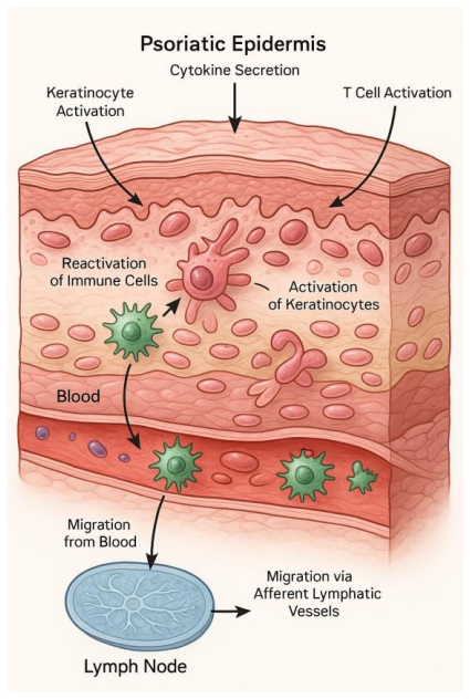
- [Figures 3 and 4] There are two figures numbered 'Figure 3' (the SEM images and the immune-cell schematic), and the Discussion refers to 'Figure 8' for the nanoparticle–protein interaction when the actual figure is numbered 4; all figure numbers should be corrected and renumbered consecutively.
- [§4.1] The subsection titled 'Pathogenesis of Psoriasis' is background material placed in the Results section; it should be moved to the Introduction or a dedicated background section.
- [References] Several reference entries are malformed or incomplete, including the Wieczorek (2017) entry and 'Kumar, V., Abbas, A. K., and Jon, C. (2018). Aster. robbins basic pathology.'; the reference list should be cleaned up.
- [Figure 6 caption] The caption states the treatment was 'ZnO and CeO nanoparticle based,' but the formulation also contains Ag nanoparticles and plant extracts; the caption should describe the full formulation.
Circularity Check
No circularity: the paper's efficacy claim is an empirical group comparison, not a derivation that reduces to its inputs.
full rationale
Walking the claimed derivation chain, there is no mathematical derivation, fitted parameter, or model-based prediction whose output is equivalent to its input by construction. The central claim—that the nanoparticle-and-extract gel accelerates healing—rests on direct in-vivo group comparisons reported in Tables 1 and 2 and narrative observations in Section 4.3. Even though the baseline imbalance (Day 0 lesion sizes differing significantly between groups) threatens the validity of the causal attribution, that is a confounding/design problem, not circularity: the conclusion is not defined in terms of the input, nor is a fitted parameter renamed as a prediction. The paper invokes no uniqueness theorem, imports no load-bearing result from the authors' own prior work, and the references are standard literature rather than self-citations carrying the argument. The asserted 'synergy' of the multiple components is not tested with omission controls, but this is a lack of mechanistic evidence rather than a circular step. Accordingly, no specific circular step can be quoted and exhibited, and the appropriate score is 0.
Assumptions & free parameters
assumptions (3)
- domain assumption Imiquimod plus full-thickness excision in the animal model is a valid surrogate for psoriatic lesion healing.
- domain assumption Nanoparticles retain their size, stability, and activity after being incorporated into the gel and applied to wounds.
- domain assumption The three experimental groups were comparable at baseline.
Cite this review
Pith. "Pith review of Advanced Nanostructured Topical Therapeutics for Psoriasis: Strategic Synthesis, Multimodal Characterization, and Preliminary Pharmacodynamic Profiling." pith.science (2026). https://pith.science/paper/UM6QLOUU
@misc{pith2026250601572,
  author       = {Pith},
  title        = {Pith review of: Advanced Nanostructured Topical Therapeutics for Psoriasis: Strategic Synthesis, Multimodal Characterization, and Preliminary Pharmacodynamic Profiling},
  year         = {2026},
  howpublished = {\url{https://pith.science/paper/UM6QLOUU}},
  note         = {Machine review of arXiv:2506.01572}
}
read the original abstract
Psoriasis is a long-term inflammatory skin disease that remains difficult to treat. In this study, we developed a new topical treatment by combining metal oxide nanoparticles: cerium oxide (CeO2), zinc oxide (ZnO), and silver (Ag), with natural plant extracts in a gel made from fish collagen and agar. The nanoparticles were characterized using UV-Vis spectroscopy, dynamic light scattering (DLS), Fourier-transform infrared spectroscopy (FTIR), and scanning electron microscopy (SEM), showing good stability and a uniform particle size distribution (ZnO averaged 66 nm). To enhance therapeutic potential, the gel was enriched with plant-derived antioxidants from bitter melon, ginger, and neem. This formulation was tested on an animal model of psoriasis. The treated group exhibited faster wound healing and reduced inflammation compared to both placebo and untreated groups, with statistically significant results (p < 0.01 to p < 0.001) observed from Day 3, becoming more pronounced by Day 14. These results indicate that the combination of nanoparticles with plant-based components in a topical gel may provide a promising new approach to psoriasis treatment. Further studies are recommended to evaluate long-term safety and therapeutic effectiveness.
Figures


Figures from the paper (4 more)


Reference graph
Works this paper leans on
-
[1]
Abdulsattar, S. A. (2015). Biological impact of gold nanoparticle on estradaiol and testosterone levels in sera of human males. Journal of the Faculty of Medicine Baghdad, 57(4):333–337. Aditya, A., Chattopadhyay, S., Jha, D., Gautam, H. K., Maiti, S., and Ganguli, M. (2018). Zinc oxide nanoparticles dispersed in ionic liquids show high antimicrobial effi...
work page 2015
-
[5]
Zhang, X.-F., Liu, Z.-G., Shen, W., and Gurunathan, S. (2016). Silver nanoparticles: synthesis, characterization, properties, applications, and therapeutic approaches. International journal of molecular sciences, 17(9):1534
work page 2016
-
[623]
Wieczorek, M. (2017). Abualrous et sticht j. ´alvaro-benito m. stolzenberg s. no´e f. freund c. Front. Immunol, 8:292. Zabriskie, J. B. (2009)
work page 2017
-
[878]
Baldim, V., Yadav, N., Bia, N., Graillot, A., Loubat, C., Singh, S., Karakoti, A
Elsevier. Baldim, V., Yadav, N., Bia, N., Graillot, A., Loubat, C., Singh, S., Karakoti, A. S., and Berret, J. -F. (2020). Polymer -coated cerium oxide nanoparticles as oxidoreductase-like catalysts. ACS Applied Materials & Interfaces , 12(37):42056–42066. Chen, S., Wang, Y., Bao, S., Yao, L., Fu, X., Yu, Y., Lyu, H., Pang, H., Guo, S., Zhang, H., et al. ...
work page 2020
-
[1268]
Grozdev, I., Korman, N., and Tsankov, N. (2014). Psoriasis as a systemic disease. Clinics in dermatology, 32(3):343–350. Henseler, T . and Christophers, E. (1995). Disease concomitance in psoriasis. Journal of the American Academy of Dermatology, 32(6):982–986. Hugh, J. M. and Weinberg, J. M. (2018). Update on the pathophysiology of psoriasis. Cutis, 102(...
work page 2014
Reviewed August 7, 2026 · model on record in the stance chip above.
Discussion (0). Sign in to comment.