REVIEW 3 major objections 5 minor 59 references
Quantum Cognition Machine Learning for Forecasting Chromosomal Instability
T0 review · 3 major / 5 minor · reviewed 2026-08-07 · deepseek-v4-flash
Pith's one-line read A quantum-inspired machine learning model predicts chromosomal instability in circulating tumor cells from microscopy features more accurately than ten classical methods.
desk verdict A legitimate first application of QCML to CTC morphology with a sound POVM extension, but the cell-level cross-validation does not support the new-patient generalization claim. read the letter →
The pith
A machine-rendered reading of the paper's core claim, the machinery that carries it, and where it could break.
The reading
What carries the argument
The central object is the error Hamiltonian $H(x_t) = \frac{1}{2}\sum_k (A_k - x_{t,k} I)^2$, where each $A_k$ is a learned Hermitian feature observable on an $N$-dimensional Hilbert space and $x_t$ is a cell's feature vector. The map from data to quantum state is the ground state $|\psi_t\rangle$ of this Hamiltonian, and a forecast observable $B$ gives predictions through $\hat{y}_t = \langle \psi_t | B | \psi_t \rangle$. The QCML POVM extension replaces point forecasts with a full probability density $p(y) = \langle \psi | \hat{Y}^\dagger(y) \hat{Y}(y) | \psi \rangle$, with the operators $\hat{Y}(y)$ parameterized by Legendre polynomials so the density integrates to one. This representation carries the argument: the parameter count grows linearly with the number of features, and the ground-state map performs an intrinsic dimensionality reduction, which the paper claims is why QCML generalizes in a high-dimensional, low-sample-size regime.
What would settle it
Run the same 5-fold cross-validation comparison with all cells from each patient held out together (patient-level splits), or on an independent cohort of CTCs; if QCML's balanced-accuracy and ROC-AUC advantages shrink or vanish, the claimed generalization advantage is an artifact of shared-patient overlap.
Extended reading notes
Core claim
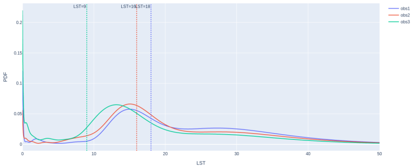
On the paper's own terms, the central discovery is that encoding each cell's 112 morphology, intensity, and texture features as a quantum state — the ground state of an error Hamiltonian built from learned observables — yields better out-of-sample classification of LST status than support vector machines, neural networks, random forests, XGBoost, and other classical models. In verification folds QCML reached balanced accuracy 70% ± 8%, sensitivity 84% ± 9%, and specificity 57% ± 10%, and QCML POVM reached ROC AUC 0.763, the best among all models tested. The authors interpret the smaller gap between in-sample and out-of-sample performance as evidence that the quantum-state representation controls overfitting without curated feature selection. They further claim that the model's feature importance ranking recovers biologically plausible correlates of chromosomal instability, including cell size, fractal nuclear shape, cross-channel colocalization, and DAPI localization.
Load-bearing premise
The load-bearing assumption is that splitting individual cells, not patients, into training and test sets measures how well the model generalizes to new patients; cells from the same patient share biological and technical factors that can inflate apparent out-of-sample performance.
Editorial extensions
If this is right
- Morphology-only pLST prediction could cut liquid-biopsy turnaround time from sequencing-scale workflows to image-analysis-scale workflows, if the reported performance holds in validation.
- QCML's gradient-based feature ranking yields a testable biological hypothesis: unstable CTCs are larger, have more fractal nuclei, and show disrupted DAPI localization and cross-channel texture alignment.
- QCML POVM's full predictive densities allow clinicians to choose operating points on specificity/sensitivity and report quantiles rather than single point predictions.
- A smaller in-sample/out-of-sample performance gap than classical models suggests QCML may generalize in other high-dimensional, low-sample-size biomedical datasets without curated feature selection.
Reading between the lines
- Editorial extension: Because the reported cross-validation splits cells rather than patients, the paper's claim of generalization to new patients is not yet fully tested; a patient-grouped split or independent cohort would be the decisive check.
- Editorial extension: The QCML distance defined by quantum fidelity is a supervised similarity measure; a natural next step the paper does not take is to use it for patient-level clustering or survival analysis in metastatic breast cancer.
- Editorial extension: The paper mentions quantum-assisted feature mapping from raw pixels as future work; that same framework could be tested against the hand-crafted 112-feature pipeline used here.
- Editorial extension: The POVM densities should be checked for calibration, since threshold-based clinical use requires trustworthy probability forecasts; the paper does not report calibration curves.
Editorial analysis
A structured set of objections, weighed in public.
Referee Report
Summary. The paper applies a quantum-inspired machine learning framework, QCML, to predict chromosomal instability (LST status) from 112 morphological and intensity features extracted from circulating tumor cells (CTCs) in metastatic breast cancer patients. The authors compare QCML regression and a new QCML POVM classification extension against 10 classical machine learning baselines using 5-fold cross-validation with 5 repeats, reporting that QCML achieves the highest out-of-sample balanced accuracy (70%) and ROC AUC (0.763) among all models. They also present a gradient-based feature importance analysis and a QCML-distance visualization. The paper concludes that QCML is a promising tool for liquid-biopsy-based real-time detection of chromosomal instability.
Significance. If the reported superiority were statistically reliable and generalizable to new patients, the paper would make a valuable contribution to high-dimensional, low-sample-size biomedical classification and to the emerging field of quantum-inspired machine learning. The manuscript is clearly written, the biological feature-importance findings are plausible and connect to known mechanisms of chromosomal instability, and the QCML POVM extension is described in detail. However, the central empirical claim rests on an evaluation protocol that does not establish generalization to unseen patients, and the reported differences are not supported by statistical testing. These issues are load-bearing for the paper's main conclusion.
major comments (3)
- [Section 2.2, Tables 1 and 2] The cross-validation procedure treats each CTC as an independent observation and splits cells, not patients. The dataset contains 227 CTCs from 73 patients (mean 3.25 cells per patient), so the same patient's cells appear in both the training and test folds. Cells from the same patient share biological state, staining batch, imaging conditions, and other technical factors, and are therefore not independent. The abstract and Discussion frame the method as enabling real-time detection for patients, so the intended use case is prediction on new patients. The current 'case-agnostic' protocol measures performance on cells with same-patient cells in the training set, which can inflate out-of-sample metrics and does not establish generalization to new patients. The authors must re-run the comparison using patient-level or group-aware cross-validation and report whether the QCML advantage persists. Without this, the headline superiority claim is not supported.
- [Tables 1 and 2] The reported performance differences lack statistical support. In Table 1, QCML's out-of-sample balanced accuracy is 70% ± 8% versus 68% ± 7% for the SVM Gaussian kernel, a difference well within the reported standard deviations. In Table 2, the QCML POVM ROC AUC of 0.763 versus XGBoost's 0.747 is presented without confidence intervals, per-fold values, or a significance test (e.g., DeLong test, bootstrap, or paired test across folds). The claim that QCML 'outperforms' the classical baselines requires formal uncertainty quantification and hypothesis tests, especially given the small absolute differences and overlapping error bars.
- [Section 3.2, Table 1] The claim that QCML shows a 'smaller disconnect between in-sample and out-of-sample performance' is made qualitatively. For balanced accuracy, the in-sample to out-of-sample drop for QCML is 78% to 70%, while for SVM Gaussian it is 74% to 68%; these gaps are similar in magnitude. If the authors wish to argue that QCML generalizes better, they should quantify the generalization gap across folds and test whether the difference is statistically significant, rather than relying on visual or qualitative comparison.
minor comments (5)
- [Section 2.3] The phrase 'LST¿12' appears to be a typographical error for 'LST > 12'; please correct it.
- [Section 2.3, Equation (2)] The description of the 'gradient-free' loss reweighting is unclear. The text says that adjusting each loss component by the corresponding gradient-free loss 'forces each component's loss to be equal to 1,' but in practice the gradient is scaled by the detached loss value, so the effective weighting is not necessarily equal. Please clarify the intended effect and the implementation.
- [Figure 4] The caption states that at lower specificity QCML POVM outperforms both XGBoost and Random Forest, but this is a qualitative claim based on visual inspection. Please provide the actual ROC curves with confidence bands or numerical comparisons at selected operating points.
- [Section 3.4, Figure 6] The feature importance table is difficult to read in the current formatting. Please present it as a proper table with clear column headers, and ensure the feature names and importance scores are legible.
- [Abstract and throughout] The phrase 'out of sample' should be hyphenated as 'out-of-sample' for consistency with standard terminology.
Circularity Check
No circularity: the QCML performance claims are empirical benchmarks against external baselines, not derivations from fitted parameters or self-citations.
full rationale
The paper's central claim—that QCML and QCML POVM outperform ten classical baselines for morphology-predicted LST status—is an empirical benchmark computed on held-out folds (Section 2.2, Tables 1-2) against external baselines such as SVM, XGBoost, and Random Forest. The QCML construction in Sections 2.3-2.4 is a mathematical encoding: data points are mapped to ground states of an error Hamiltonian, and the POVM density is normalized by construction via Legendre orthonormality; neither step defines the target y in terms of the model output, and the forecasts are scored against genomically determined LST ground truth. The main theoretical motivation cites the authors' own prior work ([25-28]) for properties such as robustness and generalization, but the reported performance does not reduce to those citations; it is independently evaluated on held-out cells. The paper itself flags its preliminary status ('Future studies will be required to validate these findings', Discussion). The cell-level 'case-agnostic' cross-validation is a generalization-validity limitation (same-patient cells can appear in both train and test), but this is a methodological concern about what 'out-of-sample' means, not a circular derivation: the comparison is still computed on data not used to fit each fold. No fitted parameter is renamed as a prediction, no uniqueness theorem is imported from the authors, and no known result is merely renamed. I find no circular step that reduces a claimed prediction to its inputs.
Assumptions & free parameters
free parameters (5)
- Hilbert space dimension N =
not reported
- positive class weight w_p =
0.5
- POVM truncation parameter K =
not reported
- classification probability threshold =
0.6
- LST threshold theta_LST =
12
assumptions (6)
- domain assumption The ground state of the error Hamiltonian H(x_t) exists and the non-differentiable optimization converges.
- standard math Born rule and POVM formalism are valid for the constructed operators.
- standard math Legendre polynomials provide an orthonormal basis on [-1,1] and the completeness relation holds.
- domain assumption Morphology features carry information about genomic LST status.
- domain assumption The LST>12 cutoff defines a clinically meaningful binary target.
- domain assumption Cells can be treated as independent observations in cross-validation.
invented entities (1)
-
QCML POVM (Positive Operator-Valued Measure forecasting extension)
Cite this review
Pith. "Pith review of Quantum Cognition Machine Learning for Forecasting Chromosomal Instability." pith.science (2026). https://pith.science/paper/DDRR3TA3
@misc{pith2026250603199,
  author       = {Pith},
  title        = {Pith review of: Quantum Cognition Machine Learning for Forecasting Chromosomal Instability},
  year         = {2026},
  howpublished = {\url{https://pith.science/paper/DDRR3TA3}},
  note         = {Machine review of arXiv:2506.03199}
}
read the original abstract
The accurate prediction of chromosomal instability from the morphology of circulating tumor cells (CTCs) enables real-time detection of CTCs with high metastatic potential in the context of liquid biopsy diagnostics. However, it presents a significant challenge due to the high dimensionality and complexity of single-cell digital pathology data. Here, we introduce the application of Quantum Cognition Machine Learning (QCML), a quantum-inspired computational framework, to estimate morphology-predicted chromosomal instability in CTCs from patients with metastatic breast cancer. QCML leverages quantum mechanical principles to represent data as state vectors in a Hilbert space, enabling context-aware feature modeling, dimensionality reduction, and enhanced generalization without requiring curated feature selection. QCML outperforms conventional machine learning methods when tested on out of sample verification CTCs, achieving higher accuracy in identifying predicted large-scale state transitions (pLST) status from CTC-derived morphology features. These preliminary findings support the application of QCML as a novel machine learning tool with superior performance in high-dimensional, low-sample-size biomedical contexts. QCML enables the simulation of cognition-like learning for the identification of biologically meaningful prediction of chromosomal instability from CTC morphology, offering a novel tool for CTC classification in liquid biopsy.
Figures


Figures from the paper (4 more)



Reference graph
Works this paper leans on
-
[1]
Breast cancer, version 3.2022, nccn clinical practice guidelines in oncology,
W. J. Gradishar et al., “Breast cancer, version 3.2022, nccn clinical practice guidelines in oncology,” J Natl Compr Canc Netw, vol. 20, no. 6, pp. 691–722, 2022. doi: 10.6004/jnccn.2022.0030 . [Online]. Available: https://www.ncbi.nlm.nih.gov/pubmed/35714673
arXiv 2022
-
[2]
Recommen- dations for her2 testing in breast cancer,
A. C. Wolff, American Society of Clinical Oncology, and College of American Pathologists, “Recommen- dations for her2 testing in breast cancer,” Arch Pathol Lab Med, vol. 138, no. 2, pp. 241–256, 2014. doi: 10.5858/arpa.2013-0953-SA
-
[3]
J. D. Schonhoft et al., “Morphology-predicted large-scale transition number in circulating tumor cells iden- tifies a chromosomal instability biomarker associated with poor outcome in castration-resistant prostate cancer,” Cancer Res, vol. 80, no. 22, pp. 4892–4903, 2020. doi: 10 . 1158 / 0008 - 5472 . CAN - 20 - 1216. [Online]. Available: https://www.ncb...
-
[4]
G. Di Caro et al., “A novel liquid biopsy assay for detection of erbb2 (her2) amplification in circulating tumor cells (ctcs),” J Circ Biomark, vol. 13, pp. 27–35, 2024. doi: 10.33393/jcb.2024.3046
-
[5]
S. Di Cosimo et al., “Low-pass whole genome sequencing of circulating tumor cells to evaluate chromosomal instability in triple-negative breast cancer,” Sci Rep, vol. 14, no. 1, p. 20 479, 2024.doi: 10.1038/s41598- 024-71378-3. [Online]. Available: https://www.ncbi.nlm.nih.gov/pubmed/39227622
arXiv 2024
-
[6]
Chromosomal instability estimation based on sequencing,
S. B. Greene and A. E. Dago, “Chromosomal instability estimation based on sequencing,” PLoS One, vol. 11, no. 11, e0165089, 2016. doi: 10.1371/journal.pone.0165089
-
[7]
Tumour heterogeneity: The key advantages of single-cell analysis,
M. Tellez-Gabriel, B. Ory, F. Lamoureux, M. F. Heymann, and D. Heymann, “Tumour heterogeneity: The key advantages of single-cell analysis,” Int J Mol Sci, vol. 17, no. 12, 2016. doi: 10.3390/ijms17122142. [Online]. Available: https://www.ncbi.nlm.nih.gov/pubmed/27999407
-
[8]
P. D. Malihi et al., “Single-cell circulating tumor cell analysis reveals genomic instability as a distinctive feature of aggressive prostate cancer,”Clin Cancer Res, vol. 26, no. 15, pp. 4143–4153, 2020.doi: 10.1158/ 1078-0432.CCR-19-4100 . [Online]. Available: https://www.ncbi.nlm.nih.gov/pubmed/32341031
Show all 59 references
-
[9]
Circulating tumor cell chromosomal instability and neuroendocrine phenotype by immunomorphology and poor outcomes in men with mcrpc treated with abiraterone or enzalutamide,
L. C. Brown et al., “Circulating tumor cell chromosomal instability and neuroendocrine phenotype by immunomorphology and poor outcomes in men with mcrpc treated with abiraterone or enzalutamide,” Clin Cancer Res, vol. 27, no. 14, pp. 4077–4088, 2021. doi: 10.1158/1078-0432.CCR...
2021
-
[10]
Genomic instability–an evolving hallmark of cancer,
S. Negrini, V. G. Gorgoulis, and T. D. Halazonetis, “Genomic instability–an evolving hallmark of cancer,” Nat Rev Mol Cell Biol, vol. 11, no. 3, pp. 220–8, 2010. doi: 10 . 1038 / nrm2858. [Online]. Available: https://www.ncbi.nlm.nih.gov/pubmed/20177397
2010
-
[11]
Determinants and clinical implications of chromoso- mal instability in cancer,
L. Sansregret, B. Vanhaesebroeck, and C. Swanton, “Determinants and clinical implications of chromoso- mal instability in cancer,” Nat Rev Clin Oncol, vol. 15, no. 3, pp. 139–150, 2018.doi: 10.1038/nrclinonc. 2017.198. [Online]. Available: https://www.ncbi.nlm.nih.gov/pubmed/29297505
2018
-
[12]
Clonal heterogeneity and tumor evolution: Past, present, and the future,
N. McGranahan and C. Swanton, “Clonal heterogeneity and tumor evolution: Past, present, and the future,” Cell, vol. 168, no. 4, pp. 613–628, 2017. doi: 10.1016/j.cell.2017.01.018. [Online]. Available: https://www.ncbi.nlm.nih.gov/pubmed/28187284
2017
-
[13]
Mechanisms of genomic instability in breast cancer,
P. H. G. Duijf, D. Nanayakkara, K. Nones, S. Srihari, M. Kalimutho, and K. K. Khanna, “Mechanisms of genomic instability in breast cancer,” Trends Mol Med, vol. 25, no. 7, pp. 595–611, 2019. doi: 10.1016/ j.molmed.2019.04.004. [Online]. Available: https://www.ncbi.nlm.nih.gov/...
2019
-
[14]
Hallmarks of cancer: New dimensions,
D. Hanahan, “Hallmarks of cancer: New dimensions,” Cancer Discov, vol. 12, no. 1, pp. 31–46, 2022. doi: 10.1158/2159- 8290.CD- 21- 1059. [Online]. Available: https://www.ncbi.nlm.nih.gov/pubmed/ 35022204. 12
2022 doi
-
[15]
Targeting cancers with chromosome instability,
A. Eccleston, “Targeting cancers with chromosome instability,” Nat Rev Drug Discov, vol. 21, no. 8, p. 556, 2022. doi: 10.1038/d41573- 022- 00111- 4. [Online]. Available: https://www.ncbi.nlm.nih. gov/pubmed/35778502
2022
-
[16]
A pan-cancer compendium of chromosomal instability,
R. M. Drews et al., “A pan-cancer compendium of chromosomal instability,” Nature, vol. 606, no. 7916, pp. 976–983, 2022. doi: 10.1038/s41586-022-04789-9 . [Online]. Available: https://www.ncbi.nlm. nih.gov/pubmed/35705807
2022
-
[17]
Oncogenic signaling pathways in the cancer genome atlas,
F. Sanchez-Vega et al., “Oncogenic signaling pathways in the cancer genome atlas,” Cell, vol. 173, no. 2, 321–337 e10, 2018. doi: 10.1016/j.cell.2018.03.035 . [Online]. Available: https://www.ncbi.nlm. nih.gov/pubmed/29625050
2018
-
[18]
Mechanisms and consequences of cancer genome instability: Lessons from genome sequencing studies,
J. K. Lee, Y. L. Choi, M. Kwon, and P. J. Park, “Mechanisms and consequences of cancer genome instability: Lessons from genome sequencing studies,” Annu Rev Pathol, vol. 11, pp. 283–312, 2016. doi: 10.1146/annurev- pathol- 012615- 044446. [Online]. Available: https://www.ncbi....
2016
-
[19]
The role of aneuploidy in cancer evolution,
L. Sansregret and C. Swanton, “The role of aneuploidy in cancer evolution,” Cold Spring Harb Perspect Med, vol. 7, no. 1, 2017. doi: 10.1101/cshperspect.a028373 . [Online]. Available: https://www.ncbi. nlm.nih.gov/pubmed/28049655
2017
-
[20]
Toward clinical implementation of next-generation sequencing- based genetic testing in rare diseases: Where are we?
Z. Liu, L. Zhu, R. Roberts, and W. Tong, “Toward clinical implementation of next-generation sequencing- based genetic testing in rare diseases: Where are we?”Trends Genet, vol. 35, no. 11, pp. 852–867, 2019.doi: 10.1016/j.tig.2019.08.006. [Online]. Available: https://www.ncbi....
2019
-
[21]
Machine learning in cell biology - teaching computers to recognize phenotypes,
C. Sommer and D. W. Gerlich, “Machine learning in cell biology - teaching computers to recognize phenotypes,” J Cell Sci, vol. 126, no. Pt 24, pp. 5529–39, 2013. doi: 10.1242/jcs.123604 . [Online]. Available: https://www.ncbi.nlm.nih.gov/pubmed/24259662
2013
-
[22]
The trifecta of single-cell, systems-biology, and machine-learning approaches,
T. M. Weiskittel et al., “The trifecta of single-cell, systems-biology, and machine-learning approaches,” Genes (Basel), vol. 12, no. 7, 2021. doi: 10.3390/genes12071098 . [Online]. Available: https://www. ncbi.nlm.nih.gov/pubmed/34356114
2021
-
[23]
Translating cancer genomics into precision medicine with artificial intelligence: Applications, challenges and future perspectives,
J. Xu et al., “Translating cancer genomics into precision medicine with artificial intelligence: Applications, challenges and future perspectives,” Hum Genet, vol. 138, no. 2, pp. 109–124, 2019.doi: 10.1007/s00439- 019-01970-5. [Online]. Available: https://www.ncbi.nlm.nih.gov...
2019
-
[24]
Informing immunotherapy with multi-omics driven machine learning,
Y. Li, X. Wu, D. Fang, and Y. Luo, “Informing immunotherapy with multi-omics driven machine learning,” NPJ Digit Med, vol. 7, no. 1, p. 67, 2024. doi: 10 . 1038 / s41746 - 024 - 01043 - 6. [Online]. Available: https://www.ncbi.nlm.nih.gov/pubmed/38486092
2024
-
[25]
Candelori et al., Robust estimation of the intrinsic dimension of data sets with quantum cognition machine learning, https://arxiv.org/abs/2409.12805, 2024
L. Candelori et al., Robust estimation of the intrinsic dimension of data sets with quantum cognition machine learning, https://arxiv.org/abs/2409.12805, 2024
2024 arXiv
-
[26]
Musaelian et al., Quantum cognition machine learning: AI Needs Quantum, https://www.qognitive
K. Musaelian et al., Quantum cognition machine learning: AI Needs Quantum, https://www.qognitive. io/papers/QCML - Qognitive, Inc.pdf, 2024
2024
-
[27]
Samson et al., Quantum cognition machine learning: Financial forecasting, Risk.net, 2024
R. Samson et al., Quantum cognition machine learning: Financial forecasting, Risk.net, 2024
2024
-
[28]
Rosaler et al., Supervised similarity for high-yield corporate bonds with quantum cognition machine learning, 2025
J. Rosaler et al., Supervised similarity for high-yield corporate bonds with quantum cognition machine learning, 2025. arXiv: 2502.01495 [q-fin.ST]. [Online]. Available: https://arxiv.org/abs/2502. 01495
2025 arXiv
-
[29]
H. C. Steinacker, Quantum Geometry, Matrix Theory, and Gravity. Cambridge University Press, 2024
2024
-
[30]
Fully automated ctc detection, segmentation and classification for multi-channel if imaging,
E. Schwab et al., “Fully automated ctc detection, segmentation and classification for multi-channel if imaging,” in Medical Optical Imaging and Virtual Microscopy Image Analysis, 2025, pp. 55–65
2025
-
[31]
[Online]
Human genome assembly grch38, Genome Reference Consortium, 2013. [Online]. Available: https : / / www.ncbi.nlm.nih.gov/datasets/genome/GCF_000001405.26/
2013
-
[32]
Visual pattern recognition by moment invariants,
H. Ming-Kuei, “Visual pattern recognition by moment invariants,” IRE Transactions on Information Theory, vol. 8, no. 2, pp. 179–187, 1962. doi: 10.1109/TIT.1962.1057692
1962
-
[33]
Laws, Textured Image Segmentation
K. Laws, Textured Image Segmentation. University of Southern California, 1980. [Online]. Available: https://books.google.com/books?id=GxvLnQEACAAJ
1980
-
[34]
Quantum cognition,
E. M. Pothos and J. R. Busemeyer, “Quantum cognition,” Annual Review of Psychology, vol. 73, no. 1, pp. 749–778, 2022. doi: 10.1146/annurev- psych- 033020- 123501. eprint: 10.1146/annurev- psych- 033020-123501. [Online]. Available: 10.1146/annurev-psych-033020-123501
2022 doi
-
[35]
M. A. Nielsen and I. L. Chuang, Quantum Computation and Quantum Information. Cambridge University Press, 2000. 13
2000
-
[36]
Weber and G
H.-J. Weber and G. B. Arfken, Mathematical methods for physicists. Elsevier Academic Cambridge, MA, USA, 2005, vol. 148
2005
-
[37]
Ai powered quantification of nuclear morphology in cancers enables prediction of genome instability and prognosis,
J. Abel, S. Jain, D. Rajan, H. Padigela, K. Leidal, et al., “Ai powered quantification of nuclear morphology in cancers enables prediction of genome instability and prognosis,” NPJ Precision Oncology, vol. 8, no. 1, p. 134, 2024. doi: 10.1038/s41698- 024- 00623- 9. [Online]. A...
2024
-
[38]
The nuclear envelope environment and its cancer connec- tions,
K. H. Chow, R. E. Factor, and K. S. Ullman, “The nuclear envelope environment and its cancer connec- tions,” Nat Rev Cancer, vol. 12, no. 3, pp. 196–209, 2012. doi: 10.1038/nrc3219 . [Online]. Available: https://www.ncbi.nlm.nih.gov/pubmed/22337151
2012
-
[39]
Blue intensity matters for cell cycle profiling in fluorescence dapi-stained images,
A. Ferro, T. Mestre, P. Carneiro, I. Sahumbaiev, R. Seruca, and J. M. Sanches, “Blue intensity matters for cell cycle profiling in fluorescence dapi-stained images,” Lab Invest, vol. 97, no. 5, pp. 615–625, 2017. doi: 10.1038/labinvest.2017.13. [Online]. Available: https://www...
2017
-
[40]
Sites of chromosomal instability in the context of nuclear architecture and function,
C. Pentzold, M. Kokal, S. Pentzold, and A. Weise, “Sites of chromosomal instability in the context of nuclear architecture and function,” Cell Mol Life Sci, vol. 78, no. 5, pp. 2095–2103, 2021. doi: 10.1007/ s00018-020-03698-2 . [Online]. Available: https://www.ncbi.nlm.nih.go...
2021
-
[41]
Nuclear envelope rupture drives genome instability in cancer,
S. Lim, R. J. Quinton, and N. J. Ganem, “Nuclear envelope rupture drives genome instability in cancer,” Mol Biol Cell, vol. 27, no. 21, pp. 3210–3213, 2016. doi: 10.1091/mbc.E16-02-0098. [Online]. Available: https://www.ncbi.nlm.nih.gov/pubmed/27799497
2016
-
[42]
A comparative study of texture measures with classification based on feature distributions,
T. Ojala, M. Pietik¨ ainen, and D. Harwood, “A comparative study of texture measures with classification based on feature distributions,” Pattern Recognition, vol. 29, pp. 51–59, 1996
1996
-
[43]
Index for rating diagnostic tests,
W. J. Youden, “Index for rating diagnostic tests,” Cancer, vol. 3, no. 1, pp. 32–35, 1950, issn: 1097-0142. doi: 10 . 1002 / 1097 - 0142(1950 ) 3 : 1<32 :: aid - cncr2820030106 > 3 . 0 . co ; 2 - 3. [Online]. Available: http://dx.doi.org/10.1002/1097-0142(1950)3:1%3C32::AID-CN...
1950 doi
-
[44]
The inconsistency of “optimal
N. J. Perkins and E. F. Schisterman, “The inconsistency of “optimal” cutpoints obtained using two criteria based on the receiver operating characteristic curve,” American Journal of Epidemiology, vol. 163, no. 7, pp. 670–675, Jan. 2006, issn: 0002-9262. doi: 10.1093/aje/kwj063...
2006 doi
-
[45]
Causes and consequences of micronuclei,
K. Krupina, A. Goginashvili, and D. W. Cleveland, “Causes and consequences of micronuclei,” Curr Opin Cell Biol, vol. 70, pp. 91–99, 2021. doi: 10 . 1016 / j . ceb . 2021 . 01 . 004. [Online]. Available: https : //www.ncbi.nlm.nih.gov/pubmed/33610905
2021
-
[46]
G2/m-phase checkpoint adaptation and micronuclei formation as mechanisms that contribute to genomic instability in human cells,
D. Kalsbeek and R. M. Golsteyn, “G2/m-phase checkpoint adaptation and micronuclei formation as mechanisms that contribute to genomic instability in human cells,” Int J Mol Sci, vol. 18, no. 11, 2017. doi: 10.3390/ijms18112344. [Online]. Available: https://www.ncbi.nlm.nih.gov/...
2017
-
[48]
Comprehensive and integrated genomic characterization of adult soft tissue sarcomas,
e. d. s. c. Cancer Genome Atlas Research Network. Electronic address and N. Cancer Genome Atlas Research, “Comprehensive and integrated genomic characterization of adult soft tissue sarcomas,” Cell, vol. 171, no. 4, 950–965 e28, 2017. doi: 10.1016/j.cell.2017.10.014 . [Online]...
2017
-
[49]
Prognostic values of cancer associated macrophage-like cells (caml) enumeration in metastatic breast cancer,
Z. Mu et al., “Prognostic values of cancer associated macrophage-like cells (caml) enumeration in metastatic breast cancer,” Breast Cancer Res Treat, vol. 165, no. 3, pp. 733–741, 2017. doi: 10.1007/s10549-017- 4372-8. [Online]. Available: https://www.ncbi.nlm.nih.gov/pubmed/28687903
2017
-
[50]
Blood-based biopsies- clinical utility beyond circulating tumor cells,
C. M. Tang, P. Zhu, S. Li, O. V. Makarova, P. T. Amstutz, and D. L. Adams, “Blood-based biopsies- clinical utility beyond circulating tumor cells,” Cytometry A, vol. 93, no. 12, pp. 1246–1250, 2018. doi: 10.1002/cyto.a.23573. [Online]. Available: https://www.ncbi.nlm.nih.gov/p...
2018
-
[51]
The value of morphometry to classic prognostica- tors in breast cancer,
J. P. Baak, H. Van Dop, P. H. Kurver, and J. Hermans, “The value of morphometry to classic prognostica- tors in breast cancer,” Cancer, vol. 56, no. 2, pp. 374–82, 1985. doi: 10.1002/1097-0142(19850715)56: 2<374::aid- cncr2820560229> 3.0.co;2- 9. [Online]. Available: https://w...
1985
-
[52]
Correlation of nuclear morphometry with progression of breast cancer,
K. J. Pienta and D. S. Coffey, “Correlation of nuclear morphometry with progression of breast cancer,” Cancer, vol. 68, no. 9, pp. 2012–6, 1991. doi: 10 . 1002 / 1097 - 0142(19911101 ) 68 : 9<2012 :: aid - cncr2820680928>3.0.co;2-c. [Online]. Available: https://www.ncbi.nlm.ni...
2012
-
[53]
Nucleus, nuclear structure, and nuclear functions: Pathogenesis of nuclear abnormalities in cancer,
A. Zimmermann, “Nucleus, nuclear structure, and nuclear functions: Pathogenesis of nuclear abnormalities in cancer,” in Springer International Publishing, 2017, pp. 3071–3087. doi: 10.1007/978-3-319-26956- 6_170. [Online]. Available: https://doi.org/10.1007/978-3-319-26956-6_170 . 14
2017 doi
-
[54]
Challenges in circulating tumour cell research,
C. Alix-Panabieres and K. Pantel, “Challenges in circulating tumour cell research,” Nat Rev Cancer, vol. 14, no. 9, pp. 623–31, 2014. doi: 10.1038/nrc3820 . [Online]. Available: https://www.ncbi.nlm. nih.gov/pubmed/25154812
2014
-
[55]
A conduit to metastasis: Circulating tumor cell biology,
D. S. Micalizzi, S. Maheswaran, and D. A. Haber, “A conduit to metastasis: Circulating tumor cell biology,” Genes Dev, vol. 31, no. 18, pp. 1827–1840, 2017. doi: 10 . 1101 / gad . 305805 . 117. [Online]. Available: https://www.ncbi.nlm.nih.gov/pubmed/29051388
2017
-
[56]
Biology, detection, and clinical implications of circulating tumor cells,
S. A. Joosse, T. M. Gorges, and K. Pantel, “Biology, detection, and clinical implications of circulating tumor cells,” EMBO Mol Med, vol. 7, no. 1, pp. 1–11, 2015. doi: 10.15252/emmm.201303698 . [Online]. Available: https://www.ncbi.nlm.nih.gov/pubmed/25398926
2015
-
[57]
Building a two-way street between cell biology and machine learning,
C. Uhler, “Building a two-way street between cell biology and machine learning,” Nat Cell Biol, vol. 26, no. 1, pp. 13–14, 2024. doi: 10.1038/s41556- 023- 01279- 6. [Online]. Available: https://www.ncbi. nlm.nih.gov/pubmed/38228823
2024
-
[58]
Machine learning for perturbational single-cell omics,
Y. Ji, M. Lotfollahi, F. A. Wolf, and F. J. Theis, “Machine learning for perturbational single-cell omics,” Cell Syst, vol. 12, no. 6, pp. 522–537, 2021. doi: 10.1016/j.cels.2021.05.016 . [Online]. Available: https://www.ncbi.nlm.nih.gov/pubmed/34139164
2021
-
[59]
Representation learning: A review and new perspectives,
Y. Bengio, A. Courville, and P. Vincent, “Representation learning: A review and new perspectives,” IEEE Trans Pattern Anal Mach Intell, vol. 35, no. 8, pp. 1798–828, 2013. doi: 10.1109/TPAMI.2013.50. [Online]. Available: https://www.ncbi.nlm.nih.gov/pubmed/23787338
2013
-
[60]
Pan-cancer computational histopathology reveals mutations, tumor composition and progno- sis,
Y. Fu et al., “Pan-cancer computational histopathology reveals mutations, tumor composition and progno- sis,” Nat Cancer, vol. 1, no. 8, pp. 800–810, 2020. doi: 10.1038/s43018-020-0085-8 . [Online]. Available: https://www.ncbi.nlm.nih.gov/pubmed/35122049. 15
2020
Reviewed August 7, 2026 · model on record in the stance chip above.
Discussion (0). Sign in to comment.