REVIEW 4 major objections 4 minor 22 references
FAMSeg: Fetal Femur and Cranial Ultrasound Segmentation Using Feature-Aware Attention and Mamba Enhancement
T0 review · 4 major / 4 minor · reviewed 2026-08-07 · deepseek-v4-flash
Pith's one-line read The paper reports 91.58 mean IoU for its FAMSeg network on fetal femur and cranial ultrasound, ahead of seven baselines.
desk verdict Plausible architecture, but the training recipe is contradictory and the evaluation is under-powered, so the headline mIoU claim is not reproducible as written. read the letter →
The pith
A machine-rendered reading of the paper's core claim, the machinery that carries it, and where it could break.
The reading
What carries the argument
The core mechanism is a staged encoder-decoder stack. In the encoder, a multi-branch deep strip convolution scans with unidirectional kernels from horizontal and vertical views instead of one $K\times K$ window, which the authors argue preserves edge and small-object texture at lower cost ($2H_oW_oK$ versus $K^2H_oW_o$ multiply-adds). Each residual shortcut in the Bottle block passes through a Mamba V2 block, whose multi-view scanning is intended to filter accumulated raw ultrasound noise. The decoder contributes the feature-aware attention module, or FAM: a $1\times1$ channel compressor, a kernel reorganizer that creates $K\times K\times C$ content-dependent weights, and a softmax normalization that weights the original features before upsampling. The final component is an alternating optimizer schedule, AdamW for the first 150 epochs and then SGD for 50 epochs, meant to converge near the optimum without overshooting.
What would settle it
Run the same models on the same 3,798 images with a fixed five-fold split and report femur-only IoU; if FAMSeg's femur margin over U-Net falls below the reported 1.91 points, or an independent boundary-jaggedness metric fails to improve, the central claim is not supported.
Extended reading notes
Core claim
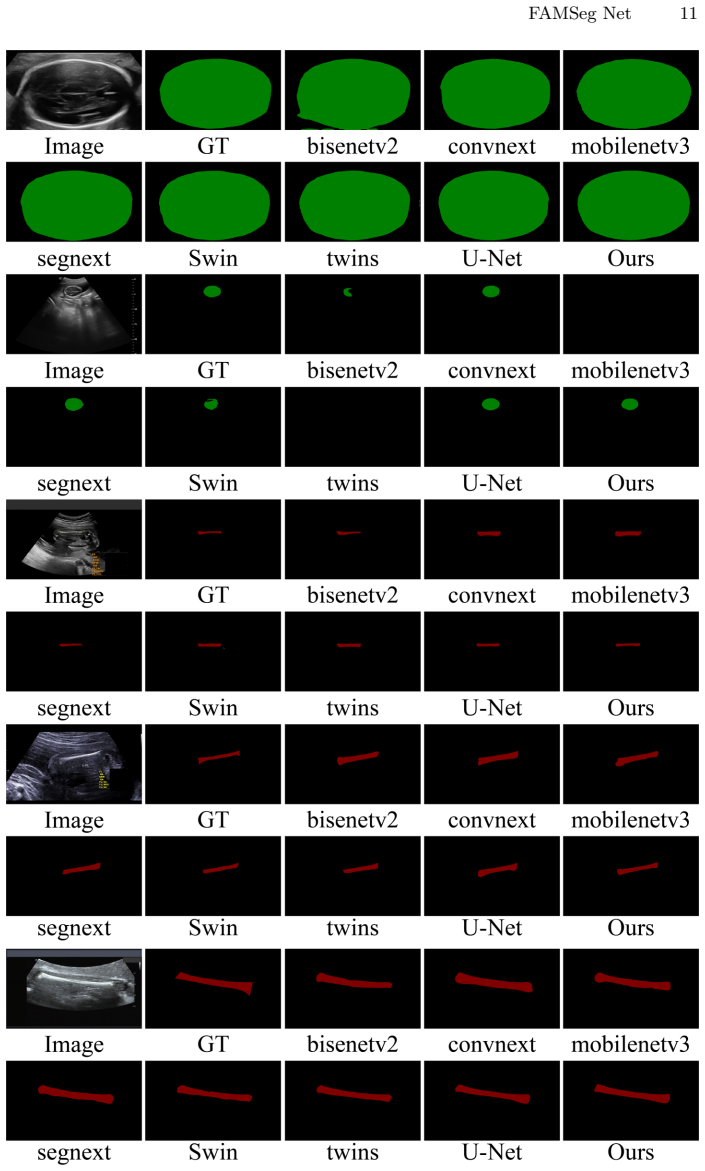
The central claim is that the combination of three mechanisms removes the jagged-edge and small-object failures typical of segmenters trained on natural scenes. In Table 1, FAMSeg reports 99.08 background IoU, 80.21 femur IoU, and 95.44 cranial IoU, the best across U-Net, Swin, SegNeXt, MobileNetV3, ConvNeXt, BiSeNetV2, and the twin version tested. The ablation attributes the largest single gain to adding the context-fusion module and the final gain to combining local and global information, with the paper further claiming the fastest reduction in training loss among the optimizer and decay combinations tried.
Load-bearing premise
The performance ordering rests on a single private dataset with no described split, annotation protocol, or inter-observer agreement, so the reported mIoU gaps may not generalize to other scanners or hospitals.
Editorial extensions
If this is right
- A cleaner femur contour would feed directly into femur-length biometry, the measurement that drives gestational-age and growth assessment.
- The strip-convolution cost equations imply the encoder needs fewer multiply-adds than a standard $7\times7$ convolution ($2H_oW_o \times 7$ versus $7H_oW_o \times 7$), so a version of the method could run on portable ultrasound hardware.
- The ablation result (91.29 mIoU without Mamba, 91.58 with it) shows the Mamba residual contributes, but the context fusion contributes more, so teams can adopt the fusion module independently.
- Switching optimizers from AdamW to SGD is a training-schedule choice, not an architectural one, so the schedule can be transferred to other segmentation backbones without changing inference code.
Reading between the lines
- Beyond the paper: re-running the comparison with five-fold cross-validation or a fixed external test set would settle whether the 91.58 mIoU is a property of the architecture or of one private dataset, since the paper reports no split or annotation-agreement metric.
- Beyond the paper: the paper does not quantify 'varying orientations,' so a natural test is to stratify femur images by rotation angle and measure whether the IoU gap to U-Net persists at extreme angles.
- Beyond the paper: the authors' closing limitations say multi-object balance is unsolved and training is time-consuming, which makes knowledge distillation or a lighter Mamba branch a concrete next experiment.
Editorial analysis
A structured set of objections, weighed in public.
Referee Report
Summary. The paper proposes FAMSeg, a deep learning model for segmenting fetal femur and cranial structures in ultrasound images. The architecture combines multi-branch strip convolutions, a Mamba V2 residual block, and a feature-aware attention upsampling module, and is trained with an alternating optimizer scheme. On a private dataset of 3,798 ultrasound images from one hospital, the authors report a mean IoU of 91.58, outperforming U-Net, Swin, SegNeXt, ConvNeXt, and other baselines, and claim the fastest loss reduction and best segmentation performance across image sizes and orientations.
Significance. If substantiated, FAMSeg would offer a practical improvement for fetal ultrasound segmentation, particularly for small anatomical structures, by combining efficient strip convolutions, Mamba-based residual filtering, and content-aware upsampling. The paper addresses a clinically relevant problem and includes comparisons with several recent methods. However, the empirical claims currently rest on a single private dataset with no cross-validation or statistical analysis, and the manuscript contains internal contradictions about the training configuration. No code or data are provided, so the results are not independently checkable. The significance of the contribution is therefore limited until the evaluation and reproducibility issues are resolved.
major comments (4)
- [§3.4, §4.4, Table 2] The training configuration behind the headline result is not uniquely specified. Section 3.4 defines the schedule as AdamW for 150 epochs followed by SGD for 50 epochs, whereas Section 4.4 and Figure 5 credit the 'AdamW and Adadelta combined optimization method', and Table 2 lists two rows (AdamW+Adadelta and AdamW+COS) that both achieve mIoU 91.58 but with different class-level IoUs (FL 80.07 vs 80.21; FB 95.58 vs 95.44). The paper therefore does not state which configuration generated the Table 1 result, making the central empirical claim non-reproducible.
- [§3.2.1, Eqs. (1)–(5)] The complexity analysis in Section 3.2.1 is not valid as written. Equation (1) defines Parameters_conv = K × H_o × W_o × K, omitting input and output channel counts, and the same equation is subsequently used as a 'cost' measure, conflating parameter count with computational cost. Equation (5) gives Cost_3×3 = 2 × H_o × W_o × 9, which is dimensionally inconsistent with the stated 3×3 case; if the intended computation is a two-direction strip convolution, the cost factor should be 3, not 9. Because these equations are used to claim that the proposed module has lower computational cost and reduced model complexity, the efficiency claim is not supported by the current derivation.
- [§4.1 and Table 1] The empirical evaluation is insufficient to establish the claimed performance ordering. The dataset is a single private collection of 3,798 images from one hospital, with no reported train-validation-test split, no annotation protocol, and no inter-observer agreement check. Table 1 reports single mIoU values without error bars or significance tests, and the differences among several baselines (e.g., SegNeXt 89.94, Swin 90.34, U-Net 90.13) are small relative to the likely random variation. The claim that FAMSeg's 91.58 mIoU is superior to all baselines is therefore not statistically supported.
- [§3.3 and §4.3–4.4] The feature-aware attention module is closely related to the content-aware upsampling method CARAFE, but no comparison with CARAFE is included in Table 1 or the ablation study; such a comparison is necessary to support the novelty and effectiveness of the proposed module. In addition, the abstract's claim of 'fastest loss reduction' is not quantified: Figure 5 shows convergence curves but no numeric comparison of epochs or wall-clock time to reach a given loss value.
minor comments (4)
- [Table 3] The ablation row named 'Apomixis' is not explained; the text refers to removing the Mamba structure and the feature fusion component, so the table should use explicit descriptions such as 'No Mamba' and 'No feature fusion' for each row.
- [§2 and §3.2.1] The term 'hamburger module' is used without definition or citation; please clarify what this component is and how it is applied.
- [Figure 2 and general] There are several typographical and formatting issues, including 'Convlution' in the Figure 2 caption, inconsistent spacing in 'F AMSeg', and the use of 'Eq. (11)' and 'Eq. (12)' without explicit variable definitions for batch size and learning-rate limits.
- [§4.2] The learning-rate adaptation formulas in Eqs. (11) and (12) are unclear; please specify the units and the meaning of 'init lr', 'min lr', 'lr limit min', and 'lr limit max'.
Circularity Check
No significant circularity: FAMSeg's central claim is an empirical benchmark comparison, not a derivation fitted to its own output.
full rationale
The paper's central claim is that the proposed FAMSeg architecture achieves the best segmentation performance (mIoU 91.58) on a private fetal ultrasound dataset, as established by comparative experiments in Table 1 and ablations in Tables 2 and 3. This claim is not derived from fitted constants or from equations whose inputs encode the claimed output; it is an empirical benchmark result. No step in the method section defines a component in terms of the final mIoU or uses the target metric as a construction input. The feature-aware decoder, strip convolution module, and Mamba residual structure are presented as architectural choices, and their contribution is tested by ablations rather than asserted through a self-referential fit. While the paper does cite prior work by authors with overlapping research groups (e.g., Lu et al. and Pu et al. in related work), those citations are contextual and not load-bearing: they do not supply the uniqueness of the architecture or the validity of the segmentation results. The noted inconsistency between the optimizer schedule in Section 3.4 (AdamW followed by SGD) and Section 4.4 / Figure 5 (AdamW with Adadelta) is a reproducibility and reporting problem, not a circularity problem, because the reported performance is not logically forced by the paper's own definitions or equations. Likewise, the absence of a CARAFE comparison and the lack of dataset split details raise evaluative risk, but they do not constitute a circular derivation. No specific reduction of the claimed result to its own inputs can be exhibited, so the honest finding is no significant circularity.
Assumptions & free parameters
free parameters (4)
- Learning rate bounds =
max 0.01, min 0.0001, limits 0.001 and 0.0001
- Optimizer switch point =
AdamW for 150 epochs then SGD for 50 epochs
- FAM kernel size k =
not reported
- Model width and channel counts =
not reported
assumptions (4)
- domain assumption The private Shenzhen dataset of 3,798 images is representative of clinical fetal femur and cranium ultrasound across equipment and populations.
- domain assumption Ground-truth segmentations are accurate enough to serve as training and evaluation labels.
- ad hoc to paper Mamba V2 residual filtering removes noise from shortcut connections and improves segmentation.
- ad hoc to paper The parameter-count formula Parameters equals K times H_o times W_o times K correctly measures model cost.
Cite this review
Pith. "Pith review of FAMSeg: Fetal Femur and Cranial Ultrasound Segmentation Using Feature-Aware Attention and Mamba Enhancement." pith.science (2026). https://pith.science/paper/5DHYLDAD
@misc{pith2026250607431,
  author       = {Pith},
  title        = {Pith review of: FAMSeg: Fetal Femur and Cranial Ultrasound Segmentation Using Feature-Aware Attention and Mamba Enhancement},
  year         = {2026},
  howpublished = {\url{https://pith.science/paper/5DHYLDAD}},
  note         = {Machine review of arXiv:2506.07431}
}
read the original abstract
Accurate ultrasound image segmentation is a prerequisite for precise biometrics and accurate assessment. Relying on manual delineation introduces significant errors and is time-consuming. However, existing segmentation models are designed based on objects in natural scenes, making them difficult to adapt to ultrasound objects with high noise and high similarity. This is particularly evident in small object segmentation, where a pronounced jagged effect occurs. Therefore, this paper proposes a fetal femur and cranial ultrasound image segmentation model based on feature perception and Mamba enhancement to address these challenges. Specifically, a longitudinal and transverse independent viewpoint scanning convolution block and a feature perception module were designed to enhance the ability to capture local detail information and improve the fusion of contextual information. Combined with the Mamba-optimized residual structure, this design suppresses the interference of raw noise and enhances local multi-dimensional scanning. The system builds global information and local feature dependencies, and is trained with a combination of different optimizers to achieve the optimal solution. After extensive experimental validation, the FAMSeg network achieved the fastest loss reduction and the best segmentation performance across images of varying sizes and orientations.
Figures


Figures from the paper (2 more)

Reference graph
Works this paper leans on
-
[1]
IEEE Transactions on Industrial Informatics 17(11), 7771–7780 (2021)
Pu, B., Li, K., Li, S., et al.: Automatic fetal ultrasound standard plane recognition based on deep learning and iiot. IEEE Transactions on Industrial Informatics 17(11), 7771–7780 (2021)
work page 2021
-
[2]
Engineer- ing Applications of Artificial Intelligence136, 108,885 (2024)
Gao, Z., Tan, G., Wang, C., et al.: Graph-enhanced ensembles of multi-scale struc- ture perception deep architecture for fetal ultrasound plane recognition. Engineer- ing Applications of Artificial Intelligence136, 108,885 (2024)
work page 2024
-
[3]
In: Forty-first International Conference on Machine Learning (2024)
Pu, B., Lv, X., Yang, J., et al.: Unsupervised domain adaptation for anatomical structure detection in ultrasound images. In: Forty-first International Conference on Machine Learning (2024)
work page 2024
-
[4]
IEEE Journal of Biomedical and Health Informatics (2024)
Zhao, L., Tan, G., Wu, Q., et al.: Farn: fetal anatomy reasoning network for detec- tion with global context semantic and local topology relationship. IEEE Journal of Biomedical and Health Informatics (2024)
work page 2024
-
[5]
Future Generation Computer Systems135, 234–243 (2022)
Zhao, L., Li, K., Pu, B., et al.: An ultrasound standard plane detection model of fetal head based on multi-task learning and hybrid knowledge graph. Future Generation Computer Systems135, 234–243 (2022)
work page 2022
-
[6]
In: Proceedings of the IEEE/CVF Conference on Computer Vision and Pattern Recognition, pp
Pu, B., Wang, L., Yang, J., et al.: M3-uda: a new benchmark for unsupervised do- main adaptive fetal cardiac structure detection. In: Proceedings of the IEEE/CVF Conference on Computer Vision and Pattern Recognition, pp. 11,621–11,630 (2024) 14 Jie He et al
work page 2024
-
[7]
Advances in Neural Information Processing Systems37, 34,800–34,823 (2025)
Yang, J., Lin, Y., Pu, B., Li, X.: Bidirectional recurrence for cardiac motion track- ing with gaussian process latent coding. Advances in Neural Information Processing Systems37, 34,800–34,823 (2025)
work page 2025
-
[8]
Computers in Biology and Medicine169, 107,898 (2024)
Chen, G., Tan, G., Duan, M., et al.: Mlmseg: A multi-view learning model for ultrasound thyroid nodule segmentation. Computers in Biology and Medicine169, 107,898 (2024)
work page 2024
Show all 22 references
-
[9]
IEEE/ACM Transactions on Computational Biology and Bioinformatics21(4), 1007–1018 (2024)
Lu, Y., Li, K., Pu, B., et al.: A yolox-based deep instance segmentation neural net- work for cardiac anatomical structures in fetal ultrasound images. IEEE/ACM Transactions on Computational Biology and Bioinformatics21(4), 1007–1018 (2024)
2024
-
[10]
IEEE Journal of Biomedical and Health Informatics26(11), 5540–5550 (2022)
Pu, B., Lu, Y., Chen, J., et al.: Mobileunet-fpn: A semantic segmentation model for fetal ultrasound four-chamber segmentation in edge computing environments. IEEE Journal of Biomedical and Health Informatics26(11), 5540–5550 (2022)
2022
-
[11]
arXiv preprint arXiv:2408.04579 (2024)
Chen, T., Lu, A., Zhu, L., et al.: Sam2-adapter: Evaluating & adapting segment anything 2 in downstream tasks: Camouflage, shadow, medical image segmentation, and more. arXiv preprint arXiv:2408.04579 (2024)
2024 arXiv
-
[12]
In: International Conference on Medi- cal Image Computing and Computer-Assisted Intervention, pp
Xing, Z., Ye, T., Yang, Y., et al.: Segmamba: Long-range sequential modeling mamba for 3d medical image segmentation. In: International Conference on Medi- cal Image Computing and Computer-Assisted Intervention, pp. 578–588. Springer (2024)
2024
-
[13]
In: International Conference on Machine Learning (ICML) (2024)
Dao, T., Gu, A.: Transformers are SSMs: Generalized models and efficient algo- rithms through structured state space duality. In: International Conference on Machine Learning (ICML) (2024)
2024
-
[14]
Advances in neural information processing systems35, 1140–1156 (2022)
Guo, M.H., Lu, C.Z., Hou, Q., et al.: Segnext: Rethinking convolutional atten- tion design for semantic segmentation. Advances in neural information processing systems35, 1140–1156 (2022)
2022
-
[15]
In: Deep learning with Python: A hands-on introduction, pp
Ketkar, N.: Stochastic gradient descent. In: Deep learning with Python: A hands-on introduction, pp. 113–132. Springer (2017)
2017
-
[17]
arXiv preprint arXiv:1711.051015, 5 (2017)
Loshchilov, I., Hutter, F., et al.: Fixing weight decay regularization in adam. arXiv preprint arXiv:1711.051015, 5 (2017)
2017 arXiv
-
[18]
In: Proceedings of the European Conference on Computer Vision (ECCV), pp
Xiao, T., Liu, Y., Zhou, B., et al.: Unified perceptual parsing for scene understand- ing. In: Proceedings of the European Conference on Computer Vision (ECCV), pp. 418–434 (2018)
2018
-
[19]
arXiv preprint arXiv:2104.13840 (2021)
Chu, X., Tian, Z., Wang, Y., et al.: Twins: Revisiting spatial attention design in vision transformers. arXiv preprint arXiv:2104.13840 (2021)
2021 arXiv
-
[20]
arXiv preprint arXiv:2103.14030 (2021)
Liu, Z., Lin, Y., Cao, Y., et al.: Swin transformer: Hierarchical vision transformer using shifted windows. arXiv preprint arXiv:2103.14030 (2021)
2021 arXiv
-
[21]
In: The IEEE Inter- national Conference on Computer Vision (ICCV), pp
Howard, A., Sandler, M., et al.: Searching for mobilenetv3. In: The IEEE Inter- national Conference on Computer Vision (ICCV), pp. 1314–1324 (2019). DOI 10.1109/ICCV.2019.00140
2019
-
[22]
Proceedings of the IEEE/CVF Conference on Computer Vision and Pattern Recognition (CVPR) (2022)
Liu, Z., Mao, H., Wu, C.Y., et al.: A convnet for the 2020s. Proceedings of the IEEE/CVF Conference on Computer Vision and Pattern Recognition (CVPR) (2022)
2022
-
[23]
International Journal of Computer Vision pp
Yu, C., Gao, C., Wang, J., et al.: Bisenet v2: Bilateral network with guided ag- gregation for real-time semantic segmentation. International Journal of Computer Vision pp. 1–18 (2021)
2021
Reviewed August 7, 2026 · model on record in the stance chip above.
Discussion (0). Sign in to comment.