REVIEW 4 major objections 4 minor 25 references
DCD: A Semantic Segmentation Model for Fetal Ultrasound Four-Chamber View
T0 review · 4 major / 4 minor · reviewed 2026-08-07 · deepseek-v4-flash
Pith's one-line read A modified DeepLabv3+ network, DCD, segments 13 fetal heart structures from four-chamber ultrasound images with a mean IoU of 76.01, the best among compared models.
desk verdict A competent but statistically unproven DeepLabv3+ variant; the architecture is prior art and the 0.46-point mIoU edge over Mask2Former needs confidence intervals before it means anything. read the letter →
The pith
A machine-rendered reading of the paper's core claim, the machinery that carries it, and where it could break.
The reading
What carries the argument
Dense ASPP and CBAM. Dense ASPP replaces the classical ASPP module with four parallel atrous convolutions whose outputs are concatenated layer by layer, allowing the receptive field to grow through inter-layer information reuse. CBAM sequentially applies channel attention and spatial attention to refine feature maps. The model is trained with a summed cross-entropy and Dice loss, and uses Xception as the encoder backbone.
What would settle it
Run the same training and test protocol on an independent multi-center dataset of fetal A4C images with annotations from several sonographers; if DCD's mean IoU does not stay above DeepLabv3+'s by more than the inter-annotator spread, the claimed superiority fails. Recomputing with cross-validation would also reveal whether the single-split 76.01 result is stable.
Extended reading notes
Core claim
The central claim is that DCD, a DeepLabv3+ encoder-decoder with Dense ASPP and CBAM, achieves the best segmentation accuracy for fetal A4C ultrasound among the compared models, with a mean IoU of 76.01 across 13 classes. Dense ASPP connects dilated convolutions with rates 3, 6, 12, and 18 through dense feature concatenation, expanding the receptive field by reusing earlier outputs; CBAM applies channel and spatial attention to shallow features. Combined with a CE+Dice loss, the model improves on small, elongated structures such as ribs and spine. The paper also reports that both modules contribute individually: ASPP+CBAM gives 75.31, Dense ASPP alone gives 75.73, and the full combination gives 76.01.
Load-bearing premise
The measured advantage rests on the assumption that 1,152 images collected at one hospital and annotated by a single experienced sonographer represent fetal four-chamber ultrasound truth across machines, gestational ages, and abnormalities.
Editorial extensions
If this is right
- If DCD's accuracy holds, sonographers can replace much of the manual delineation of the 13 A4C structures with a one-shot automatic outline.
- The ablation numbers indicate that adding CBAM to ASPP and swapping in Dense ASPP each yield measurable gains, so the two changes are complementary.
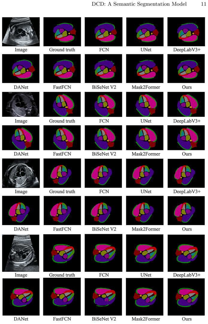
- DCD outperforms all compared baselines on the overall mIoU and on several fine structures, including ribs, spine, and left atrium.
- Because the training data came from multiple ultrasound machine brands, the model is at least exposed to vendor variation within the same imaging protocol.
Reading between the lines
- A natural next test is to measure inter-observer variability: if two sonographers disagree by more than the 1.06-point mIoU gap between DCD and DeepLabv3+, the reported advantage may be within annotation noise.
- The same dense multi-scale plus attention recipe could transfer to other fetal views, such as outflow tracts, where the 13-class labeling would need to be redefined.
- Reporting per-class IoU with confidence intervals across several random seeds would clarify whether the small margins over Mask2Former are stable.
Editorial analysis
A structured set of objections, weighed in public.
Referee Report
Summary. The paper proposes DCD, a DeepLabv3+-based semantic segmentation model for 13 anatomical structures in fetal ultrasound apical four-chamber (A4C) views. The two claimed contributions are replacing ASPP with Dense ASPP and adding CBAM to shallow features. Experiments are carried out on 1,152 images from a single hospital, split 7:2:1 into training, validation, and test sets. The authors report mIoU 76.01 for DCD, compared with 74.95 for DeepLabv3+, 75.55 for Mask2Former, and lower values for four other baselines. They conclude that DCD achieves the best segmentation accuracy and improves segmentation of fine structures such as ribs and spine.
Significance. If the reported results are statistically robust, DCD would be a modest but useful incremental contribution to fetal ultrasound segmentation, one of the few efforts to segment 13 distinct A4C structures across multiple ultrasound vendors. The ablation study is logically ordered and the comparison includes several standard baselines. However, the central 'best accuracy' claim currently rests on a single split with no confidence intervals, significance tests, or repeated runs; the margin over Mask2Former is only 0.46 mIoU, and Mask2Former wins on 5 of 13 classes. The paper also provides no code, no detailed architecture specifications for the Dense ASPP module, and no external validation, all of which limit the reproducibility and generalizability of the conclusions. The significance is therefore conditional: the method is plausible and the experiments are internally consistent, but the headline claim is not yet established at the standard expected for a clinical imaging venue.
major comments (4)
- [Section 4.3, Tables 2 and 3] The central claim that DCD 'achieves the best segmentation accuracy' is not supported by the reported statistics. The test set contains only about 115 images (7:2:1 split of 1,152), and Tables 2 and 3 report single point estimates with no standard deviations, no repeated runs, and no significance tests. DCD's mIoU of 76.01 is only 0.46 points above Mask2Former's 75.55, and Mask2Former is better on 5 of 13 classes (RA 89.18 vs 88.48, RV 81.46 vs 80.66, LA 83.76 vs 83.45, LV 84.16 vs 82.95, VS 72.94 vs 72.47). With this test-set size, the observed differences are within the range of seed-to-seed variation. The authors should either provide repeated-run statistics with confidence intervals and a paired significance test, or soften the claim to 'comparable performance'.
- [Section 4.1] The generalizability claim is unsupported by the data description. All 1,152 images come from a single hospital, and the text states that all images were annotated by an experienced sonographer without reporting inter-observer variability or a second annotation pass. With no external validation cohort and no cross-validation, the 76.01 mIoU may reflect properties of this specific dataset rather than a general capability across gestational ages, pathologies, and ultrasound vendors. The authors claim robustness across 'various challenging imaging conditions' (end of Section 4.4), but this is not demonstrated beyond the internal test set. I recommend adding an external test set or at least k-fold cross-validation plus an annotation-agreement measure.
- [Section 3.2, Eq. (1)] The Dense ASPP module is not specified precisely enough for a reader to reproduce it. Equation (1) writes F_l = f0(Conv_{K,d_l}([F0, F1, ..., F_{l-1}])), but the text does not state how the concatenated feature maps are projected to a common channel dimension, what the output channel count of each dilated branch is, or how the final module output is formed (e.g., concatenation followed by a 1x1 convolution). Since Dense ASPP is one of the two primary architectural contributions, this lack of implementation detail is a load-bearing omission, especially in the absence of released code.
- [Section 4.4] The comparison protocol is underspecified. The text does not state whether all baselines were trained with the same loss function, optimizer, learning-rate schedule, input size, training epochs, or backbone initialization, nor how the final checkpoint was selected (e.g., best validation mIoU or last epoch). Without this information, the reported differences between DCD and the baselines could be due to training details rather than architectural improvements. The authors should describe the common training recipe and ideally report results over multiple seeds for all methods.
minor comments (4)
- [Section 3.3, Eq. (10)] Equation (10) uses δ (ReLU) to compute the spatial attention map, but the text after Eq. (6) defines σ as the Sigmoid function that produces values between 0 and 1. Spatial attention weights in CBAM should be produced by a Sigmoid; Eq. (10) appears to be a typo and should read σ.
- [Tables 2 and 3] The proposed method is called DCD in the text but is labeled 'DC-DeepLabv3+ (Ours)' in Table 3 and 'DCD' in Table 2. Please use one consistent name throughout.
- [Section 3.2] The sentence 'The traditional ASPP has dilation rates of 6, 12, and 18, resulting in receptive fields of 13, 25, and 37' is incomplete because DeepLabv3+ ASPP also includes a 1x1 convolution branch and global average pooling; this description should be updated for accuracy.
- [Throughout] Several typographical issues need editorial cleanup, including 'Equation Eq. (5)', 'in Equation in Eq. (12)', and the incomplete sentence 'the DCD model demonstrates in multi-class anatomical structure segmentation' in Section 5.
Circularity Check
No circularity: DCD's improvements are empirically evaluated on a held-out test split, with no fitted constants or self-citation chains used to manufacture the result.
full rationale
This paper is an empirical architecture-comparison study rather than a derivation-based contribution. The claimed improvement comes from replacing ASPP with Dense ASPP and adding CBAM to shallow features in a DeepLabv3+ encoder-decoder, and the paper reports mIoU on a fixed 70/20/10 split of 1,152 fetal A4C images (Section 4.1). Tables 2 and 3 report test-set mIoU values for the proposed method and baselines, and the ablation study (Table 2) isolates the effect of each module. No fitted parameter is relabeled as a prediction: the model is trained once on the training split and evaluated on held-out images, so the reported numbers are genuine test-set measurements, not quantities forced by construction. The Dense ASPP and CBAM modules are standard components cited from external literature, and no equation in the paper defines the reported mIoU in terms of the loss function, the training data, or the authors' own prior claims. The only self-citation (reference [6], which includes an overlapping author) is used to support a general statement that deep learning has advanced ultrasound segmentation; it is not load-bearing for the central result. Concerns about the small test set, lack of significance testing, or annotation representativeness are legitimate evidence-strength criticisms, but they are not circularity. There is no self-definitional step, no fitted input called a prediction, and no uniqueness theorem or ansatz imported through self-citation. The central claim therefore has independent empirical content, and the circularity score is 0.
Assumptions & free parameters
free parameters (2)
- Dense ASPP dilation rates =
[3, 6, 12, 18]
- CBAM channel reduction ratio r =
not specified
assumptions (5)
- domain assumption Dilated convolutions with dense concatenation expand receptive field and improve multi-scale feature extraction without increasing parameters excessively (Eq. 1).
- domain assumption The private dataset of 1,152 images from one hospital, annotated by experienced sonographers, is representative of fetal A4C ultrasound across vendors and clinical conditions.
- domain assumption Manual annotations by sonographers are treated as ground truth for training and evaluation.
- domain assumption The 7:2:1 train/validation/test split on the internal dataset is a valid estimator of generalization.
- standard math Standard CNN building blocks (Xception, CBAM, decoders, CE plus Dice loss) are taken as given from prior literature.
Cite this review
Pith. "Pith review of DCD: A Semantic Segmentation Model for Fetal Ultrasound Four-Chamber View." pith.science (2026). https://pith.science/paper/U4NTMYW2
@misc{pith2026250608534,
  author       = {Pith},
  title        = {Pith review of: DCD: A Semantic Segmentation Model for Fetal Ultrasound Four-Chamber View},
  year         = {2026},
  howpublished = {\url{https://pith.science/paper/U4NTMYW2}},
  note         = {Machine review of arXiv:2506.08534}
}
read the original abstract
Accurate segmentation of anatomical structures in the apical four-chamber (A4C) view of fetal echocardiography is essential for early diagnosis and prenatal evaluation of congenital heart disease (CHD). However, precise segmentation remains challenging due to ultrasound artifacts, speckle noise, anatomical variability, and boundary ambiguity across different gestational stages. To reduce the workload of sonographers and enhance segmentation accuracy, we propose DCD, an advanced deep learning-based model for automatic segmentation of key anatomical structures in the fetal A4C view. Our model incorporates a Dense Atrous Spatial Pyramid Pooling (Dense ASPP) module, enabling superior multi-scale feature extraction, and a Convolutional Block Attention Module (CBAM) to enhance adaptive feature representation. By effectively capturing both local and global contextual information, DCD achieves precise and robust segmentation, contributing to improved prenatal cardiac assessment.
Figures

Reference graph
Works this paper leans on
-
[1]
In: 2019 Computing in Cardiology (CinC), pp
Vullings, R.: Fetal electrocardiography and deep learning for prenatal detection of congenital heart disease. In: 2019 Computing in Cardiology (CinC), pp. Page–1. IEEE (2019)
work page 2019
-
[2]
Current Pediatric Reviews17(2), 120–126 (2021)
Taksande, A., Jameel, P.Z.: Critical congenital heart disease in neonates: a review article. Current Pediatric Reviews17(2), 120–126 (2021)
work page 2021
-
[3]
Circulation149(13), e997–e1022 (2024) DCD: A Semantic Segmentation Model 13
Sood, E., Newburger, J.W., Anixt, J.S., Cassidy, A.R., Jackson, J.L., Jonas, R.A., Lisanti, A.J., Lopez, K.N., Peyvandi, S., Marino, B.S., et al.: Neurodevelopmental outcomes for individuals with congenital heart disease: updates in neuroprotection, risk-stratification, evaluation, and management: a scientific statement from the american heart association...
work page 2024
-
[4]
IEEE Access9, 54,310–54,324 (2021)
Wang, Y., Ge, X., Ma, H., Qi, S., Zhang, G., Yao, Y.: Deep learning in medical ultrasound image analysis: a review. IEEE Access9, 54,310–54,324 (2021)
work page 2021
-
[5]
Journal of Ultrasound in Medicine26(11), 1601–1610 (2007)
Jeanty, P., Chaoui, R., Tihonenko, I., Grochal, F.: A review of findings in fetal cardiac section drawings: Part 1: The 4-chamber view. Journal of Ultrasound in Medicine26(11), 1601–1610 (2007)
work page 2007
-
[6]
IEEE/ACM Transactions on Computational Biology and Bioinformatics (2022)
Lu, Y., Li, K., Pu, B., Tan, Y., Zhu, N.: A yolox-based deep instance segmenta- tion neural network for cardiac anatomical structures in fetal ultrasound images. IEEE/ACM Transactions on Computational Biology and Bioinformatics (2022)
2022
-
[7]
Expert Systems with Applications263, 125,699 (2025)
Zhou, Z., Lu, Y., Bai, J., Campello, V.M., Feng, F., Lekadir, K.: Segment anything model for fetal head-pubic symphysis segmentation in intrapartum ultrasound im- age analysis. Expert Systems with Applications263, 125,699 (2025)
work page 2025
-
[8]
Computers in Biology and Medicine169, 107,898 (2024)
Chen, G., Tan, G., Duan, M., Pu, B., Luo, H., Li, S., Li, K.: Mlmseg: a multi-view learning model for ultrasound thyroid nodule segmentation. Computers in Biology and Medicine169, 107,898 (2024)
2024
Show all 25 references
-
[9]
Biomedical Signal Processing and Control104, 107,499 (2025)
Wang, Q., Zhao, D., Ma, H., Liu, B.: Fb-zwunet: A deep learning network for corpus callosum segmentation in fetal brain ultrasound images for prenatal diagnostics. Biomedical Signal Processing and Control104, 107,499 (2025)
2025
-
[10]
Neural Computing and Applications pp
Wu, X., Tan, G., Pu, B., Duan, M., Cai, W.: Dh-gac: Deep hierarchical context fu- sion network with modified geodesic active contour for multiple neurofibromatosis segmentation. Neural Computing and Applications pp. 1–16 (2022)
2022
-
[11]
Biomedical Signal Processing and Control103, 107,434 (2025)
Hekal, A.A., Amer, H.M., Moustafa, H.E.D., Elnakib, A.: Automatic measurement of head circumference in fetal ultrasound images using a squeeze atrous pooling unet. Biomedical Signal Processing and Control103, 107,434 (2025)
2025
-
[12]
IEEE Journal of Biomedical and Health Informatics 26(11), 5540–5550 (2022)
Pu, B., Lu, Y., Chen, J., Li, S., Zhu, N., Wei, W., Li, K.: Mobileunet-fpn: A semantic segmentation model for fetal ultrasound four-chamber segmentation in edge computing environments. IEEE Journal of Biomedical and Health Informatics 26(11), 5540–5550 (2022)
2022
-
[13]
IEEE Journal of Biomedical and Health Informatics (2023)
Zhao, L., Tan, G., Pu, B., Wu, Q., Ren, H., Li, K.: Transfsm: Fetal anatomy segmentation and biometric measurement in ultrasound images using a hybrid transformer. IEEE Journal of Biomedical and Health Informatics (2023)
2023
-
[14]
Acta electronica sinica50(10), 1–10 (2022)
Zhang, S., Peng, Z., Li, H.: Sau-net: Medical image segmentation method based on u-net and self-attention. Acta electronica sinica50(10), 1–10 (2022)
2022
-
[15]
In: 2023 45th Annual International Conference of the IEEE Engineering in Medicine & Biology Society (EMBC), pp
Rajamani, S.T., Rajamani, K., Schuller, B.W.: A novel and simple approach to regularise attention frameworks and its efficacy in segmentation. In: 2023 45th Annual International Conference of the IEEE Engineering in Medicine & Biology Society (EMBC), pp. 1–4. IEEE (2023)
2023
-
[16]
IEEE Signal Processing Letters (2024)
Zhou, W., Guan, G., Cui, W., Yi, Y.: Biasam: Bidirectional-attention guided seg- ment anything model for very few-shot medical image segmentation. IEEE Signal Processing Letters (2024)
2024
-
[17]
IET Image Processing18(1), 77–87 (2024)
Tong, S., Zuo, Z., Liu, Z., Sun, D., Zhou, T.: Hybrid attention mechanism of feature fusion for medical image segmentation. IET Image Processing18(1), 77–87 (2024)
2024
-
[18]
IEEE Access (2025)
Cao, Y., Cheng, Y.: Sacu-net: Shape-aware u-net for biomedical image segmenta- tion with attention mechanism and context extraction. IEEE Access (2025)
2025
-
[19]
In: Proceedings of the European conference on computer vision (ECCV), pp
Chen, L.C., Zhu, Y., Papandreou, G., Schroff, F., Adam, H.: Encoder-decoder with atrous separable convolution for semantic image segmentation. In: Proceedings of the European conference on computer vision (ECCV), pp. 801–818 (2018)
2018
-
[20]
In: Medical image computing and computer-assisted intervention–MICCAI 2015: 18th international conference, Munich, Germany, Oc- tober 5-9, 2015, proceedings, part III 18, pp
Ronneberger, O., Fischer, P., Brox, T.: U-net: Convolutional networks for biomed- ical image segmentation. In: Medical image computing and computer-assisted intervention–MICCAI 2015: 18th international conference, Munich, Germany, Oc- tober 5-9, 2015, proceedings, part III 18,...
2015
-
[21]
In: Proceedings of the IEEE/CVF conference on computer vision and pattern recognition, pp
Cheng, B., Misra, I., Schwing, A.G., Kirillov, A., Girdhar, R.: Masked-attention mask transformer for universal image segmentation. In: Proceedings of the IEEE/CVF conference on computer vision and pattern recognition, pp. 1290–1299 (2022)
2022
-
[22]
In: Proceedings of the IEEE conference on computer vision and pattern recognition, pp
Long, J., Shelhamer, E., Darrell, T.: Fully convolutional networks for semantic segmentation. In: Proceedings of the IEEE conference on computer vision and pattern recognition, pp. 3431–3440 (2015)
2015
-
[23]
In: Proceedings of the IEEE/CVF conference on computer vision and pattern recognition, pp
Fu, J., Liu, J., Tian, H., Li, Y., Bao, Y., Fang, Z., Lu, H.: Dual attention network for scene segmentation. In: Proceedings of the IEEE/CVF conference on computer vision and pattern recognition, pp. 3146–3154 (2019)
2019
-
[24]
arXiv preprint arXiv:1903.11816 (2019)
Wu, H., Zhang, J., Huang, K., Liang, K., Yu, Y.: Fastfcn: Rethinking dilated convo- lution in the backbone for semantic segmentation. arXiv preprint arXiv:1903.11816 (2019)
2019 arXiv
-
[25]
International journal of computer vision129, 3051–3068 (2021)
Yu, C., Gao, C., Wang, J., Yu, G., Shen, C., Sang, N.: Bisenet v2: Bilateral network with guided aggregation for real-time semantic segmentation. International journal of computer vision129, 3051–3068 (2021)
2021
Reviewed August 7, 2026 · model on record in the stance chip above.
Discussion (0). Sign in to comment.