REVIEW 3 major objections 5 minor 76 references
Sparse Autoencoders Bridge The Deep Learning Model and The Brain
T0 review · 3 major / 5 minor · reviewed 2026-08-07 · deepseek-v4-flash
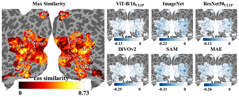
Pith's one-line read Sparse autoencoder units trained without brain data match human visual cortex fMRI responses, with cosine similarity up to 0.76.
desk verdict Genuinely new SAE-to-fMRI alignment method whose headline 0.76 similarity lacks any null baseline, so the core claim overreaches but the tool is worth referee time. read the letter →
The pith
A machine-rendered reading of the paper's core claim, the machinery that carries it, and where it could break.
The reading
What carries the argument
The central object is a sparse autoencoder trained per layer with tied encoder-decoder weights, reconstructing activations as sparse positive combinations of interpretable feature units: $Z = \mathrm{ReLU}(WA+b)$, with loss $\|\hat{A}-A\|_2^2 + \alpha\|Z\|_1$. The load-bearing comparison is the cosine similarity between z-scored SAE unit activations and z-scored voxel fMRI responses across the shared image set, $M^{(j)}_{\mathrm{simi}} = \cos(B^{\top}/\|B\|_2, Z^{(j)}_{\mathrm{zscore}}/\|Z^{(j)}_{\mathrm{zscore}}\|_2)$, normalized over images. This similarity matrix drives two constructions: a voxel dictionary $D^{(j)}[x] = W[\arg\max_y M_{x,y}]$ assigning each voxel its most similar feature, and a voxel-wise layer alignment $C[x] = \arg\max_j \max_y M^{(j)}_{x,y}$. Projecting dictionary activations $S^{(j)} = A^{(j)}D^{(j)}$ onto the cortex lets the brain act as a spatial template for tracing information flow through the model.
What would settle it
A permutation test that shuffles the voxel-unit correspondence, or replaces trained SAE features with random features, and recomputes the maximum cosine similarities; if random features still yield maxima around 0.7, the claimed strong correspondence would reflect shared stimulus statistics rather than learned representations.
Extended reading notes
Core claim
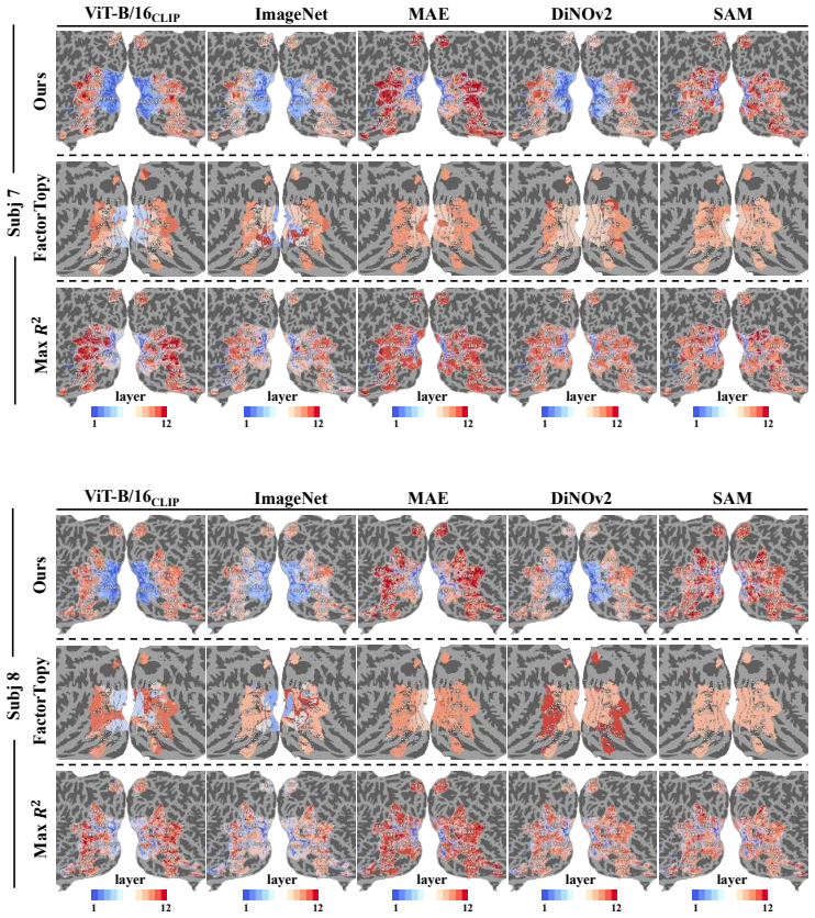
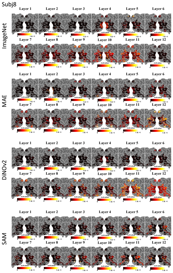
Training layer-wise sparse autoencoders on model activations from natural images yields features whose z-scored activation patterns correlate strongly with z-scored fMRI responses of individual voxels, with cosine similarity up to 0.76. The most similar SAE feature per voxel forms a 'voxel dictionary' that preserves ROI functional structure, and the best-matching layer per voxel reconstructs the classic hierarchy, with early layers aligning to early visual cortex and deeper layers to higher-level visual areas. For CLIP ViT-B/16, the voxel-dictionary activation path suggests low-level features are transformed into abstract semantic information in early layers and later reconstructed alongside high-level information. The paper claims this constitutes a direct, downstream-task-free alignment between deep neural network representations and the human visual cortex.
Load-bearing premise
The load-bearing premise is that cosine similarity between activation profiles in response to the same images measures functional correspondence; the paper reports maxima and averages without a null distribution, so chance alignment due to shared image statistics is not ruled out.
Editorial extensions
If this is right
- SAE features learned from images alone can be matched to individual fMRI voxels without any supervised brain encoder, providing a training-free voxel-wise alignment procedure.
- The voxel dictionary inherits known ROI structure, so model features can be probed for face, body, scene, word, and food selectivity in a data-driven way.
- Layer-to-cortex mapping reproduces the ventral-stream hierarchy, with early layers near V1 and later layers near high-level visual areas, across several architectures and training objectives.
- For CLIP ViT-B/16, early layers convert low-level information into abstract semantic content while later layers re-integrate low-level details, a pattern the paper links to the residual stream.
- The framework extends to any vision model with per-layer activations, making it a general tool for comparing model internals against human visual cortex.
Reading between the lines
- The reported 0.76 maximum is selected across thousands of voxel-unit-layer comparisons, and the paper gives no permutation or null baseline; a natural test is whether randomly initialized SAEs or shuffled voxel labels produce similar maxima driven by shared stimulus statistics.
- If the alignment survives such nulls, the method becomes a zero-shot cortical mapper: any new model layer could be localized on the cortex without training a brain encoder, as long as paired image-fMRI data are available.
- The ROI-consistent selectivity of SAE units could be turned into a generative stimulus-search tool, using units tied to face, scene, or word regions to synthesize images and probe cortical selectivity directly.
- Applying the same framework to recurrent or multi-scale architectures might reveal whether the bidirectional low- and high-level information flow observed in CLIP ViT-B/16 is specific to transformer residual streams or a general property of deep visual processing.
Editorial analysis
A structured set of objections, weighed in public.
Referee Report
Summary. The paper introduces SAE-BrainMap, a framework that trains sparse autoencoders (SAEs) on the layer activations of several vision models (ViT-B/16 CLIP, ImageNet, MAE, DiNOv2, SAM, ResNet50 CLIP) without any brain data, and then correlates the SAE unit activation profiles with voxel-level fMRI responses from the Natural Scenes Dataset using cosine similarity. The authors report a maximum similarity of 0.76, construct a voxel dictionary by assigning to each voxel the most similar SAE unit, and use this dictionary to argue that SAE units preserve the functional structure of visual ROIs, to establish a layer-wise brain-model alignment, and to visualize model information processing on the cortical surface. The main claims are that SAE features provide a direct, downstream-task-free bridge between deep network representations and the human ventral visual pathway.
Significance. If the central similarity result is statistically validated, the paper offers a conceptually attractive approach: features extracted by SAEs trained purely on model activations, with no supervised brain-encoding objective, might align voxel-wise with cortical fMRI responses. The work is also broad in scope, covering multiple architectures, public data, and several analyses, and the SAEs themselves are trained without brain data, which avoids the most obvious form of circularity. However, the key quantitative claims currently lack the statistical baselines needed to distinguish genuine structure from shared stimulus statistics and selection effects. The paper is therefore potentially interesting and publishable in a revised form, but the headline 'strong activation correspondence' is not yet supported by the evidence as presented.
major comments (3)
- [Section 3.2, Eq. (4); Table 1] The headline maximum similarity of 0.76 is the maximum over subjects, voxels, SAE units, layers, and models, and Table 1 reports no null distribution, p-values, or error bars for either the maximum or the per-voxel average (~0.26). Because natural images share low-level statistics (luminance, edges, spatial frequency), positive cosine similarities between model features and voxel responses are expected even with no true model-brain correspondence, and the maximum of an enormous search is especially uninterpretable. The paper's own NeurIPS checklist states that statistical significance is illustrated in Tables 1-3, but those tables contain no error bars or significance tests. The authors should provide a permutation or surrogate baseline (e.g., shuffling image labels, using units from untrained models, or an analytic null for the maximum of many correlations) and report selection-corrected statistics; without this, the abstract's 'strong activation correspondence' and 'direct bridge' overstate what Eq. (4) demonstrates.
- [Section 4.3, Eq. (6); Table 2] The claim that SAE units preserve the functional structure of ROIs is validated by computing an RSA score between the voxel dictionary and 'brain encoder weights' that were themselves trained to predict fMRI responses from the same model activations (Appendix C). Since the voxel dictionary is constructed by an argmax over the same similarity matrix used throughout the paper, and since both the dictionary and the ground-truth encoder weights derive from the same model activation statistics, the reported RSA values (highest 0.516) may reflect shared ancestry rather than an independent correspondence between SAE features and brain function. The authors should validate the dictionary against held-out fMRI data, or against an independently defined functional atlas, and should also report a null distribution for the RSA scores.
- [Section 4.4, Eq. (5); Table 3] The brain-model layer alignment C is obtained by taking an argmax over layers of the per-layer maximum over SAE units, and the 'slope' metric is computed on this selected alignment without any null model. The similarities to FactorTopy and Max R2 are presented without confidence intervals, and the text states that the results are 'well correlated with previous methods' without a statistical comparison. The authors should report bootstrap intervals across subjects and voxels, and compare the slope and alignment similarity against a permutation baseline in which layer labels are shuffled, before claiming a 'clear and consistent hierarchical correspondence' between model layers and the human visual cortex.
minor comments (5)
- [Throughout] There are numerous typos and grammatical errors that should be corrected: 'Relate Work' (Section 2), 'constrainted' (Introduction), 'dosen't' (checklist item 3), 'V oxels' (multiple occurrences), 'V oxel Dictionary' (figure captions), and inconsistent capitalization throughout.
- [Section 3.1, Eqs. (1)-(4)] The notation is inconsistent: Eq. (1) defines Z with a ReLU nonlinearity, but Eq. (4) computes Z_zscore = Zscore(W A + b) without the ReLU. The definition of the norm in Eq. (4) is also ambiguous (Frobenius norm vs. column-wise norms), and the matrix dimensions in the cosine expression should be stated explicitly.
- [Section 4.3, Figure 5 and Figure 4] The text refers to 'Figure 4' for the unit selectivity visualization and for the voxel dictionary / brain encoder similarity matrix, but the relevant figures appear to be Figures 4 and 5 in the paper; the references should be corrected and the figures should be cross-checked.
- [Section 4.4, Table 3] The text says 'Table 2 presents the cosine similarity between our alignment results and two voxel-wise mapping methods,' but the table referenced is Table 3. The similarity columns are also not clearly defined: the reader should be told whether the values are correlations over voxel labels, IoU-like scores, or something else.
- [Section 5, Limitations] The limitations section mentions only the fixed SAE hyperparameters. It does not mention the lack of statistical baselines, the selection over many comparisons, or the fact that the brain-encoder validation is model-dependent. These are important limitations that should be acknowledged.
Circularity Check
Partial circularity: the ROI-preservation claim and the 'ground-truth' brain-encoder RSA validation reuse the same similarity/fitted data; the headline 0.76 SAE-fMRI similarity itself is not circular.
-
self definitional
[Section 3.2 Equation 6 and Section 4.3]
"D(j)[x] = W[argmax_y M_{x,y}], x in {0,1,...,v} (Eq. 6). ... 'By computing the representation correlation matrix based on pairwise cosine similarities, we find that the voxel dictionary preserves functional separability across ROIs and shares a high RSA score with the ground truth (highest: 0.516, see Table 2).'"
The voxel dictionary is defined by assigning to each voxel the SAE unit whose activation profile is most similar to that voxel's fMRI profile, using the same cosine-similarity matrix M computed from B and Z. Voxels inside a predefined ROI share similar fMRI response profiles by functional definition, so the argmax assignment tends to select the same or similar SAE dictionary rows for voxels in the same ROI. The reported pairwise similarity structure of D, and hence the 'preserved functional separability across ROIs,' is inherited from the construction input B rather than being an independent confirmation that SAE units discovered the brain's ROI organization.
-
fitted input called prediction
[Table 2 caption / Section 4.3 / Appendix C]
"'RSA Similarity Score between the brain encoder weights of the last layer and the voxel dictionary. This indicates that SAE units maintain a strong structural correspondence with the brain encoder weights, which are regarded as the ground truth representation of cortical functional structure.' Appendix C: 'training a linear layer with equation: A(j)W + b = B'."
The 'ground truth' brain encoder weights are themselves fitted by linear regression to predict the same voxel fMRI responses B from the same model activations. The voxel dictionary is built from M, the voxel-unit similarity matrix computed from the same B and model activations. Both sides of the RSA comparison therefore share their target data and are aligned to B; the high RSA score is a consistency check between two quantities fitted to the same signal, not validation against an independent ground truth. Naming the fitted encoder weights 'ground truth' makes the claimed structural correspondence partially circular.
full rationale
The paper's central alignment step is not circular: layer-wise SAEs are trained on ImageNet activations (Equations 1-3) without any brain data, so the reported cosine similarities between SAE unit activation profiles and voxel fMRI profiles (Equation 4) are a genuine out-of-sample comparison. The core 0.76 maximum similarity is not forced by construction, though it lacks a null baseline. However, two load-bearing validation claims reduce to their own inputs. First, the voxel dictionary D(j)[x] = W[argmax_y M_{x,y}] is defined by the very similarity matrix M that already encodes each voxel's fMRI profile; voxels in the same predefined ROI share fMRI response structure, so the dictionary's within-ROI pairwise similarity (the 'preserved functional separability' in Section 4.3) is a restatement of the construction rather than an independent discovery. Second, the RSA validation in Table 2 treats a linear brain encoder fitted to the same B (Appendix C: A(j)W + b = B) as 'ground truth'; both the dictionary and the encoder weights are derived from the same model-brain association data, so their high RSA is a shared-input consistency check, not external confirmation. The lack of a permutation null for the maximum 0.76 is a statistical-significance concern rather than a circularity, and is not scored here. Overall, the 'direct bridge' similarity claim is non-circular, but the ROI-preservation and ground-truth correspondence claims are partially circular, giving a score of 6.
Assumptions & free parameters
free parameters (4)
- SAE expansion factor R =
16
- SAE sparsity coefficient alpha =
0.00086
- SAE training schedule =
3 epochs, lr 5e-5, AdamW
- brain encoder linear weights =
per model, per layer, per subject
assumptions (6)
- domain assumption SAE features are interpretable, monosemantic units that capture the model's functional processing.
- domain assumption fMRI z-scored voxel responses are stable, meaningful measures of cortical function.
- domain assumption Cosine similarity between per-image activation vectors is a valid measure of brain-model correspondence.
- domain assumption The CLS token (or averaged patch tokens for SAM) is a sufficient model representation for voxel-level alignment.
- ad hoc to paper Brain encoder weights constitute ground truth brain function.
- ad hoc to paper Voxel dictionary argmax assignment preserves ROI functional structure.
invented entities (1)
-
Voxel dictionary D^(j)
Cite this review
Pith. "Pith review of Sparse Autoencoders Bridge The Deep Learning Model and The Brain." pith.science (2026). https://pith.science/paper/W4KJJDZD
@misc{pith2026250611123,
  author       = {Pith},
  title        = {Pith review of: Sparse Autoencoders Bridge The Deep Learning Model and The Brain},
  year         = {2026},
  howpublished = {\url{https://pith.science/paper/W4KJJDZD}},
  note         = {Machine review of arXiv:2506.11123}
}
abstract
We present SAE-BrainMap, a novel framework that directly aligns deep learning visual model representations with voxel-level fMRI responses using sparse autoencoders (SAEs). First, we train layer-wise SAEs on model activations and compute the correlations between SAE unit activations and cortical fMRI signals elicited by the same natural image stimuli with cosine similarity, revealing strong activation correspondence (maximum similarity up to 0.76). Depending on this alignment, we construct a voxel dictionary by optimally assigning the most similar SAE feature to each voxel, demonstrating that SAE units preserve the functional structure of predefined regions of interest (ROIs) and exhibit ROI-consistent selectivity. Finally, we establish fine-grained hierarchical mapping between model layers and the human ventral visual pathway, also by projecting voxel dictionary activations onto individual cortical surfaces, we visualize the dynamic transformation of the visual information in deep learning models. It is found that ViT-B/16$_{CLIP}$ tends to utilize low-level information to generate high-level semantic information in the early layers and reconstructs the low-dimension information later. Our results establish a direct, downstream-task-free bridge between deep neural networks and human visual cortex, offering new insights into model interpretability.
Figures


Figures from the paper (37 more)






Reference graph
Works this paper leans on
-
[1]
James J. DiCarlo and David D. Cox. Untangling invariant object recognition. Trends in Cognitive Sciences, 11(8):333–341, August 2007. ISSN 13646613. doi: 10.1016/j.tics.2007.06.010
-
[2]
Brain Decodes Deep Nets, March 2024
Huzheng Yang, James Gee, and Jianbo Shi. Brain Decodes Deep Nets, March 2024
work page 2024
-
[3]
David D. Coggan, Luke A. Allen, Oliver R. H. Farrar, et al. Differences in selectivity to natural images in early visual areas (v1–v3). Scientific Reports, 7:2444, 2017. doi: 10.1038/s41598-017-02569-4
-
[4]
CLIP-Dissect: Automatic Description of Neuron Representations in Deep Vision Networks, June 2023
Tuomas Oikarinen and Tsui-Wei Weng. CLIP-Dissect: Automatic Description of Neuron Representations in Deep Vision Networks, June 2023
work page 2023
-
[5]
Byun, Zifan Wang, Alex Mallen, Steven Basart, Sanmi Koyejo, Dawn Song, Matt Fredrikson, J
Andy Zou, Long Phan, Sarah Chen, James Campbell, Phillip Guo, Richard Ren, Alexander Pan, Xuwang Yin, Mantas Mazeika, Ann-Kathrin Dombrowski, Shashwat Goel, Nathaniel Li, Michael J. Byun, Zifan Wang, Alex Mallen, Steven Basart, Sanmi Koyejo, Dawn Song, Matt Fredrikson, J. Zico Kolter, and Dan Hendrycks. Representation Engineering: A Top-Down Approach to A...
work page 2023
-
[6]
Diego García Cerdas, Christina Sartzetaki, Magnus Petersen, Gemma Roig, Pascal Mettes, and Iris Groen. BrainACTIV: Identifying visuo-semantic properties driving cortical selectivity using diffusion-based image manipulation, October 2024
work page 2024
-
[8]
Yaoda Xu and Maryam Vaziri-Pashkam. Limits to visual representational correspondence between convolutional neural networks and the human brain. Nature Communications, 12(1):2065, April 2021. ISSN 2041-1723. doi: 10.1038/s41467-021-22244-7
-
[9]
Seyed-Mahdi Khaligh-Razavi, Linda Henriksson, Kendrick Kay, and Nikolaus Kriegeskorte. Fixed versus mixed RSA: Explaining visual representations by fixed and mixed feature sets from shallow and deep computational models. Journal of Mathematical Psychology, 76:184–197, February 2017. ISSN 0022-2496. doi: 10.1016/j.jmp.2016.10.007
Show all 76 references
-
[10]
Neural tuning and representational geometry
Nikolaus Kriegeskorte and Xue-Xin Wei. Neural tuning and representational geometry. Nature Re- views Neuroscience, 22(11):703–718, November 2021. ISSN 1471-003X, 1471-0048. doi: 10.1038/ s41583-021-00502-3
2021
-
[12]
Luo, Margaret M
Andrew F. Luo, Margaret M. Henderson, Michael J. Tarr, and Leila Wehbe. BrainSCUBA: Fine-Grained Natural Language Captions of Visual Cortex Selectivity, May 2024
2024
-
[13]
Luo, Jacob Yeung, Rushikesh Zawar, Shaurya Dewan, Margaret M
Andrew F. Luo, Jacob Yeung, Rushikesh Zawar, Shaurya Dewan, Margaret M. Henderson, Leila Wehbe, and Michael J. Tarr. Brain Mapping with Dense Features: Grounding Cortical Semantic Selectivity in Natural Images With Vision Transformers, October 2024
2024
-
[14]
Wang, Kendrick Kay, Thomas Naselaris, Michael J
Aria Y . Wang, Kendrick Kay, Thomas Naselaris, Michael J. Tarr, and Leila Wehbe. Better models of human high-level visual cortex emerge from natural language supervision with a large and diverse dataset. Nature Machine Intelligence, 5(12):1415–1426, November 2023. ISSN 2522-58...
2023
-
[15]
Brain Diffusion for Visual Exploration: Cortical Discovery using Large Scale Generative Models
Andrew F Luo, Margaret M Henderson, Leila Wehbe, and Michael J Tarr. Brain Diffusion for Visual Exploration: Cortical Discovery using Large Scale Generative Models. 10
-
[16]
Do Vision Transformers See Like Convolutional Neural Networks? In Advances in Neural Information Processing Systems, volume 34, pages 12116–12128
Maithra Raghu, Thomas Unterthiner, Simon Kornblith, Chiyuan Zhang, and Alexey Dosovitskiy. Do Vision Transformers See Like Convolutional Neural Networks? In Advances in Neural Information Processing Systems, volume 34, pages 12116–12128. Curran Associates, Inc., 2021
2021
-
[17]
CLIP-MSM: A Multi-Semantic Mapping Brain Representation for Human High-Level Visual Cortex
Guoyuan Yang, Mufan Xue, Ziming Mao, Haofang Zheng, Jia Xu, Dabin Sheng, Ruotian Sun, Ruoqi Yang, and Xuesong Li. CLIP-MSM: A Multi-Semantic Mapping Brain Representation for Human High-Level Visual Cortex. Proceedings of the AAAI Conference on Artificial Intelligence, 39(9):91...
-
[18]
Sparse Autoencoders Find Highly Interpretable Features in Language Models, October 2023
Hoagy Cunningham, Aidan Ewart, Logan Riggs, Robert Huben, and Lee Sharkey. Sparse Autoencoders Find Highly Interpretable Features in Language Models, October 2023
2023
-
[19]
Michaud, Yonatan Belinkov, David Bau, and Aaron Mueller
Samuel Marks, Can Rager, Eric J. Michaud, Yonatan Belinkov, David Bau, and Aaron Mueller. Sparse Feature Circuits: Discovering and Editing Interpretable Causal Graphs in Language Models, March 2025
2025
-
[20]
Allen, Ghislain St-Yves, Yihan Wu, Jesse L
Emily J. Allen, Ghislain St-Yves, Yihan Wu, Jesse L. Breedlove, Jacob S. Prince, Logan T. Dowdle, Matthias Nau, Brad Caron, Franco Pestilli, Ian Charest, J. Benjamin Hutchinson, Thomas Naselaris, and Kendrick Kay. A massive 7T fMRI dataset to bridge cognitive neuroscience and ...
2022 doi
-
[21]
Sparse Autoencoders Do Not Find Canonical Units of Analysis, February 2025
Patrick Leask, Bart Bussmann, Michael Pearce, Joseph Bloom, Curt Tigges, Noura Al Moubayed, Lee Sharkey, and Neel Nanda. Sparse Autoencoders Do Not Find Canonical Units of Analysis, February 2025
2025
-
[22]
Scaling and evaluating sparse autoencoders, June 2024
Leo Gao, Tom Dupré la Tour, Henk Tillman, Gabriel Goh, Rajan Troll, Alec Radford, Ilya Sutskever, Jan Leike, and Jeffrey Wu. Scaling and evaluating sparse autoencoders, June 2024
2024
-
[23]
Jumping Ahead: Improving Reconstruction Fidelity with JumpReLU Sparse Autoencoders, August 2024
Senthooran Rajamanoharan, Tom Lieberum, Nicolas Sonnerat, Arthur Conmy, Vikrant Varma, János Kramár, and Neel Nanda. Jumping Ahead: Improving Reconstruction Fidelity with JumpReLU Sparse Autoencoders, August 2024
2024
-
[24]
Prince, Matthew Kowal, Victor Boutin, Isabel Papadimitriou, Binxu Wang, Martin Wattenberg, Demba Ba, and Talia Konkle
Thomas Fel, Ekdeep Singh Lubana, Jacob S. Prince, Matthew Kowal, Victor Boutin, Isabel Papadimitriou, Binxu Wang, Martin Wattenberg, Demba Ba, and Talia Konkle. Archetypal SAE: Adaptive and Stable Dictionary Learning for Concept Extraction in Large Vision Models, February 2025
2025
-
[25]
Universal Sparse Autoencoders: Interpretable Cross-Model Concept Alignment, February 2025
Harrish Thasarathan, Julian Forsyth, Thomas Fel, Matthew Kowal, and Konstantinos Derpanis. Universal Sparse Autoencoders: Interpretable Cross-Model Concept Alignment, February 2025
2025
-
[26]
Interpreting CLIP with Hierarchical Sparse Autoencoders, February 2025
Vladimir Zaigrajew, Hubert Baniecki, and Przemyslaw Biecek. Interpreting CLIP with Hierarchical Sparse Autoencoders, February 2025
2025
-
[27]
Sparse Autoencoder as a Zero-Shot Classifier for Concept Erasing in Text-to-Image Diffusion Models, March 2025
Zhihua Tian, Sirun Nan, Ming Xu, Shengfang Zhai, Wenjie Qu, Jian Liu, Kui Ren, Ruoxi Jia, and Jiaheng Zhang. Sparse Autoencoder as a Zero-Shot Classifier for Concept Erasing in Text-to-Image Diffusion Models, March 2025
2025
-
[28]
The Missing Curve Detectors of InceptionV1: Applying Sparse Autoencoders to InceptionV1 Early Vision, September 2024
Liv Gorton. The Missing Curve Detectors of InceptionV1: Applying Sparse Autoencoders to InceptionV1 Early Vision, September 2024
2024
-
[29]
Toy Models of Superposition, September 2022
Nelson Elhage, Tristan Hume, Catherine Olsson, Nicholas Schiefer, Tom Henighan, Shauna Kravec, Zac Hatfield-Dodds, Robert Lasenby, Dawn Drain, Carol Chen, Roger Grosse, Sam McCandlish, Jared Kaplan, Dario Amodei, Martin Wattenberg, and Christopher Olah. Toy Models of Superposi...
2022
-
[30]
[Interim research report] Taking features out of superposition with sparse autoencoders
Lee Sharkey, Dan Braun, and beren. [Interim research report] Taking features out of superposition with sparse autoencoders. December 2022
2022
-
[31]
Olshausen, and Yann LeCun
Zeyu Yun, Yubei Chen, Bruno A. Olshausen, and Yann LeCun. Transformer visualization via dictionary learning: Contextualized embedding as a linear superposition of transformer factors, April 2023
2023
-
[32]
A Survey on Sparse Autoencoders: Interpreting the Internal Mechanisms of Large Language Models, March 2025
Dong Shu, Xuansheng Wu, Haiyan Zhao, Daking Rai, Ziyu Yao, Ninghao Liu, and Mengnan Du. A Survey on Sparse Autoencoders: Interpreting the Internal Mechanisms of Large Language Models, March 2025
2025
-
[34]
A Convolutional Neural Network Interpretable Framework for Human Ventral Visual Pathway Representation
Mufan Xue, Xinyu Wu, Jinlong Li, Xuesong Li, and Guoyuan Yang. A Convolutional Neural Network Interpretable Framework for Human Ventral Visual Pathway Representation. Proceedings of the AAAI Conference on Artificial Intelligence, 38(6):6413–6421, March 2024. ISSN 2374-3468. do...
2024 doi
-
[35]
Goodale, John Paul Meenan, Heinrich H
Melvyn A. Goodale, John Paul Meenan, Heinrich H. Bülthoff, David A. Nicolle, Kelly J. Murphy, and Carolynn I. Racicot. Separate neural pathways for the visual analysis of object shape in perception and prehension. Current Biology, 4(7):604–610, July 1994. ISSN 0960-9822. doi: ...
1994 doi
-
[36]
Kalanit Grill-Spector and Kevin S. Weiner. The functional architecture of the ventral temporal cortex and its role in categorization. Nature Reviews Neuroscience, 15(8):536–548, August 2014. ISSN 1471-0048. doi: 10.1038/nrn3747
2014 doi
-
[37]
Selectivity for the shape, size, and orientation of objects for grasping in neurons of monkey parietal area aip
Akira Murata, Vittorio Gallese, Giuseppe Luppino, Masakazu Kaseda, and Hideo Sakata. Selectivity for the shape, size, and orientation of objects for grasping in neurons of monkey parietal area aip. Journal of Neurophysiology, 83(5):2580–2601, 2000. doi: 10.1152/jn.2000.83.5.2580
-
[38]
Dumoulin and Brian A
Serge O. Dumoulin and Brian A. Wandell. Population receptive field estimates in human visual cortex. NeuroImage, 39(2):647–660, January 2008. ISSN 1053-8119. doi: 10.1016/j.neuroimage.2007.09.034
2008 doi
-
[39]
D. H. Hubel and T. N. Wiesel. Receptive fields, binocular interaction and functional architecture in the cat’s visual cortex. The Journal of Physiology, 160(1):106–154.2, January 1962. ISSN 0022-3751
1962
-
[40]
Demb, Valerio Mante, David J
Matteo Carandini, Jonathan B. Demb, Valerio Mante, David J. Tolhurst, Yang Dan, Bruno A. Olshausen, Jack L. Gallant, and Nicole C. Rust. Do we know what the early visual system does? Journal of Neuroscience, 25(46):10577–10597, 2005. ISSN 0270-6474. doi: 10.1523/JNEUROSCI.3726-05.2005
2005 doi
-
[41]
Levitt, David C
Jonathan B. Levitt, David C. Kiper, and J. Anthony Movshon. Receptive fields and functional architecture of macaque v2. Journal of Neurophysiology, 71(6):2517–2542, June 1994. doi: 10.1152/jn.1994.71.6.2517
1994 doi
-
[42]
Ziemba, David J
Jeremy Freeman, Corey M. Ziemba, David J. Heeger, Eero P. Simoncelli, and J. Anthony Movshon. A functional and perceptual signature of the second visual area in primates. Nature Neuroscience, 16(7): 974–981, July 2013. ISSN 1546-1726. doi: 10.1038/nn.3402
2013 doi
-
[43]
Nancy Kanwisher, Josh McDermott, and Marvin M. Chun. The fusiform face area: A module in human extrastriate cortex specialized for face perception. Journal of Neuroscience, 17(11):4302–4311, 1997. ISSN 0270-6474. doi: 10.1523/JNEUROSCI.17-11-04302.1997
1997
-
[44]
Epstein and N
R. Epstein and N. Kanwisher. A cortical representation of the local visual environment. Nature, 392: 598–601, 1998. doi: 10.1038/33402
1998 doi
-
[45]
P. E. Downing, Y . Jiang, M. Shuman, and N. Kanwisher. A cortical area selective for visual processing of the human body. Science, 293(5539):2470–2473, September 2001. doi: 10.1126/science.1063414
2001 doi
-
[46]
The visual word form area: spatial and temporal characterization of an initial stage of reading in normal subjects and posterior split-brain patients
Laurent Cohen, Stanislas Dehaene, Lionel Naccache, Stéphane Lehéricy, Ghislaine Dehaene-Lambertz, Marie-Anne Hénaff, and Franck Michel. The visual word form area: spatial and temporal characterization of an initial stage of reading in normal subjects and posterior split-brain ...
-
[47]
Apurva Ratan Murty, and Nancy Kanwisher
Meenakshi Khosla, N. Apurva Ratan Murty, and Nancy Kanwisher. A highly selective response to food in human visual cortex revealed by hypothesis-free voxel decomposition. Current Biology, 32(19): 4159–4171.e9, 2022. ISSN 0960-9822. doi: 10.1016/j.cub.2022.08.009
2022 doi
-
[48]
Prince, Kendrick N
Colin Conwell, Jacob S. Prince, Kendrick N. Kay, George A. Alvarez, and Talia Konkle. A large-scale examination of inductive biases shaping high-level visual representation in brains and machines. Nature Communications, 15(1):9383, October 2024. ISSN 2041-1723. doi: 10.1038/s4...
2024 doi
-
[49]
Bandettini
Nikolaus Kriegeskorte, Marieke Mur, and Peter A. Bandettini. Representational similarity analysis - connecting the branches of systems neuroscience. Frontiers in Systems Neuroscience, 2, November 2008. ISSN 1662-5137. doi: 10.3389/neuro.06.004.2008
2008 doi
-
[50]
Lawrence Zitnick, and Piotr Dollár
Tsung-Yi Lin, Michael Maire, Serge Belongie, Lubomir Bourdev, Ross Girshick, James Hays, Pietro Perona, Deva Ramanan, C. Lawrence Zitnick, and Piotr Dollár. Microsoft COCO: Common Objects in Context, February 2015
2015
-
[51]
Gao, Alexander G
James S. Gao, Alexander G. Huth, Mark D. Lescroart, and Jack L. Gallant. Pycortex: An interactive surface visualizer for fMRI. Frontiers in Neuroinformatics, 9, September 2015. ISSN 1662-5196. doi: 10.3389/fninf.2015.00023
2015
-
[52]
ImageNet: A Large-Scale Hierarchical Image Database
Jia Deng, Wei Dong, Richard Socher, Li-Jia Li, Kai Li, and Li Fei-Fei. ImageNet: A Large-Scale Hierarchical Image Database. 12
-
[53]
Learning Transferable Visual Models From Natural Language Supervision
Alec Radford, Jong Wook Kim, Chris Hallacy, Aditya Ramesh, Gabriel Goh, Sandhini Agarwal, Girish Sastry, Amanda Askell, Pamela Mishkin, Jack Clark, Gretchen Krueger, and Ilya Sutskever. Learning Transferable Visual Models From Natural Language Supervision. In Proceedings of th...
2021
-
[54]
Deep Residual Learning for Image Recognition, December 2015
Kaiming He, Xiangyu Zhang, Shaoqing Ren, and Jian Sun. Deep Residual Learning for Image Recognition, December 2015
2015
-
[55]
Masked Autoencoders Are Scalable Vision Learners, December 2021
Kaiming He, Xinlei Chen, Saining Xie, Yanghao Li, Piotr Dollár, and Ross Girshick. Masked Autoencoders Are Scalable Vision Learners, December 2021
2021
-
[56]
DINOv2: Learning Robust Visual Features without Supervision, February 2024
Maxime Oquab, Timothée Darcet, Théo Moutakanni, Huy V o, Marc Szafraniec, Vasil Khalidov, Pierre Fernandez, Daniel Haziza, Francisco Massa, Alaaeldin El-Nouby, Mahmoud Assran, Nicolas Ballas, Wojciech Galuba, Russell Howes, Po-Yao Huang, Shang-Wen Li, Ishan Misra, Michael Rabb...
2024
-
[57]
Berg, Wan-Yen Lo, Piotr Dollár, and Ross Girshick
Alexander Kirillov, Eric Mintun, Nikhila Ravi, Hanzi Mao, Chloe Rolland, Laura Gustafson, Tete Xiao, Spencer Whitehead, Alexander C. Berg, Wan-Yen Lo, Piotr Dollár, and Ross Girshick. Segment Anything, April 2023
2023
-
[58]
An Image is Worth 16x16 Words: Transformers for Image Recognition at Scale, June 2021
Alexey Dosovitskiy, Lucas Beyer, Alexander Kolesnikov, Dirk Weissenborn, Xiaohua Zhai, Thomas Unterthiner, Mostafa Dehghani, Matthias Minderer, Georg Heigold, Sylvain Gelly, Jakob Uszkoreit, and Neil Houlsby. An Image is Worth 16x16 Words: Transformers for Image Recognition at...
2021
-
[59]
PyTorch: An Imperative Style, High-Performance Deep Learning Library, December 2019
Adam Paszke, Sam Gross, Francisco Massa, Adam Lerer, James Bradbury, Gregory Chanan, Trevor Killeen, Zeming Lin, Natalia Gimelshein, Luca Antiga, Alban Desmaison, Andreas Köpf, Edward Yang, Zach DeVito, Martin Raison, Alykhan Tejani, Sasank Chilamkurthy, Benoit Steiner, Lu Fan...
2019
-
[60]
Network Dissection: Quantify- ing Interpretability of Deep Visual Representations, April 2017
David Bau, Bolei Zhou, Aditya Khosla, Aude Oliva, and Antonio Torralba. Network Dissection: Quantify- ing Interpretability of Deep Visual Representations, April 2017
2017
-
[61]
Similarity of Neural Network Representations Revisited
Simon Kornblith, Mohammad Norouzi, Honglak Lee, and Geoffrey Hinton. Similarity of Neural Network Representations Revisited. In Proceedings of the 36th International Conference on Machine Learning, pages 3519–3529. PMLR, May 2019. 13 A Broader impact Our work introduces the si...
2019
-
[63]
CLS" token for all models except SAM. We take the average of all patches in SAM’s target layer output as the
introduces CKA method, a kind of representation analysis method similar to Representa- tion Similarities to explore two deep learning models’ activation similarity. For model A and B, Let Kij = k(xi, xj), L ij = l(yi, yj), where k, lare kernel functions and xi and yi are the i...
-
[64]
Guidelines: • The answer NA means that the abstract and introduction do not include the claims made in the paper
Claims Question: Do the main claims made in the abstract and introduction accurately reflect the paper’s contributions and scope? Answer: [Yes] Justification: We describe the contributions in the last paragraph of introduction 1. Guidelines: • The answer NA means that the abst...
-
[65]
Limitations
Limitations Question: Does the paper discuss the limitations of the work performed by the authors? Answer: [Yes] Justification: We discuss the limitations in Section 5. Guidelines: • The answer NA means that the paper has no limitation while the answer No means that the paper ...
-
[66]
Guidelines: • The answer NA means that the paper does not include theoretical results
Theory assumptions and proofs Question: For each theoretical result, does the paper provide the full set of assumptions and a complete (and correct) proof? Answer: [NA] 48 Justification: The article dosen’t include theoretical results. Guidelines: • The answer NA means that th...
-
[67]
Guidelines: • The answer NA means that the paper does not include experiments
Experimental result reproducibility Question: Does the paper fully disclose all the information needed to reproduce the main ex- perimental results of the paper to the extent that it affects the main claims and/or conclusions of the paper (regardless of whether the code and da...
-
[68]
• Please see the NeurIPS code and data submission guidelines ( https://nips.cc/ public/guides/CodeSubmissionPolicy) for more details
Open access to data and code Question: Does the paper provide open access to the data and code, with sufficient instruc- tions to faithfully reproduce the main experimental results, as described in supplemental material? 49 Answer: [Yes] Justification: We provide code in suppl...
-
[69]
Guidelines: • The answer NA means that the paper does not include experiments
Experimental setting/details Question: Does the paper specify all the training and test details (e.g., data splits, hyper- parameters, how they were chosen, type of optimizer, etc.) necessary to understand the results? Answer: [Yes] Justification: Dataset splits are mentioned ...
-
[70]
Experiment statistical significance Question: Does the paper report error bars suitably and correctly defined or other appropriate information about the statistical significance of the experiments? Answer: [Yes] Justification: We illustrate the statistical information in Table...
-
[71]
Guidelines: • The answer NA means that the paper does not include experiments
Experiments compute resources Question: For each experiment, does the paper provide sufficient information on the com- puter resources (type of compute workers, memory, time of execution) needed to reproduce the experiments? Answer: [Yes] Justification: We provide computer res...
-
[72]
Guidelines: • The answer NA means that the authors have not reviewed the NeurIPS Code of Ethics
Code of ethics Question: Does the research conducted in the paper conform, in every respect, with the NeurIPS Code of Ethics https://neurips.cc/public/EthicsGuidelines? Answer: [Yes] Justification: We follow the NeurIPS Code of Ethics strictly. Guidelines: • The answer NA mean...
-
[73]
Guidelines: • The answer NA means that there is no societal impact of the work performed
Broader impacts Question: Does the paper discuss both potential positive societal impacts and negative societal impacts of the work performed? Answer: [NA] Justification: We describe the alignment between deep learning model and brain, provide an insight rather than particular...
-
[74]
Guidelines: • The answer NA means that the paper poses no such risks
Safeguards Question: Does the paper describe safeguards that have been put in place for responsible release of data or models that have a high risk for misuse (e.g., pretrained language models, image generators, or scraped datasets)? Answer: [NA] Justification: Our work poses ...
-
[75]
Guidelines: • The answer NA means that the paper does not use existing assets
Licenses for existing assets Question: Are the creators or original owners of assets (e.g., code, data, models), used in the paper, properly credited and are the license and terms of use explicitly mentioned and properly respected? Answer: [Yes] Justification: We respect the l...
-
[76]
Guidelines: • The answer NA means that the paper does not release new assets
New assets Question: Are new assets introduced in the paper well documented and is the documentation provided alongside the assets? Answer: [NA] Justification: Our paper does not release new assets. Guidelines: • The answer NA means that the paper does not release new assets. ...
-
[77]
Guidelines: • The answer NA means that the paper does not involve crowdsourcing nor research with human subjects
Crowdsourcing and research with human subjects Question: For crowdsourcing experiments and research with human subjects, does the paper include the full text of instructions given to participants and screenshots, if applicable, as well as details about compensation (if any)? A...
-
[78]
All ethical regulations relevant to human research participants were followed
Institutional review board (IRB) approvals or equivalent for research with human subjects Question: Does the paper describe potential risks incurred by study participants, whether such risks were disclosed to the subjects, and whether Institutional Review Board (IRB) approvals...
-
[79]
Answer: [NA] Justification: The core method in the research does not involve LLMs
Declaration of LLM usage Question: Does the paper describe the usage of LLMs if it is an important, original, or non-standard component of the core methods in this research? Note that if the LLM is used only for writing, editing, or formatting purposes and does not impact the ...
2025
- [2025]
Reviewed August 7, 2026 · model on record in the stance chip above.
Discussion (0). Sign in to comment.