REVIEW 1 major objections 5 minor 38 references
Neutron Reflectometry Reveals Conformational Changes in a Mechanosensitive Protein Induced by an Antimicrobial Peptide in Tethered Lipid Bilayers
T0 review · 1 major / 5 minor · reviewed 2026-08-15 · deepseek-v4-flash
Pith's one-line read Pexiganan shortens the C-terminal protrusion of the mechanosensitive channel MscL by about 8 Å, the first direct evidence that an antimicrobial peptide triggers a gating-like conformational change.
desk verdict Promising new tethered-bilayer PNR platform shows a real 46-to-38 Å protrusion change in MscL on pexiganan, but the peptide is left out of the model, so the attribution to a protein conformational change needs one more control. read the letter →
The pith
A machine-rendered reading of the paper's core claim, the machinery that carries it, and where it could break.
The reading
What carries the argument
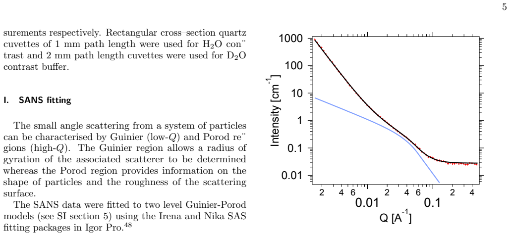
The load-bearing measurement is polarized neutron reflectivity (PNR) from a polymer-tethered bilayer, modelled as a stack of slabs from the silicon substrate out to solution: oxide, permalloy, gold, PDP anchor, PEG brush, inner and outer lipid leaflets, and a final slab representing the MscL protrusion. The lipid leaflets are parametrized by area per molecule and water molecules per lipid head, the transmembrane protein enters as a volume fraction, and the protrusion slab has its own fitted thickness; Bayesian posterior sampling is used to estimate parameter uncertainties and to show that the protrusion-thickness posterior shifts after pexiganan addition. Supporting characterization comes from small-angle neutron scattering of MscL-containing vesicles, where a two-level Guinier-Porod fit yields a protein radius of gyration of 26–29 Å, matching the crystal structure of individual MscL channels and indicating that the channels are incorporated as unclustered monomers rather than large aggregates.
What would settle it
A protein-free POPC:POPG tethered bilayer measured by polarized neutron reflectivity before and after 1.6 µM pexiganan would settle the point: if the apparent protrusion-layer thickness or scattering length density changes by roughly the same 8 Å, the MscL conformational assignment is not supported. A complementary check is patch-clamp recording of MscL proteoliposomes at 1.6 µM pexiganan, which should show channel opening if the structural change is gating.
Extended reading notes
Core claim
The central claim is that pexiganan induces a conformational change in MscL that appears as a decrease in the fitted thickness of the channel's C-terminus-containing protrusion in a tethered lipid bilayer: from 46 ± 3 Å in the pristine bilayer to 38 ± 3 Å at 1.6 µM and 38 ± 5 Å at 3.2 µM pexiganan, with the Bayesian posterior for this parameter shifting to lower values. The accompanying changes, protein coverage falling from 14% to 9–11% and bilayer coverage rising from 0.66 to 0.71–0.73, are shown by the authors to be the expected signature of open channels occluding a larger area than closed channels. The authors present this as the first direct experimental evidence of an antimicrobial peptide triggering a structural change in the C-terminus of MscL, consistent with a gating motion previously predicted by molecular dynamics and inferred from EPR and FRET measurements.
Load-bearing premise
The load-bearing assumption is that the fitted protrusion thickness decrease is caused by protein movement, not by pexiganan's own scattering contribution, since pexiganan is not included in the reflectivity model and the paper reports no protein-free bilayer-plus-pexiganan control.
Editorial extensions
If this is right
- If the protrusion shortening is genuine, PNR on tethered bilayers provides a direct structural readout of MscL gating that does not require detergents or protein clustering.
- The correlated rise in bilayer coverage and fall in protein coverage can serve as a secondary fingerprint of channel opening in future screens of antimicrobial peptides.
- The result places a structural constraint on models of MscL gating: the C-terminus retracts by roughly 8 Å toward the transmembrane domain rather than moving outward.
- Because the change saturates between 1.6 and 3.2 µM pexiganan, the effect occurs at the lower end of reported minimum inhibitory concentrations, suggesting channel gating precedes membrane disruption.
- The same tethered-bilayer platform can be applied to other amphipathic drugs and MscL mutants to test whether gating-like conformational changes are a general antimicrobial-peptide mechanism.
Reading between the lines
- Beyond the paper: if pexiganan gates MscL open at sub-MIC concentrations, combination therapies pairing antimicrobial peptides with antibiotics that enter through the MscL pore, such as streptomycin-like aminoglycosides, should show synergistic killing; this is testable in bacterial viability assays.
- Beyond the paper: the protrusion thickness being 38 Å at both 1.6 and 3.2 µM pexiganan suggests a two-state transition rather than a graded response, and patch-clamp recordings at intermediate concentrations would reveal whether the structural change is all-or-none.
- Beyond the paper: because pexiganan is absent from the reflectivity model, a protein-free bilayer control exposed to pexiganan would determine how much of the apparent protrusion change is peptide contrast; without that control, the quantitative 8 Å number is tied to the model's assumption.
- Beyond the paper: using deuterated pexiganan would change the peptide's scattering length density and allow the peptide's own location to be fitted rather than omitted, providing a direct test of where the peptide acts.
Signed reviews
Editorial analysis
A structured set of objections, weighed in public.
Referee Report
Summary. The manuscript reports a cell-free, detergent-free expression of the mechanosensitive channel MscL into POPC:POPG vesicles, SANS characterization of the vesicle-embedded protein (Rg = 26-29 Å), formation of PEG-tethered lipid bilayers on gold, and polarized neutron reflectivity (PNR) analysis of the bilayer before and after exposure to the antimicrobial peptide pexiganan. The fitted thickness of the MscL protrusion decreases from 46 ± 3 Å to 38 ± 3 Å at 1.6 μM pexiganan and to 38 ± 5 Å at 3.2 μM (Table II), with a shifted posterior distribution (Fig. 7). The authors interpret this as the first direct experimental evidence of an AMP-induced conformational change in the C-terminal protrusion of MscL, consistent with channel gating.
Significance. If the attribution holds, this is a valuable structural observation linking antimicrobial-peptide action to MscL gating, with potential implications for antibiotic mechanisms and drug design. The experimental platform is a genuine advance: cell-free expression avoids detergents, the polymeric tether provides a flexible membrane mimetic, and the PNR analysis uses three solvent contrasts and two spin contrasts with Bayesian posterior sampling. The authors provide the fitting code and data repository, which strengthens reproducibility. The two peptide concentrations give consistent thickness values, and the model selection for protrusion orientation is based on log-evidence. However, the central interpretation rests on an unvalidated assumption that pexiganan contributes negligibly to the reflectivity; the reported 'direct experimental evidence' is therefore not yet secured.
major comments (1)
- [Section II.G and SI Section 2] The central claim—that the 46-to-38 Å protrusion decrease reflects an MscL conformational change—relies on a model in which pexiganan is not explicitly included. As stated in Section II.G and SI Section 2: 'Pexiganan is not explicitly included in the model.' Pexiganan is an amphipathic cationic peptide known to insert at the lipid headgroup/tail interface (ref. 9), and after the buffer rinse it will remain in the bilayer. This peptide contributes to the scattering length density in the very layers (outer headgroup and protrusion region) whose thickness and SLD are being fitted. Because the model contains no peptide component, all post-peptide SLD changes are absorbed into the MscL protrusion thickness, the protein coverage, and the bilayer coverage parameters. A thin peptide-rich layer at the outer leaflet or at the base of the protrusion could plausibly mimic a reduced protrusion thickness. The justification for omitting the peptide is a prior thesis measurement (ref. 46) rather than a measurement in the present tethered-bilayer system, and no protein-free POPC:POPG tethered bilayer challenged with pexiganan is reported. To secure the attribution, the authors should either (i) add an explicit pexiganan layer to the model with the peptide:lipid ratio constrained by an independent assay, or (ii) measure a protein-free control bilayer under the same pexiganan challenge. Without one of these, the claim of 'first direct experimental evidence' is overstated.
minor comments (5)
- [SI Section 2B and Table II] The argument that the increase in bilayer coverage from 0.66 to 0.73 is 'a signature of channel opening' is a consistency check rather than an independent prediction: the predicted value of 0.72 is derived from the fitted pre-peptide protein coverage (13.7%) and an assumed open-channel radius of 35 Å, while the post-peptide protein coverage is itself a fitted parameter that decreases to 9-11%. This supporting evidence should be rephrased as model-dependent consistency, not as confirmation.
- [Section III.D] The PNR experiment was performed on a single bilayer preparation. The agreement between the 1.6 and 3.2 μM pexiganan concentrations is encouraging, but an independent bilayer replicate would strengthen the generality of the reported 8 Å protrusion decrease.
- [SI Section 2 (Python code, sum_mscl)] The protein scattering length density depends on assumed H/D exchange fractions (0.7 for the transmembrane region and 0.9 for the cytoplasmic region). The authors do not report a sensitivity analysis of these values; a brief test over a plausible range would clarify how robust the fitted protrusion thickness and its peptide-induced shift are to this assumption.
- [SI Section 5B] The SANS experiments with pexiganan show a 62.5% decrease in scattered intensity and creaming of lipid/protein, indicating vesicle destabilization. The authors should discuss whether analogous peptide-induced effects on the tethered bilayer could influence the fitted bilayer coverage or roughness parameters in the PNR analysis.
- [Throughout] A few typographical errors should be corrected, including 'deterimental' in the Introduction and 'Bicinchronic acid' in SI Section 4 (should be 'bicinchoninic acid').
Circularity Check
No circularity: the protrusion thickness is a directly fitted PNR parameter, and the SI coverage calculation is an independent prediction, not an input to the post-peptide fit.
full rationale
The paper's central claim is that the fitted MscL protrusion thickness decreases from 46 ± 3 Å to 38 ± 3 Å / 38 ± 5 Å after pexiganan addition (Table II), with the posterior shift in Fig. 7. This quantity is a free parameter of the reflectivity model, optimized against the measured reflectivity; it is not derived from the model inputs or from the hypothesis that pexiganan gates MscL. The SI Section 2B 'Justification of observed variation in bilayer coverage' combines pre-peptide fitted bilayer coverage (0.66) and protein coverage (0.137) with assumed open-channel dimensions from the literature to predict a post-gating coverage of 0.72, and then compares this with the independently fitted post-peptide value of 0.73. That is a genuine prediction from separate inputs, not an equation that reproduces the fitted value by construction. The model's omission of pexiganan is justified by a prior thesis measurement of a low peptide:lipid ratio (ref 46); this is an external empirical input rather than a self-referential derivation, and it does not make the central structural result equivalent to an input. No uniqueness theorem from the same authors is invoked, and no known result is merely renamed. While the absence of a protein-free peptide control and the single-bilayer replication are legitimate scientific concerns, they are correctness or robustness issues, not circularity. Therefore the paper contains no significant circular step.
Assumptions & free parameters
free parameters (5)
- MscL protrusion thickness =
46 ± 3 Å; 38 ± 3 Å (1.6 μM PXG); 38 ± 5 Å (3.2 μM PXG)
- Protein coverage in the transmembrane region =
0.14 ± 0.01; 0.09 ± 0.01; 0.11 ± 0.01
- Bilayer coverage =
0.66 ± 0.02; 0.73 ± 0.02; 0.71 ± 0.02
- H/D exchange fraction in protein SLD =
0.7 (transmembrane), 0.9 (cytoplasmic)
- Lipid area per molecule (inner/outer leaflet) and waters per lipid head =
79/98 to 99 Ų; 6 WPLH
assumptions (7)
- standard math Optical matrix formalism and Nevot-Croce roughness describe the reflectivity of the layered interface.
- domain assumption Scattering length densities are volume-fraction averages of components with literature molecular volumes and atomic scattering lengths.
- ad hoc to paper The C-terminal protrusion of MscL is represented as a single homogeneous slab whose volume fraction derives from crystal-structure volume ratios.
- ad hoc to paper Pexiganan contributes negligibly to the layer scattering length density and is excluded from the model.
- ad hoc to paper The H/D exchange fractions used in the protein scattering length (0.7 transmembrane, 0.9 cytoplasmic regions) are correct.
- domain assumption The open-state channel geometry (radius increasing from 25 to 35 Å, C-terminus retraction) from the Perozo/Bavi literature is used to predict bilayer coverage changes on gating.
- domain assumption A two-level Guinier-Porod model can separate vesicle-level and protein-level SANS contributions with a fixed vesicle Rg.
Cite this review
Pith. "Pith review of Neutron Reflectometry Reveals Conformational Changes in a Mechanosensitive Protein Induced by an Antimicrobial Peptide in Tethered Lipid Bilayers." pith.science (2026). https://pith.science/paper/HHY3L5J5
@misc{pith2026250616972,
  author       = {Pith},
  title        = {Pith review of: Neutron Reflectometry Reveals Conformational Changes in a Mechanosensitive Protein Induced by an Antimicrobial Peptide in Tethered Lipid Bilayers},
  year         = {2026},
  howpublished = {\url{https://pith.science/paper/HHY3L5J5}},
  note         = {Machine review of arXiv:2506.16972}
}
abstract
Membrane proteins serve a wide range of vital roles in the functioning of living organisms. Compared to other classes of proteins, determining membrane protein structures remains a challenge, in large part due to the difficulty in establishing experimental conditions that can preserve the correct conformation and function of the protein in isolation from its native environment. We investigated the ion channel in lipid vesicles and in a planar lipid bilayer. By using a polymeric tether our planar membrane mimetic was not constrained by the underlying solid substrate, making it sufficiently flexible to allow for increases in bilayer curvature and changes in membrane tension. We used quartz crystal microbalance with dissipation (QCM-D), and polarised neutron reflectivity (PNR) to show the formation of MscL containing phospholipid bilayers, tethered with a high density PEG layer onto gold substrates from vesicle rupture. The MscL containing vesicles were separately characterised with small angle neutron scattering (SANS). MscL was expressed into vesicles using cell free protein expression. Analysing these vesicles with small angle neutron scattering, the radius of gyration of the protein was determined to be between 26-29~\AA{}, consistent with the crystal structure of individual MscL channels. The MscL composition of the formed bilayer was 14\%$v/v$, close to the initial composition of the vesicles, and a protein protrusion extending ca. 46~\AA{} into the solvent was determined by PNR. Addition of 1.6 and 3.2 $\mu$M pexiganan resulted in a decrease in the protrusion of MscL (from $\sim$46 to $\sim$38~\AA{}). To our knowledge, these findings represent the first direct experimental evidence of a structural change in the C-terminus containing protrusion of MscL, triggered by an antimicrobial peptide.
Figures


Figures from the paper (5 more)


Reference graph
Works this paper leans on
-
[1]
QUAR TZ-CR YST AL MICROBALANCE WITH DISSIPA TION (QCM-D) A typical QCM-D protocol followed the following steps:
-
[2]
After testing that the frequency is stable for 30 min with on ly clean buffer in the cell, i.e. there is no desorption of tether occurring, adsorption of contaminants or bubbles in the system, then a solution of vesicles was passed through the cells at 1 mL/min using the peristaltic pump. 10 ml of vesicle solution is pass ed through each QCM cell
-
[3]
Buffer solution (10 mL) was passed through each of the flow cells and the measurement started with the sensors in HEPES buffer
-
[4]
This was immediately followed by a salt rupture step, push ing 5 mL of lower salt concentration 150 mM NaCl HEPES buffer through the QCM cell. Since tethered bilayers are viscoelastic it was inappropriate to analyse the results quanti- tatively using the Sauerbrey equation. The system has high water content in the tether layer and also in any tethered vesi...
-
[5]
After 1 h, 300 mM NaCl HEPES buffer was passed through the cells t o remove excess vesicles
-
[6]
REFLECTIVITY MODEL IMPLEMENTED IN PYTHON USING REFNX Listing 1. Python code for fitting polarized neutron reflectivity data measured from the tethered bilayer in three sub-phase contrasts. The full set of Jupyter note books used for the analysis of PNR can be found in the data repository for the manuscript [27]. #! / u s r / b i n / env p y t h o n # c o d ...
work page 1990
-
[7]
NEUTRON REFLECTIVITY DA T A ANAL YSIS While the layers from silicon oxide to poly(ethylene glycol) (PEG2000) were parame- terised as simple slabs, the DSPE and lipid bilayer were para meterised in terms of area per molecule (APM) and water molecules per lipid head (WPLH) using l iterature values for molecular volumes and atomic scattering lengths.[3–6] Th...
-
[8]
The Bayesian approach is useful for examining parameter covariances
ERROR ANAL YSIS POSTERIOR DISTRIBUTIONS RefNX uses a Bayesian approach employing a Markov-chain Mont e Carlo algorithm to investigate the posterior probability distribution of the model parameters. The Bayesian approach is useful for examining parameter covariances. The figures below (Figs. S2, S3 and S4) show the corner plots for the conditions without PX...
Show all 38 references
-
[9]
13.5 mg of POPC and 4.5 mg of POPC were dissolved in the minimum amount of chlo- roform and the chloroform removed under a stream of nitrogen to produce a dried lipid film
CELL-FREE PROTEIN EXPRESSION First a concentrated solution (18 mg/mL) of 3:1 POPC:POPG lip ids was prepared. 13.5 mg of POPC and 4.5 mg of POPC were dissolved in the minimum amount of chlo- roform and the chloroform removed under a stream of nitrogen to produce a dried lipid fi...
-
[10]
SMALL ANGLE NEUTRON SCA TTERING ANAL YSIS Hammouda et al. proposed a generalized Guinier-Porod approach to fitting the scattering from a mixture of arbitrary shapes or fractal structures for which it is difficult to build analytical models.[15] For each particulate type in the sy...
-
[11]
coli HY Kit — leap and lead — biotec hrab- bit.com, https://www.biotechrabbit.com/rts-500-proteomaster-e-coli-hy-kit-405
biotechrabbit — RTS 500 ProteoMaster E. coli HY Kit — leap and lead — biotec hrab- bit.com, https://www.biotechrabbit.com/rts-500-proteomaster-e-coli-hy-kit-405. html, [Accessed 15-Aug-2024] (2023)
2023
-
[12]
F. Inci, U. Celik, B. Turken, H. ¨Ozg¨ ur¨Ozer, F. N. Kok, Construction of p-glycoprotein incorporated tethered lipid bilayer membranes, Biochemistry an d Biophysics Reports 2 (2015) 115–122. doi:https://doi.org/10.1016/j.bbrep.2015.05.012. 39 URL https://www.sciencedirect.com...
2015 doi
-
[13]
H¨ opfner, U
M. H¨ opfner, U. Rothe, G. B. and, Biosensor-based evaluation of liposomal b ehav- ior in the target binding process, Journal of Liposome Research 18 (1) (2008) 71–82, pMID: 18348073. arXiv:https://doi.org/10.1080/08982100801894000, doi:10.1080/ 08982100801894000. URL https://...
2008 doi
-
[14]
R. S. Armen, O. D. Uitto, S. E. Feller, Phospholipid component volum es: Determination and application to bilayer structure calculations, Biophysical Journal 75 (1998) 734–744. doi: 10.1016/s0006-3495(98)77563-0
1998 doi
-
[15]
A. P. Dabkowska, D. J. Barlow, A. V. Hughes, R. A. Campbell, P. J. Quinn, M. J. Lawrence, The effect of neutral helper lipids on the structure of cationic lipi d monolayers, J. R. Soc. Interface 9 (2012) 548–561. doi:10.1098/rsif.2011.0356
2012
-
[16]
J. Pan, F. A. Heberle, S. Tristram-Nagle, M. Szymanski, M. Koepfinger , J. Katsaras, N. Kuˇ cerka, Molecular structures of fluid phase phosphatidylglycerol bilayers as determined by small angle neutron and x-ray scattering, Biochimica et Biophysic a Acta (BBA) - Biomem- branes 1...
2012 doi
-
[17]
V. F. Sears, Neutron scattering lengths and cross sections, Neutron New s 3 (1992) 26–37. doi:10.1080/10448639208218770
1992 doi
-
[18]
A. R. J. Nelson, S. W. Prescott, refnx: neutron and X-ray reflectometry analysis in Python, Journal of Applied Crystallography 52 (1) (2019) 193–200. doi:10.1107/ S1600576718017296. URL https://doi.org/10.1107/S1600576718017296
2019 doi
-
[19]
S. Chiu, E. Jakobsson, S. Subramaniam, H. Scott, Combined monte carlo an d molecular dynamics simulation of fully hydrated dioleyl and palmitoyl-oleyl ph osphatidylcholine lipid bilayers, Biophysical Journal 77 (5) (1999) 2462–2469. doi:https://doi.org/10.1016/ S0006-3495(99)7...
1999
-
[20]
Pabst, S
G. Pabst, S. Danner, S. Karmakar, G. Deutsch, V. A. Raghunathan, On the p ropensity of phosphatidylglycerols to form interdigitated phases, Biophys J 93 (2) ( 2007) 513–525
2007
-
[21]
McKinley, Neutron reflectivity studies of bacterial membranes , peptides and proteins, Ph.D
L. McKinley, Neutron reflectivity studies of bacterial membranes , peptides and proteins, Ph.D. thesis, Edinburgh University (2017). 40
2017
-
[22]
Perozo, D
E. Perozo, D. M. Cortes, P. Sompornpisut, A. Kloda, B. Martinac, Open channel structure 41 of mscl and the gating mechanism of mechanosensitive channels, Nature 418 ( 6901) (2002) 942–948
2002
-
[23]
J. S. Najem, M. D. Dunlap, I. D. Rowe, E. C. Freeman, J. W. Grant, S . Sukharev, D. J. Leo, Activation of bacterial channel mscl in mechanically stimulated drople t interface bilayers, Scientific Reports 5 (1) (2015) 13726. URL https://doi.org/10.1038/srep13726
2015 doi
-
[24]
Martinac, P
B. Martinac, P. R. Rohde, A. R. Battle, E. Petrov, P. Pal, A. F. W. Foo, V. V´ asquez, T. Huynh, A. Kloda, Studying Mechanosensitive Ion Channels Using Liposomes, Human a Press, Totowa, NJ, 2010, pp. 31–53. doi:10.1007/978-1-60761-447-0_4 . URL https://doi.org/10.1007/978-1-60...
2010 doi
-
[25]
Takeda, M
M. Takeda, M. Kainosho, Cell-free protein production for nmr studi es, Methods in Molecular Biology 831 (2012) 71–84
2012
-
[26]
Hammouda, A new guinier–porod model, Journal of Applied Crystallograph y 43 (4) (2010) 716–719
B. Hammouda, A new guinier–porod model, Journal of Applied Crystallograph y 43 (4) (2010) 716–719
2010
-
[27]
Irena, nika Manuals 1.5.1, Guinier Porod irena and nika manuals 1.5.1 do cumentation, https: //saxs-igorcodedocs.readthedocs.io/en/stable/Irena/GuinierPorod.html, accessed: 2020-09-14 (2020)
2020
-
[28]
Delhi, Radius of Gyration, http://www.scfbio-iitd.res.in/software/proteomics/rg
I. Delhi, Radius of Gyration, http://www.scfbio-iitd.res.in/software/proteomics/rg. jsp, accessed: 2020-10-14
2020
-
[29]
Van Der Spoel, E
D. Van Der Spoel, E. Lindahl, B. Hess, G. Groenhof, A. E. Mark, H. J. Be rendsen, Gromacs: fast, flexible, and free, Journal of computational chemistry 26 (16) (2005) 1701–1718
2005
-
[30]
Chang, R
G. Chang, R. H. Spencer, A. T. Lee, M. T. Barclay, D. C. Rees, Struct ure of the mscl homolog from mycobacterium tuberculosis: a gated mechanosensitive ion channe l, Science 282 (5397) (1998) 2220–2226
1998
-
[31]
Xie, G.-H
J.-Y. Xie, G.-H. Ding, Studies on sensitivity to tension and gating p athway of mscl by molec- ular dynamic simulation, Acta Mechanica Sinica 29 (2) (2013) 256–266
2013
-
[32]
P. W. Rose, B. Beran, C. Bi, W. F. Bluhm, D. Dimitropoulos, D. S. Go odsell, A. Prli´ c, M. Quesada, G. B. Quinn, J. D. Westbrook, et al., The rcsb protein dat a bank: redesigned web site and web services, Nucleic acids research 39 (suppl
-
[34]
N. Bavi, A. D. Martinac, D. M. Cortes, O. Bavi, P. Ridone, T. Nomura, A. P . Hill, B. Martinac, E. Perozo, Structural dynamics of the mscl c-terminal domain, Scient ific reports 7 (1) (2017) 17229
2017
-
[35]
Martinac, J
B. Martinac, J. Adler, C. Kung, Mechanosensitive ion channels of e. c oli activated by am- phipaths, Nature 348 (6298) (1990) 261–263
1990
-
[36]
Strutt, J
R. Strutt, J. W. Hindley, J. Gregg, P. J. Booth, J. D. Harling, R. V. Law , M. S. Friddin, O. Ces, Activating mechanosensitive channels embedded in droplet interface bilayers using membrane asymmetry., Chem Sci 12 (6) (2021) 2138–2145. doi:10.1039/d0sc03889j
2021 doi
-
[37]
S. L. Grage, A. M. Keleshian, T. Turdzeladze, A. R. Battle, W. C. Tay, R. P. May, S. A. Holt, S. A. Contera, M. Haertlein, M. Moulin, et al., Bilayer-mediated clus tering and functional interaction of mscl channels, Biophysical journal 100 (5) (2011) 1252–1260
2011
-
[38]
S. E. Ayscough, M. W. A. Skoda, J. Doutch, A. Caruana, C. Kinane, L. Clifton , S. Titmuss, Tethered Lipid Bilayers as a Platform for Studying the Mechanosensi tive Ion Channel MscL (Mar. 2025). doi:10.5281/zenodo.14975055. URL https://github.com/maxskoda/MscL_tether_NR 42
2025 doi
-
[122]
URL https://www.sciencedirect.com/science/article/pii/ S240558081500031X 55M
doi:https://doi.org/10.1016/j.bbrep.2015.05.012. URL https://www.sciencedirect.com/science/article/pii/ S240558081500031X 55M. Kowalska, M. Broniatowski, M. Mach, /suppress Lukasz P/suppress lachta, P. Wydro, The effect of the polyethylene glycol chain length of a lipopolymer (...
2021
Reviewed August 15, 2026 · model on record in the stance chip above.
Discussion (0). Continue with ORCID to comment.