REVIEW 4 major objections 5 minor 35 references
X-SiT: Inherently Interpretable Surface Vision Transformers for Dementia Diagnosis
T0 review · 4 major / 5 minor · reviewed 2026-08-06 · deepseek-v4-flash
Pith's one-line read The paper presents X-SiT, an inherently interpretable surface vision transformer that classifies Alzheimer's disease and frontotemporal dementia with accuracy matching non-interpretable models, explaining each prediction through…
desk verdict A genuinely novel spatial-prototype decoder for cortical surfaces, but the 'inherently interpretable' claim needs stronger faithfulness evidence and an honest abstract. read the letter →
The pith
A machine-rendered reading of the paper's core claim, the machinery that carries it, and where it could break.
The reading
What carries the argument
The central mechanism is the prototypical surface patch (PSP) decoder attached to a Surface Vision Transformer encoder. The input cortical surface is assumed pre-registered to a common template and partitioned into a fixed set of non-overlapping triangular patches, so patch index $i$ denotes the same anatomical region across subjects and prototypes. The encoder maps each patch to a latent embedding; a prototypical sequence $\xi=(\xi_1,\dots,\xi_N)$ is obtained by encoding learned prototype patches. For each index the decoder computes the cosine similarity $\cos(x_i, \xi_i)$ between the input embedding $x_i$ and the prototype embedding $\xi_i$, rectifies all embeddings with ReLU so that absent features cannot increase class probability, scales each similarity by a sparse weight $w_i$ (with $\sum_i w_i=1$ and weights below $1/N$ set to zero), and sums to form $P(c|x)=\sum_i w_i\cos(x_i,\xi_i)$. Every five epochs each prototype's latent representation is replaced by the most similar patch from the training set at the same location, keeping prototypes interpretable as real anatomy. This pipeline is what makes the explanation identical to the decision rule.
What would settle it
Register the same set of brains to a common template twice, for example with different template choices or surface-processing settings, and check whether the same patch indices still correspond to the same anatomy; if the cosine similarities, the sparse weights, or the prototypes change substantially under re-registration, the interpretability claim is tied to the registration pipeline rather than to cortex structure. Alternatively, replace the template with an independently built average surface and see whether the learned prototypes still land on the known disease regions, namely temporal and temporoparietal areas for Alzheimer's disease and frontal and temporal areas for frontotemporal dementia.
Extended reading notes
Core claim
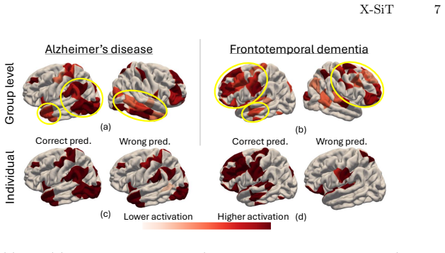
On the paper's own terms, the central discovery is that case-based reasoning can replace the opaque classification head of a surface vision transformer without a performance penalty. X-SiT's decoder compares each input patch embedding to a learned prototype embedding at the same spatial index, applies ReLU so that only positive evidence counts, and forms the class probability as a sparse weighted sum of cosine similarities. The prototypes are real training patches refreshed during training, so predictions are literally "this region resembles that region of a patient in the training set." In experiments on two dementia tasks, X-SiT's test balanced accuracy (80.2% for AD, 79.6% for FTD) is competitive with the non-interpretable Surface Vision Transformer (80.5% and 80.6%), and its learned prototypes activate in the temporal and temporoparietal regions for Alzheimer's disease and in frontal and temporal regions for frontotemporal dementia, matching published cortical-thinning patterns. The paper further reports that prototype sets are stable across random initializations (76.3% overlap for AD, 71.7% for FTD) and that individual explanations of misclassified cases deviate from the group-level pattern, suggesting the explanations can flag errors.
Load-bearing premise
The whole explanation pipeline depends on the assumption that after registration to a common template, patch index $i$ names the same anatomical location in every subject's cortex and every prototype, so a cosine similarity at that index is a comparison of matching brain regions; residual registration error or folding variability would make the similarities and the resulting explanations anatomically meaningless.
Editorial extensions
If this is right
- X-SiT delivers per-prediction explanations that are the model's actual decision rule, not post-hoc attributions, so a clinician can inspect the exact cortical regions that drove a dementia diagnosis.
- The learned prototypes double as global disease maps: the same weights and similarities that classify a patient can be averaged across a cohort to show which regions the model finds characteristic of Alzheimer's disease or frontotemporal dementia.
- Because prototypes are replaced with real training patches, every explanation can be traced to concrete patient anatomy, making the model's reasoning auditable against clinical knowledge.
- The method matches the balanced accuracy of the non-interpretable surface transformer while outperforming the adapted neural-tree interpretable decoder, indicating that interpretability need not be bought with accuracy in this setting.
Reading between the lines
- If the anatomical-correspondence assumption is satisfied across larger, multi-site datasets, the same prototype decoder could be applied to differential diagnosis among several dementia subtypes, where the spatial pattern of atrophy is the key discriminator.
- The sparse weighting scheme implies the model deliberately ignores below-average regions; a testable extension would be to compare sparse against dense weighting to see whether diffuse but real disease patterns are being discarded for interpretability's sake.
- A natural stress test is to feed X-SiT surfaces registered with different templates or from different preprocessing pipelines; if the prototypes and explanations do not remain stable, the interpretability claim would be tied to a specific registration pipeline rather than to the cortex itself.
Editorial analysis
A structured set of objections, weighed in public.
Referee Report
Summary. The manuscript introduces X-SiT, an interpretable transformer for cortical surface classification. A Surface Vision Transformer (SiT) encoder produces patch-wise latent embeddings, and a prototypical surface patch (PSP) decoder computes the class probability as a weighted sum of cosine similarities between encoded patches and learned prototypes that are kept in spatial correspondence. Prototypes are periodically replaced with the most similar patches from training data, so the model is claimed to use case-based reasoning on interpretable cortical features. The method is evaluated for Alzheimer's disease and frontotemporal dementia diagnosis on ADNI and NIFD data, with balanced accuracy comparable to prior non-interpretable models, and qualitative group-level and individual-level visualizations are provided as interpretability evidence.
Significance. The architectural idea is timely and potentially valuable: extending prototype-based interpretability to cortical surface data with template correspondence is a natural and clinically relevant direction, and the sparse weighted decision rule is transparent in structure. The authors report competitive accuracy on two clinical tasks using public datasets, and the prototype-stability overlap statistic is a useful addition. However, the paper's core contribution, "inherent interpretability," is not yet established: the visualizations and evaluations do not demonstrate that the latent-space comparisons underlying the decision rule reflect the raw interpretable features shown to the user. The performance claim in the abstract is also stronger than the test results support. Provided these points are addressed with additional analysis, the method would be a solid contribution to the interpretable medical imaging literature.
major comments (4)
- [Section 2.2, Eqs. (1)-(2); Section 2.3; Figure 4] The decision rule is defined entirely on latent embeddings xi and ξi, while the interpretability visualizations (Figure 4) show raw cortical thickness, sulcal depth, and curvature. The encoder is trained with binary cross-entropy only, and there is no loss term (e.g., reconstruction, clustering, or similarity-alignment) that forces high cosine similarity in latent space to correspond to high similarity of the visualized raw features. Therefore, the prototypes may be illustrative rather than faithful: two raw patches that appear very different could be close in latent space if that helps classification. To support the central interpretability claim, the authors must connect latent-space similarity to raw-feature similarity, for example by measuring agreement between latent nearest neighbors and raw-feature nearest neighbors on held-out patches, or by adding an explicit alignment or clustering regularizer. Without such evidence, "inherently interpretable" is not established.
- [Section 3.4] The interpretability evaluation is exclusively qualitative. Figure 3 shows mean activation maps and two individual examples, and Figure 4 shows selected prototypes. There is no user study, no quantitative explanation metric (e.g., fidelity, consistency, or agreement with expert-defined regions), and no statistical test that the prototypes align with known disease patterns beyond visual inspection. The claim that prototypes "reveal classification errors" rests on anecdotal cases. As a result, the paper does not demonstrate that X-SiT explanations are more faithful or more useful than the attention weights or saliency maps it criticizes in the introduction. The authors should add a quantitative evaluation of explanation quality, or at minimum an inter-rater or expert-agreement study.
- [Abstract; Section 3.2; Table 2] The abstract's claim of "state-of-the-art performance" is not supported by Table 2. On both test sets, the non-interpretable SiT achieves equal or higher balanced accuracy (AD: 80.5±0.5 vs 80.2±2.0; FTD: 80.6±1.8 vs 79.6±1.6), and X-SiT is second to SiT in both cases. The differences are within one standard deviation, so a fair statement is "competitive" or "matches accuracy," which is the phrasing used in the introduction and conclusion. The abstract should be revised to avoid an unsupported claim. Additionally, in the FTD task, cognitively normal subjects are pooled from ADNI and NIFD while patients come only from NIFD, so scanner and protocol differences are a potential confound that should be discussed or mitigated.
- [Section 2.1; Section 2.2] The interpretability mechanism relies on patch index i denoting the same anatomical region for every subject and every prototype, stated as an assumption in Section 2.1. The paper does not verify this correspondence after FreeSurfer registration to the FsAverage template. Residual registration error or folding variability could make "corresponding prototypes" and the group-level average maps in Figure 3 anatomically misaligned, which would undermine the clinical meaning of the explanations. A demonstration of registration quality (e.g., overlap of cortical labels on a validation population, or within-subject repeat-scan correspondence) would strengthen the central claim. If correspondence is not reliable, the cosine similarities and the resulting explanations are not anatomically meaningful.
minor comments (5)
- [Related work and Table 2] The naming is inconsistent: the related work uses "ViT-Net" while the adapted surface model is called "SiT-NeT" in Table 2 and in the text; please use a single consistent name.
- [Section 3.2] There is a typo in the first sentence of Section 3.2: "T able 1" should be "Table 1."
- [Section 3.4] The prototype robustness overlap (76.3% and 71.7%) is reported without a definition of how the overlap is computed across random initializations; please specify the metric.
- [Figure 3] The yellow ellipses indicating hallmark regions are described in the caption but are difficult to identify in a print version of the figure; consider adding explicit region labels to the figure itself.
- [Section 3.4] The statement that the temporal lobe received comparatively low weight in the FTD experiments is somewhat confusing in light of the preceding reference to frontal and temporal regions as FTD hallmarks; please clarify whether the model's focus is expected to be incomplete.
Circularity Check
No significant circularity found: X-SiT's predictions are empirical classification outputs on held-out data, and the prototype/interpretability claims are externally evaluated rather than restatements of model inputs.
full rationale
I examined the derivation chain: the surface encoder maps input patches to latent embeddings; the PSP decoder computes cosine similarities between encoded input patches and learned prototypes (Eq. 1); the class probability is a weighted sparse sum of those similarities (Eq. 2). The prototypes are learned end-to-end with binary cross-entropy and periodically replaced by the most similar training patches at each location. No predicted quantity is defined in terms of the quantity it is said to predict: test-set balanced accuracy is computed on held-out subjects, and the alignment of prototype activations with temporal/frontal disease patterns is checked against external neuroimaging references (e.g., [9,30]), not against the model's own training labels. The only self-citation is reference [33] in a related-work sentence listing prior explainable-medical-imaging efforts; it is not load-bearing for the method, the architecture, or the performance claims. The latent-space-to-raw-feature interpretability question raised by the skeptic is a validity concern, not a circularity: nothing in the paper's equations makes 'prototype looks like input' true by construction. Therefore the derivation is self-contained with respect to circularity.
Assumptions & free parameters
free parameters (4)
- Sparse scaling weights w_1,...,w_N =
Learned during end-to-end training
- Prototype latent embeddings ξ_1,...,ξ_N =
Learned and periodically replaced by training patches
- SiT encoder network parameters =
Learned end-to-end
- Architectural and training hyperparameters =
Tuned on validation, not reported
assumptions (4)
- domain assumption Cortical surfaces from all subjects are pre-registered to FsAverage so that patch i is the same anatomical region across subjects.
- domain assumption Cosine similarity in the ReLU-rectified latent space is a valid proxy for 'looks like' reasoning about disease patterns.
- domain assumption FreeSurfer-derived thickness, sulcal depth, and curvature are informative for dementia classification.
- domain assumption Pooling cognitively normal subjects from ADNI and NIFD for the FTD task does not introduce a systematic site or scanner confound.
Cite this review
Pith. "Pith review of X-SiT: Inherently Interpretable Surface Vision Transformers for Dementia Diagnosis." pith.science (2026). https://pith.science/paper/GVREBGRY
@misc{pith2026250620267,
  author       = {Pith},
  title        = {Pith review of: X-SiT: Inherently Interpretable Surface Vision Transformers for Dementia Diagnosis},
  year         = {2026},
  howpublished = {\url{https://pith.science/paper/GVREBGRY}},
  note         = {Machine review of arXiv:2506.20267}
}
read the original abstract
Interpretable models are crucial for supporting clinical decision-making, driving advances in their development and application for medical images. However, the nature of 3D volumetric data makes it inherently challenging to visualize and interpret intricate and complex structures like the cerebral cortex. Cortical surface renderings, on the other hand, provide a more accessible and understandable 3D representation of brain anatomy, facilitating visualization and interactive exploration. Motivated by this advantage and the widespread use of surface data for studying neurological disorders, we present the eXplainable Surface Vision Transformer (X-SiT). This is the first inherently interpretable neural network that offers human-understandable predictions based on interpretable cortical features. As part of X-SiT, we introduce a prototypical surface patch decoder for classifying surface patch embeddings, incorporating case-based reasoning with spatially corresponding cortical prototypes. The results demonstrate state-of-the-art performance in detecting Alzheimer's disease and frontotemporal dementia while additionally providing informative prototypes that align with known disease patterns and reveal classification errors.
Figures


Figures from the paper (1 more)

Reference graph
Works this paper leans on
-
[1]
Azcona, E.A., Besson, P., Wu, Y., Punjabi, A., Martersteck, A., Dravid, A., Par- rish, T.B., Bandt, S.K., Katsaggelos, A.K.: Interpretation of brain morphology in association to alzheimer’s disease dementia classification using graph convolutional networks on triangulated meshes. In: Shape in Medical Imaging. p. 95–107 (2020)
work page 2020
-
[2]
Cerebral Cortex33(10), 6474–6485 (2023)
de Chastelaine, M., Srokova, S., Hou, M., Kidwai, A., Kafafi, S.S., Racenstein, M.L., Rugg, M.D.: Cortical thickness, gray matter volume, and cognitive perfor- mance: a crosssectional study of the moderating effects of age on their interrela- tionships. Cerebral Cortex33(10), 6474–6485 (2023)
work page 2023
-
[3]
Advances in neural information processing systems 32 (2019)
Chen, C., Li, O., Tao, D., Barnett, A., Rudin, C., Su, J.K.: This looks like that: deep learning for interpretable image recognition. Advances in neural information processing systems 32 (2019)
work page 2019
-
[4]
In: Machine Learning in Medical Imaging
Cheng, J., Zhang, X., Zhao, F., Wu, Z., Yuan, X., Gilmore, J.H., Wang, L., Lin, W., Li, G.: Spherical transformer on cortical surfaces. In: Machine Learning in Medical Imaging. p. 406–415 (2022)
work page 2022
-
[5]
Molecular Psychiatry28(10), 4084–4097 (2023)
Chouliaras, L., O’Brien, J.T.: The use of neuroimaging techniques in the early and differential diagnosis of dementia. Molecular Psychiatry28(10), 4084–4097 (2023)
work page 2023
-
[6]
In: Medical Imaging with Deep Learn- ing
Dahan, S., Fawaz, A., Williams, L.Z.J., Yang, C., Coalson, T.S., Glasser, M.F., Ed- wards, A.D., Rueckert, D., Robinson, E.C.: Surface vision transformers: Attention- based modelling applied to cortical analysis. In: Medical Imaging with Deep Learn- ing. Proceedings of Machine Learning Research, vol. 172, pp. 282–303 (2022)
work page 2022
-
[7]
NeuroImage 9(2), 179–194 (1999)
Dale, A.M., Fischl, B., Sereno, M.I.: Cortical surface-based analysis i. NeuroImage 9(2), 179–194 (1999)
work page 1999
-
[8]
In: International Conference on Learn- ing Representations (2021)
Dosovitskiy, A., Beyer, L., Kolesnikov, A., et al.: An image is worth 16x16 words: Transformers for image recognition at scale. In: International Conference on Learn- ing Representations (2021)
work page 2021
Show all 35 references
-
[9]
Brain130(4), 1159–1166 (2006)
Du, A.T., Schuff, N., Kramer, J.H., Rosen, H.J., Gorno-Tempini, M.L., Rankin, K., Miller, B.L., Weiner, M.W.: Different regional patterns of cortical thinning in alzheimer’s disease and frontotemporal dementia. Brain130(4), 1159–1166 (2006)
2006
-
[10]
NeuroImage62(2), 774–781 (2012)
Fischl, B.: FreeSurfer. NeuroImage62(2), 774–781 (2012)
2012
-
[11]
NeuroImage 9(2), 195–207 (1999) 10 F
Fischl, B., Sereno, M.I., Dale, A.M.: Cortical surface-based analysis ii. NeuroImage 9(2), 195–207 (1999) 10 F. Bongratz, T.N. Wolf, et al
1999
-
[12]
Gallée, L., Beer, M., Götz, M.: Interpretable medical image classification using pro- totypelearningandprivilegedinformation.In:InternationalConferenceonMedical Image Computing and Computer-Assisted Intervention. pp. 435–445 (2023)
2023
-
[13]
In: 2019 IEEE/CVF International Conference on Computer Vision Workshop (ICCVW)
Gong, S., Chen, L., Bronstein, M., Zafeiriou, S.: Spiralnet++: A fast and highly efficient mesh convolution operator. In: 2019 IEEE/CVF International Conference on Computer Vision Workshop (ICCVW). p. 4141–4148 (2019)
2019
-
[14]
IEEE Transactions on Pattern Analysis and Machine Intelligence44(2), 864–876 (2022)
Gopinath, K., Desrosiers, C., Lombaert, H.: Learnable pooling in graph convolu- tional networks for brain surface analysis. IEEE Transactions on Pattern Analysis and Machine Intelligence44(2), 864–876 (2022)
2022
-
[15]
Advances in Neural Information Processing Systems33, 4211–4222 (2020)
Jeyakumar, J.V., Noor, J., Cheng, Y.H., Garcia, L., Srivastava, M.: How can i ex- plain this to you? an empirical study of deep neural network explanation methods. Advances in Neural Information Processing Systems33, 4211–4222 (2020)
2020
-
[16]
Kashefi, R., Barekatain, L., Sabokrou, M., Aghaeipoor, F.: Explainability of vision transformers: A comprehensive review and new perspectives. arxiv. arXiv preprint arXiv:2311.06786 (2023)
2023 arXiv
-
[17]
In: International Conference on Machine Learning
Kim,S.,Nam,J.,Ko,B.C.:ViT-NeT:Interpretablevisiontransformerswithneural tree decoder. In: International Conference on Machine Learning. Proceedings of Machine Learning Research, vol. 162, pp. 11162–11172 (2022)
2022
-
[18]
help me help the ai
Kim, S.S., Watkins, E.A., Russakovsky, O., Fong, R., Monroy-Hernández, A.: "help me help the ai": Understanding how explainability can support human-ai interac- tion. In: Conference on Human Factors in Computing Systems. pp. 1–17 (2023)
2023
-
[19]
In: Proceedings of the IEEE/CVF conference on computer vision and pattern recognition
Komorowski, P., Baniecki, H., Biecek, P.: Towards evaluating explanations of vision transformers for medical imaging. In: Proceedings of the IEEE/CVF conference on computer vision and pattern recognition. pp. 3726–3732 (2023)
2023
-
[20]
In: International Conference on Learning Representations (2019)
Loshchilov, I., Hutter, F.: Decoupled weight decay regularization. In: International Conference on Learning Representations (2019)
2019
-
[21]
In: Canadian AI (2021)
Mohammadjafari, S., Cevik, M., Thanabalasingam, M., Basar, A.: Using protopnet for interpretable alzheimer’s disease classification. In: Canadian AI (2021)
2021
-
[22]
npj Digital Medicine7(1) (2024)
Pahud de Mortanges, A., Luo, H., Shu, S.Z., Kamath, A., Suter, Y., Shelan, M., Pöllinger, A., Reyes, M.: Orchestrating explainable artificial intelligence for multi- modal and longitudinal data in medical imaging. npj Digital Medicine7(1) (2024)
2024
-
[23]
Imaging Neuroscience2, 1–37 (2024)
Munroe, L., da Silva, M., Heidari, F., Grigorescu, I., Dahan, S., Robinson, E.C., Deprez, M., So, P.W.: Applications of interpretable deep learning in neuroimaging: A comprehensive review. Imaging Neuroscience2, 1–37 (2024)
2024
-
[24]
Proceedings of the IEEE/CVF Conference on Computer Vision and Pattern Recognition (2023)
Nauta, M., Schlötterer, J., van Keulen, M., Seifert, C.: Pip-net: Patch-based intuitive prototypes for interpretable image classification. Proceedings of the IEEE/CVF Conference on Computer Vision and Pattern Recognition (2023)
2023
-
[25]
Advances in Neural Informa- tion Processing Systems34, 26422–26436 (2021)
Nguyen, G., Kim, D., Nguyen, A.: The effectiveness of feature attribution methods and its correlation with automatic evaluation scores. Advances in Neural Informa- tion Processing Systems34, 26422–26436 (2021)
2021
-
[26]
IEEE Transactions on Medical Imaging19(2), 143–150 (2000)
Nyul, L., Udupa, J., Zhang, X.: New variants of a method of mri scale standard- ization. IEEE Transactions on Medical Imaging19(2), 143–150 (2000)
2000
-
[27]
Nature 618(7965), 566–574 (2023)
Pang, J.C., Aquino, K.M., Oldehinkel, M., Robinson, P.A., Fulcher, B.D., Break- spear, M., Fornito, A.: Geometric constraints on human brain function. Nature 618(7965), 566–574 (2023)
2023
-
[28]
Nature machine intelligence1(5), 206–215 (2019)
Rudin, C.: Stop explaining black box machine learning models for high stakes decisions and use interpretable models instead. Nature machine intelligence1(5), 206–215 (2019)
2019
-
[29]
Statistic Sur- veys 16, 1–85 (2022) X-SiT 11
Rudin, C., Chen, C., Chen, Z., Huang, H., Semenova, L., Zhong, C.: Interpretable machine learning: Fundamental principles and 10 grand challenges. Statistic Sur- veys 16, 1–85 (2022) X-SiT 11
2022
-
[30]
Brain 129(11), 2885–2893 (2006)
Singh, V., Chertkow, H., Lerch, J.P., Evans, A.C., Dorr, A.E., Kabani, N.J.: Spatial patterns of cortical thinning in mild cognitive impairment and alzheimer’s disease. Brain 129(11), 2885–2893 (2006)
2006
-
[31]
Brain 145(11), 3859–3871 (2022)
Spitzer, H., Ripart, M., Whitaker, K., et al.: Interpretable surface-based detection of focal cortical dysplasias: a multi-centre epilepsy lesion detection study. Brain 145(11), 3859–3871 (2022)
2022
-
[32]
Journal of the Royal Statistical Society Series B: Statistical Methodology58(1), 267–288 (1996)
Tibshirani, R.: Regression shrinkage and selection via the lasso. Journal of the Royal Statistical Society Series B: Statistical Methodology58(1), 267–288 (1996)
1996
-
[33]
In: International Conference on Information Processing in Medical Imaging
Wolf, T.N., Pölsterl, S., Wachinger, C.: Don’t panic: Prototypical additive neu- ral network for interpretable classification of alzheimer’s disease. In: International Conference on Information Processing in Medical Imaging. pp. 82–94 (2023)
2023
-
[34]
Information Fusion77, 29–52 (2022)
Yang, G., Ye, Q., Xia, J.: Unbox the black-box for the medical explainable ai via multi-modal and multi-centre data fusion: A mini-review, two showcases and beyond. Information Fusion77, 29–52 (2022)
2022
-
[35]
Zhao, F., Xia, S., Wu, Z., Duan, D., Wang, L., Lin, W., Gilmore, J.H., Shen, D., Li, G.: Spherical U-Net on Cortical Surfaces: Methods and Applications, p. 855–866. Springer International Publishing (2019)
2019
Reviewed August 6, 2026 · model on record in the stance chip above.
Discussion (0). Continue with ORCID to comment.