REVIEW 3 major objections 6 minor 81 references
Coercivity-size map of magnetic nanoflowers: spin disorder tunes the vortex reversal mechanism and tailors the hyperthermia sweet spot
T0 review · 3 major / 6 minor · reviewed 2026-08-06 · deepseek-v4-flash
Pith's one-line read Disordered iron-oxide nanoflowers reach a maximum coercivity at a diameter of about 110 nm, because grain anisotropy pins the vortex core until the flux-closure region takes over.
desk verdict A genuinely useful simulation study of vortex reversal in disordered nanoflowers, with a clear secondary coercivity peak and a clean reversal-mode criterion; the hyperthermia sweet-spot claims overreach because no AC loop area or SAR is computed. read the letter →
The pith
A machine-rendered reading of the paper's core claim, the machinery that carries it, and where it could break.
The reading
What carries the argument
The load-bearing object is the disordered vortex state in a simulated nanoflower: a near-spherical cluster of grains with randomly oriented uniaxial easy axes, coupled across grain boundaries by an exchange stiffness reduced by a factor $k$. The reversal analysis tracks the polar angle of the net magnetization and the relative vortex-core volume $V_{\mathrm{core}}/V_{\mathrm{NF}}$. The key structural threshold is $V_{\mathrm{core}}/V_{\mathrm{NF}} \approx 1/3$, the point at which flux-closure moments outnumber core moments and the reversal mode changes from immediate core reversal to a gradual perpendicular rotation. The competing energy terms are demagnetizing energy, which favors the vortex; grain anisotropy, which creates pinning potentials for the core; and inter-grain exchange, which keeps the vortex profile coherent—too little coupling lets grains switch independently and kills the peak.
What would settle it
Measure the specific absorption rate (heating power per gram) of monodisperse iron-oxide nanoflowers with diameters from about 70 nm to 150 nm under a clinical alternating field of roughly 20–30 mT at 100–300 kHz: if the SAR maximum is not at or near the diameter where the zero-temperature coercivity peaks (about 110 nm for the calibrated microstructure), the coercivity-proxy assumption fails.
Extended reading notes
Core claim
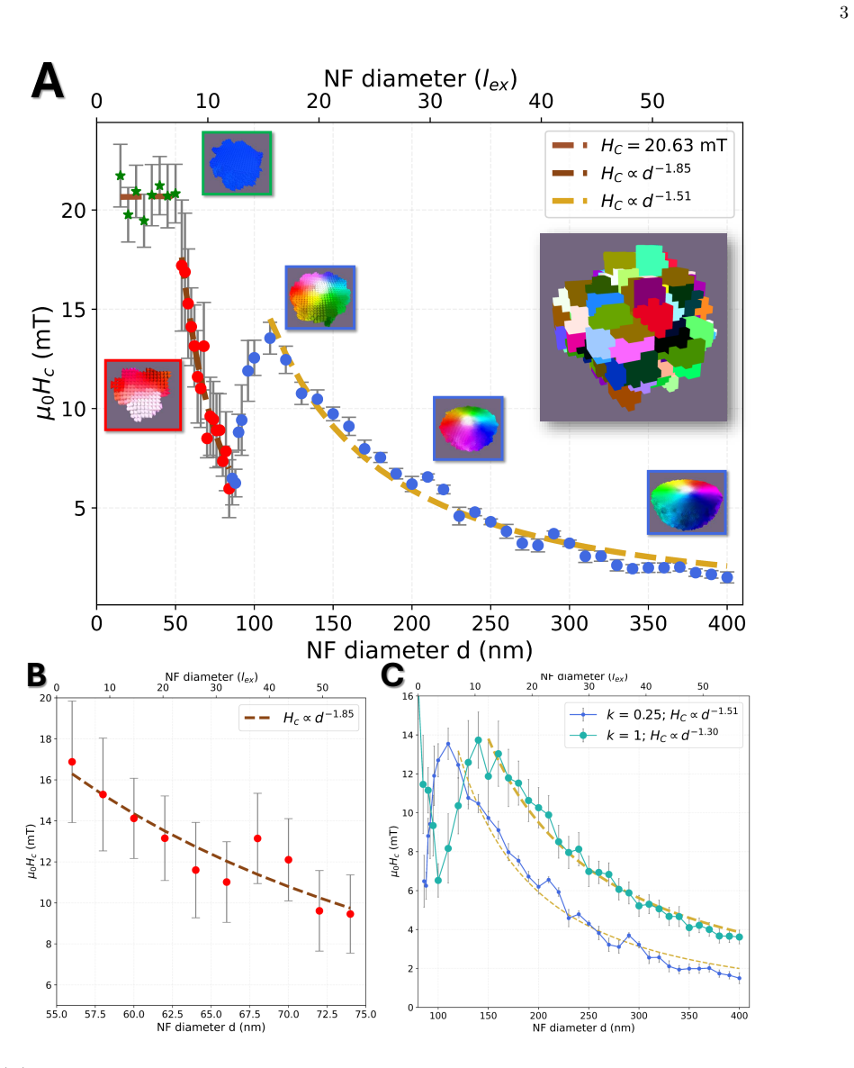
The central discovery is a mechanism-based coercivity–size map for realistic iron-oxide nanoflowers. Above the single-domain threshold the flower hosts a magnetic vortex: an inhomogeneous spin texture in which moments circulate around a small, strongly magnetized core. In the disordered flower the coercivity rises with size just after vortex nucleation because the randomly oriented easy axes of the grains pin the vortex core, then reaches a maximum of about 14 mT at $d \approx 110$ nm for inter-grain exchange factor $k = 0.25$, and decays as $d^{-1.5}$ as the core shrinks. The reversal switches at a vortex-core volume fraction $V_{\mathrm{core}}/V_{\mathrm{NF}} \approx 1/3$: below that threshold the core reverses immediately along the field, above it the surrounding flux-closure moments rotate the core perpendicular to the field before switching. The same threshold in a perfect sphere leads to vanishing coercivity, so the peak is a signature of grain anisotropy and grain-boundary pinning, not of geometry alone. The location of the peak moves to larger sizes when grains are fully exchange-coupled (about 150 nm for $k = 1$), showing that disorder tunes the sweet spot while the peak height comes from the grains themselves.
Load-bearing premise
The load-bearing premise is that coercivity from static hysteresis is a valid proxy for heating under clinical alternating fields, so that the size maximizing coercivity also maximizes heat release, together with the modelling premise that a random-grained particle with one reduced inter-grain exchange factor represents a real nanoflower.
Editorial extensions
If this is right
- At the calibrated microstructure (15 nm grains, $k = 0.25$), nanoflowers near $d \approx 110$ nm should display a coercivity maximum of roughly 14 mT, bracketed by lower values on both sides.
- Tuning the grain size shifts the sweet spot: fully exchange-coupled grains place the peak at about 150 nm, so a larger number of weaker grain-boundary links behaves like a smaller number of stronger ones.
- Defect-free nanospheres of the same material should show a monotonic coercivity decline beyond the single-domain limit, with no secondary peak, because they lack the random easy axes that pin the core.
- Beyond the peak, the coercivity decays as a power law: approximately $d^{-1.5}$ at $k = 0.25$ and $d^{-1.3}$ at $k = 1$, consistent with earlier vortex-state scalings.
- The switch between reversal modes is controlled by the vortex-core volume fraction $V_{\mathrm{core}}/V_{\mathrm{NF}} \approx 1/3$, a geometric threshold that applies to both disordered and ideal spheres, while only the pinning landscape decides whether that threshold yields a coercivity peak.
Reading between the lines
- The paper's coercivity proxy could be tested directly by a finite-temperature alternating-field simulation that computes hysteresis-loop area, and by calorimetry on monodisperse nanoflowers near 70–150 nm; nothing in the text guarantees the loop area peaks at exactly the coercivity peak.
- The $V_{\mathrm{core}}/V_{\mathrm{NF}} \approx 1/3$ threshold may serve as a cheap screening rule for other soft-magnetic particle geometries, since it is derived from the vortex profile rather than from the specific grain model; this universality is not claimed by the paper.
- Because the peak height is nearly independent of the exchange-reduction factor while the peak position is not, grain-size control offers an experimentally accessible dial for the sweet spot; this suggests a similar tuning strategy for other multicore particle architectures such as chains or clusters, a transfer the paper leaves implicit.
Editorial analysis
A structured set of objections, weighed in public.
Referee Report
Summary. The paper presents large-scale micromagnetic simulations of iron-oxide nanoflowers from d = 10 nm to 400 nm, using a Voronoi grain structure with random anisotropy axes and a reduced inter-grain exchange factor k to model spin disorder. It reports a coercivity-versus-size map with three regimes: a Stoner-Wohlfarth coherent-rotation regime below roughly 50 nm, a domain-wall-nucleation regime between about 50 and 75 nm, and a vortex regime above about 70 nm. In the vortex regime the authors find a non-monotonic coercivity with a secondary maximum near d = 110 nm for k = 0.25, which is absent in defect-free nanospheres. They attribute the peak to the interplay of grain anisotropy and grain-boundary pinning, identify a Vcore/VNF = 1/3 threshold separating core-dominated from flux-closure-dominated reversal, and conclude that the grain size can tailor this coercivity 'sweet spot' for magnetic hyperthermia.
Significance. If the central mechanism is correct, the work is a valuable step beyond macrospin descriptions of nanoflowers: it provides a coercivity-size map over a wide diameter range, uses 15-25 random realizations per size, includes a clear baseline comparison with fully coupled nanoflowers and defect-free nanospheres, and proposes a concrete geometric threshold (Vcore/VNF = 1/3) for the reversal-mode crossover. The separation of reversal modes and the demonstration that grain disorder rather than mere size creates the secondary coercivity peak are physically interesting and testable. However, the practical claim that the coercivity peak marks a 'hyperthermia sweet spot' is not supported by the simulations as presented, because heating performance in magnetic hyperthermia depends on the hysteresis-loop area under the applied AC field and frequency, not on the zero-temperature DC coercivity.
major comments (3)
- [Section II.A and Conclusion] The paper's central practical claim — that the coercivity peak located at the reversal-mode transition tailors the 'hyperthermia sweet spot' — is unsupported because heating performance is identified with zero-temperature DC coercivity without computing the AC hysteresis-loop area or specific absorption rate. The Introduction states that heating performance is 'represented by the coercivity,' but MHT losses are proportional to ∮M·dH at the applied field amplitude and frequency, and clinical fields (typically μ0H0 ≈ 10–30 mT at 100–500 kHz) may be below the reported peak coercivity of about 14 mT, so minor-loop and loop-shape effects can decouple coercivity from dissipated power. Please compute loop areas or SAR at relevant AC conditions (or at least at finite temperature for representative sizes), or restrict the conclusions to coercivity rather than hyperthermia efficiency.
- [Section II.B and Fig. 2(E)] The Vcore/VNF = 1/3 threshold is asserted for nanoflowers, but the quantitative core-volume data shown in Fig. 2(E) are for the ideal nanosphere only. The sentence 'In the NF, we find the same Vcore/VNF = 1/3 threshold' is not backed by a corresponding plot or tabulated NF core-volume analysis in the main text or the available supplement. Because this threshold is load-bearing for the explanation of the coercivity peak location, the authors should present NF core-volume fractions versus diameter, or explicitly state where the supporting data appear.
- [Section II.A and Methods] The absolute coercivity scale and the location of the sweet spot depend on the inter-grain coupling k = 0.25, which is selected to match experimental coercivity values [19,44], but no quantitative comparison between simulated and measured coercivities is shown, and no sensitivity analysis for k is reported. Since the abstract and Fig. 1 emphasize 'excellent agreement' and the practical field-amplitude comparisons use the simulated peak height, please provide the experimental comparison plot, the statistical spread over the 15-25 random realizations, and at least a brief statement of how the peak height and location vary over the plausible range of k and material parameters.
minor comments (6)
- [Section II.A] The text contains a typo: 'Stoner-Wolhfarth' should be 'Stoner-Wohlfarth'.
- [Fig. 1(A) caption] The caption contains 'nmnm' and should read 'nm'.
- [Conclusion] The word 'offeres' should be 'offers'.
- [Section II.A] The d^-1.85 power law is fit to a very small number of points in the 50-75 nm range; please report the fit range, R^2, and standard error, and avoid overinterpreting the difference from the d^-1.5 expectation.
- [Fig. 2 caption and Section II.B] The text describes Fig. 2(C) for d = 120 nm, whereas the caption states d = 110 nm; this inconsistency should be corrected.
- [Data Availability] The statement that 'all data supporting the findings are included' would be more useful if simulation scripts or a repository link were provided, given that the results depend on specific random Voronoi tessellations and Mumax3 settings.
Circularity Check
The coercivity map and reversal-mechanism analysis are genuine simulation outputs, but the advertised 'hyperthermia sweet spot' is defined as the coercivity maximum, so the practical MHT prediction reduces to the simulated input by definition.
-
self definitional
[Section I (Introduction) and Section IIA, Fig. 1C caption]
""heating performance, represented by the coercivity" (Sec. I); "the maximum of the coercivity (and thus, of the hyperthermia sweet spot)" (Fig. 1C caption)."
The paper equates heating performance with coercivity and then labels the coercivity maximum the 'hyperthermia sweet spot.' The conclusion that tuning the coercivity peak tailors hyperthermia is therefore a restatement of the coercivity-size map, not an independent heating result. No hysteresis-loop area, SAR, or AC finite-frequency loss is computed anywhere in the study. Since MHT heating is set by the loop area at applied field amplitude and frequency, a lower-coercivity configuration can enclose more area; the 'sweet spot' claim is valid only relative to the paper's own definitional proxy, making the practical prediction equivalent to its input by construction.
full rationale
The central coercivity-vs-size map is a self-contained micromagnetic simulation result. The secondary maximum, the two reversal modes, and the Vcore/VNF=1/3 threshold are extracted from the simulations and are not used to set any model constant; the perfect-sphere (NS) comparison provides an independent control showing that the peak is absent without grain disorder. The choice k=0.25 is calibrated against experimental coercivity data (refs. [19,44], partly the authors' own), which makes the absolute coercivity magnitudes and the k-dependent shift of the peak position partially fitted; however, the non-monotonic shape and the reversal-mode transition are not forced by this calibration. The only step that reduces by construction is the identification of the coercivity maximum with the 'hyperthermia sweet spot,' which follows from the stated representation of heating performance by coercivity. That definitional reduction affects the practical MHT guidance rather than the underlying micromagnetic physics, so the circularity is partial rather than total. The missing link—loop area or SAR under clinical AC fields—is a correctness risk separate from circularity, and the paper does not claim to compute it.
Assumptions & free parameters
free parameters (2)
- Inter-grain exchange coupling factor k =
0.25 (default); also 0, 0.15, 0.4, 0.5, 0.75, 1
- Grain (Voronoi cell) size =
15 nm (and 7 nm in Fig. 3)
assumptions (5)
- domain assumption A nanoflower can be represented as a quasi-spherical Voronoi cluster of single-domain grains with independently random uniaxial easy axes.
- domain assumption Inter-grain exchange disorder is captured by a single scalar k that multiplies the exchange stiffness at all grain boundaries.
- domain assumption Coercivity is a valid proxy for magnetic hyperthermia heating performance (larger HC implies larger hysteresis losses).
- domain assumption Material parameters Ms = 400 kA/m, A = 10 pJ/m, Ku = 10^4 J/m^3 are representative of the maghemite nanoflowers under study.
- domain assumption Nanoflowers below 50 nm diameter are well approximated by a single grain with coherent Stoner-Wohlfarth rotation.
Cite this review
Pith. "Pith review of Coercivity-size map of magnetic nanoflowers: spin disorder tunes the vortex reversal mechanism and tailors the hyperthermia sweet spot." pith.science (2026). https://pith.science/paper/ON277Q7H
@misc{pith2026250702154,
  author       = {Pith},
  title        = {Pith review of: Coercivity-size map of magnetic nanoflowers: spin disorder tunes the vortex reversal mechanism and tailors the hyperthermia sweet spot},
  year         = {2026},
  howpublished = {\url{https://pith.science/paper/ON277Q7H}},
  note         = {Machine review of arXiv:2507.02154}
}
read the original abstract
Iron-oxide nanoflowers (NFs) are one of the most efficient nanoheaters for magnetic hyperthermia therapy (MHT). However, the physics underlying the spin texture of disordered iron-oxide nanoparticles beyond the single-domain limit remains still poorly understood. Using large-scale micromagnetic simulations we completely map the magnetization of NFs over an unprecedented size range, from 10 to 400 nm in diameter, connecting their microstructure to their macroscopic magnetic response. Above the single domain (d > 50 nm), the magnetization folds into a vortex state, within which the coercivity describes a secondary maximum, not present for non-disordered nanoparticles. We have extended our understanding by resolving also the NF magnetization dynamics, capturing the physics of the magnetization reversal. Within the vortex regime, two distinct reversal modes exist: i) A core-dominated one, in which the core immediately switches along the direction of the applied field, resulting in an increasing coercivity for larger sizes; and ii) a flux-closure dominated reversal mode, going through the perpendicular alignment of the vortex core to the field, resulting in a decreasing coercivity-size dependence. The coercivity maximum is located at the transition between both reversal modes, and results from the combination of grain anisotropy and grain-boundary pinning: weak (but non-negligible) inter-grain exchange keeps the vortex profile coherent, yet allows the core to be pinned by the random anisotropy easy axes of the single grains, maximizing magnetic losses. Our results provide the first full description of spin textures in iron oxide NFs beyond the macrospin framework, and clarify the role of internal spin disorder in magnetic hyperthermia heating. By adjusting the grain size, the coercivity "sweet spot" can be tailored, offering a practical route to next-generation, high-efficiency nanoheaters.
Figures


Reference graph
Works this paper leans on
-
[1]
Material parameters typical for iron oxide were used based on literature
We find good agreement with the experimental results for k = 0.25 [19, 44, 48, 49]. Material parameters typical for iron oxide were used based on literature. These include saturation magnetization Ms = 400× 103 A/m [74, 75], exchange stiffness A = 10 pJ /m [76], and uniaxial magnetocrystalline anisotropy Ku = 104 J/m3 [59, 74, 77, 78]. Each simulated size...
work page 2020
-
[2]
hyperthermia sweet spot
The fitting of our simulated results, included in Figure 1(B), yields HC ∝ d−1.85, indicating a reasonable agreement with this simplified model. We attribute the slightly steeper decline than the -1.5 power law expected for perfect spheres to disorder associated with the irregular edges of the nanoflowers. The third regime (blue symbols in Figure 1A) star...
-
[3]
As observed in Figure 2(E), this happens for diametersd >107 nm, when the vortex core is no longer the main volume and the flux-closure mo- ments along the x and y directions carry more volume. The NS thus minimizes its energy by rotating the mag- netization and aligning about half (a bit more due to de- formation of the profile) of these flux-closure spi...
-
[4]
K. Wu, D. Su, J. Liu, R. Saha, and J.-P. Wang, Nan- otechnology 30, 502003 (2019)
work page 2019
- [5]
- [6]
- [7]
- [8]
Show all 81 references
-
[9]
Coene and J
A. Coene and J. Leliaert, Journal of Applied Physics131 (2022)
2022
-
[10]
Rubia-Rodríguez, A
I. Rubia-Rodríguez, A. Santana-Otero, S. Spassov, E. Tombácz, C. Johansson, P. De La Presa, F. J. Teran, M. d. P. Morales, S. Veintemillas-Verdaguer, N. T. Thanh, et al., Materials14, 706 (2021). 10
2021
-
[11]
R. D. Issels, European journal of cancer44, 2546 (2008)
2008
-
[12]
Z. V. Díaz-Riascos, M. Llaguno-Munive, N. Lafuente- Gómez, Y. Luengo, S. Holmes, J. Volatron, O. Ibarrola, S. Mancilla, F. Sarno, J. J. Aguirre,et al., ACS Applied Materials & Interfaces (2025)
2025
-
[13]
Durando, F
G. Durando, F. Vurro, F. Saba, A. M. Ivory, R. de Melo Baesso, P. Miloro, and A. E. Spinelli, Sci- entific Reports14, 19878 (2024)
2024
-
[14]
Wu, J.-P
K. Wu, J.-P. Wang, N. A. Natekar, S. Ciannella, C. González-Fernández, J. Gomez-Pastora, Y. Bao, J. Liu, S. Liang, X. Wu,et al., Nanotechnology (2024)
2024
-
[15]
A. C. Anselmo and S. Mitragotri, Bioengineering & translational medicine1, 10 (2016)
2016
-
[16]
A. C. Anselmo and S. Mitragotri, Bioengineering & translational medicine4, e10143 (2019)
2019
-
[17]
A. C. Anselmo and S. Mitragotri, Bioengineering & translational medicine6, e10246 (2021)
2021
-
[18]
Soetaert, P
F. Soetaert, P. Korangath, D. Serantes, S. Fiering, and R. Ivkov, Advanced drug delivery reviews163, 65 (2020)
2020
-
[19]
Berns, U
A. Berns, U. Ringborg, J. E. Celis, M. Heitor, N. K. Aaronson, N. Abou-Zeid, H.-O. Adami, K. Apostolidis, M. Baumann, A. Bardelli,et al., Molecular oncology14, 1589 (2020)
2020
-
[20]
A. G. Roca, L. Gutiérrez, H. Gavilán, M. E. F. Brollo, S. Veintemillas-Verdaguer, and M. del Puerto Morales, Advanced drug delivery reviews138, 68 (2019)
2019
-
[21]
Gavilán, S
H. Gavilán, S. K. Avugadda, T. Fernández-Cabada, N. Soni, M. Cassani, B. T. Mai, R. Chantrell, and T. Pel- legrino, Chemical Society Reviews50, 11614 (2021)
2021
-
[22]
E. M. Jefremovas, L. Gandarias, I. Rodrigo, L. Marcano, C. Grüttner, J. Á. García, E. Garayo, I. Orue, A. García- Prieto, A. Muela,et al., IEEE Access9, 99552 (2021)
2021
-
[23]
Gandia, L
D. Gandia, L. Gandarias, I. Rodrigo, J. Robles-García, R.Das, E.Garaio, J.Á.García, M.-H.Phan, H.Srikanth, I. Orue, et al., Small15, 1902626 (2019)
2019
-
[24]
Mekseriwattana, N
W. Mekseriwattana, N. Silvestri, R. Brescia, E. Tiryaki, J. Barman, F. G. Mohammadzadeh, N. Jarmouni, and T. Pellegrino, Advanced Functional Materials 35, 2413514 (2025)
2025
-
[25]
Lappas, G
A. Lappas, G. Antonaropoulos, K. Brintakis, M. Vasi- lakaki, K. N. Trohidou, V. Iannotti, G. Ausanio, A. Kostopoulou, M. Abeykoon, I. K. Robinson, et al. , Physical Review X9, 041044 (2019)
2019
-
[26]
Serantes and D
D. Serantes and D. Baldomir, Nanomaterials11, 2786 (2021)
2021
-
[27]
M. L. Etheridge, K. R. Hurley, J. Zhang, S. Jeon, H. L. Ring, C. Hogan, C. L. Haynes, M. Garwood, and J. C. Bischof, Technology2, 214 (2014)
2014
-
[28]
D. Ho, X. Sun, and S. Sun, Accounts of chemical research 44, 875 (2011)
2011
-
[29]
Goiriena-Goikoetxea, A
M. Goiriena-Goikoetxea, A. García-Arribas, M. Rouco, A. Svalov, and J. Barandiaran, Nanotechnology 27, 175302 (2016)
2016
-
[30]
Goiriena-Goikoetxea, D
M. Goiriena-Goikoetxea, D. Muñoz, I. Orue, M. Fernández-Gubieda, J. Bokor, A. Muela, and A. García-Arribas, Applied Physics Reviews7 (2020)
2020
-
[31]
N. Usov, M. Nesmeyanov, and V. Tarasov, Scientific re- ports 8, 1224 (2018)
2018
-
[32]
Di Fratta, C
G. Di Fratta, C. Serpico, and M. d’Aquino, Physica B: Condensed Matter407, 1368 (2012)
2012
-
[33]
Tauxe, H
L. Tauxe, H. N. Bertram, and C. Seberino, Geochemistry, Geophysics, Geosystems3, 1 (2002)
2002
-
[34]
W. F. Brown Jr, Annals of the New York Academy of Sciences 147, 463 (1969)
1969
-
[35]
W. F. Brown Jr, Journal of Applied Physics49, 1937 (1978)
1978
-
[36]
M. E. Schabes and H. N. Bertram, Journal of Applied Physics 64, 1347 (1988)
1988
-
[37]
Hertel and H
R. Hertel and H. Kronmüller, Journal of magnetism and magnetic materials238, 185 (2002)
2002
-
[38]
W. Rave, K. Fabian, and A. Hubert, Journal of Mag- netism and Magnetic Materials190, 332 (1998)
1998
-
[39]
A. R. Muxworthy, W. Williams, A. P. Roberts, M. Win- klhofer, L. Chang, and M. Posfai, Geochemistry, Geo- physics, Geosystems14, 2430 (2013)
2013
-
[40]
Betto and J
D. Betto and J. Coey, Journal of Applied Physics115 (2014)
2014
-
[41]
W. Gan, M. Chandra Sekhar, D. Wong, I. Purnama, S. Chiam, L. Wong, and W. Lew, Applied Physics Letters 105 (2014)
2014
-
[42]
Kákay and L
A. Kákay and L. Varga, Journal of applied physics97 (2005)
2005
-
[43]
A. Witt, K. Fabian, and U. Bleil, Earth and Planetary Science Letters233, 311 (2005)
2005
-
[44]
H. Gao, T. Zhang, Y. Zhang, Y. Chen, B. Liu, J. Wu, X. Liu, Y. Li, M. Peng, Y. Zhang,et al., Journal of Ma- terials Chemistry B8, 515 (2020)
2020
-
[45]
G. R. Lewis, J. C. Loudon, R. Tovey, Y.-H. Chen, A. P. Roberts, R. J. Harrison, P. A. Midgley, and E. Ringe, Nano letters20, 7405 (2020)
2020
-
[46]
G. C. Concas, W. B. Jalil, R. J. Caraballo-Vivas, V. L. Gomes, M. A. Camarena, M. B. Fontes, S. K. Sharma, T. P. Almeida, E. C. Santos, and F. Garcia, Journal of Alloys and Compounds , 177022 (2024)
2024
-
[47]
C. Moya, M. Escoda-Torroella, J. Rodríguez-Álvarez, A. I. Figueroa, Í. García, I. B. Ferrer-Vidal, A. Gallo- Cordova, M. P. Morales, L. Aballe, A. F. Rodríguez, et al., Nanoscale16, 1942 (2024)
2024
-
[48]
Bender, J
P. Bender, J. Fock, C. Frandsen, M. F. Hansen, C. Bal- ceris, F. Ludwig, O. Posth, E. Wetterskog, L. K. Bogart, P. Southern,et al., The Journal of Physical Chemistry C 122, 3068 (2018)
2018
-
[49]
L. G. Vivas, R. Yanes, D. Berkov, S. Erokhin, M. Ber- sweiler, D. Honecker, P. Bender, and A. Michels, Physical Review Letters125, 117201 (2020)
2020
-
[50]
Hugounenq, M
P. Hugounenq, M. Levy, D. Alloyeau, L. Lartigue, E. Dubois, V. Cabuil, C. Ricolleau, S. Roux, C. Wilhelm, F. Gazeau, et al., The Journal of Physical Chemistry C 116, 15702 (2012)
2012
-
[51]
Storozhuk, M
L. Storozhuk, M. O. Besenhard, S. Mourdikoudis, A. P. LaGrow, M. R. Lees, L. D. Tung, A. Gavriilidis, and N. T. K. Thanh, ACS Applied Materials & Interfaces 13, 45870 (2021)
2021
-
[52]
Gavilán, E
H. Gavilán, E. H. Sánchez, M. E. Brollo, L. Asín, K. K. Moerner, C. Frandsen, F. J. Lázaro, C. J. Serna, S. Veintemillas-Verdaguer, M. P. Morales, et al. , ACS omega 2, 7172 (2017)
2017
-
[53]
B.G.Prajapati, K.Verma, S.Sharma,andD.U.Kapoor, Medical Oncology41, 295 (2024)
2024
-
[54]
Gatel, F
C. Gatel, F. J. Bonilla, A. Meffre, E. Snoeck, B. Warot- Fonrose, B. Chaudret, L.-M. Lacroix, and T. Blon, Nano Letters 15, 6952 (2015)
2015
-
[55]
Pratami Sinaga, M
E. Pratami Sinaga, M. P. Adams, M. Bersweiler, L. G. Vivas, E. H. Hasdeo, J. Leliaert, P. Bender, D. Honecker, and A. Michels, Physical Review B107, 014416 (2023)
2023
-
[56]
A. Lak, S. Disch, and P. Bender, Advanced Science8, 2002682 (2021). 11
2021
-
[57]
E. Lima, A. Brandl, A. Arelaro, and G. Goya, Journal of Applied Physics99 (2006)
2006
-
[58]
Khurshid, W
H. Khurshid, W. Li, M.-H. Phan, P. Mukherjee, G. C. Hadjipanayis, and H. Srikanth, Applied Physics Letters 101 (2012)
2012
-
[59]
Zákutná, D
D. Zákutná, D. Nižňansk` y, L. C. Barnsley, E. Babcock, Z. Salhi, A. Feoktystov, D. Honecker, and S. Disch, Phys- ical Review X10, 031019 (2020)
2020
-
[60]
Poon and G
K. Poon and G. Singh, Applied Materials Today 44, 102787 (2025)
2025
-
[61]
Gavilán, K
H. Gavilán, K. Simeonidis, E. Myrovali, E. Mazarío, O. Chubykalo-Fesenko, R. Chantrell, L. Balcells, M. An- gelakeris, M. P. Morales, and D. Serantes, Nanoscale13, 15631 (2021)
2021
-
[62]
Sharma, H
J.Borchers, K.Krycka, B.Bosch-Santos, E.deLimaCor- rea, A. Sharma, H. Carlton, Y. Dang, M. Donahue, C. Grüttner, R. Ivkov,et al., Small Structures6, 2400410 (2025)
2025
-
[63]
Tannous and J
C. Tannous and J. Gieraltowski, European journal of physics 29, 475 (2008)
2008
-
[64]
Carrey, B
J. Carrey, B. Mehdaoui, and M. Respaud, Journal of ap- plied physics109 (2011)
2011
-
[65]
S.-h. Noh, W. Na, J.-t. Jang, J.-H. Lee, E. J. Lee, S. H. Moon, Y. Lim, J.-S. Shin, and J. Cheon, Nano letters12, 3716 (2012)
2012
-
[66]
Sung Lee, J
J. Sung Lee, J. Myung Cha, H. Young Yoon, J.-K. Lee, and Y. Keun Kim, Scientific reports5, 12135 (2015)
2015
-
[67]
Demortiere, P
A. Demortiere, P. Panissod, B. Pichon, G. Pourroy, D. Guillon, B. Donnio, and S. Bégin-Colin, Nanoscale 3, 225 (2011)
2011
-
[68]
Kneller and F
E. Kneller and F. Luborsky, Journal of Applied Physics 34, 656 (1963)
1963
-
[69]
Hergt, S
R. Hergt, S. Dutz, and M. Röder, Journal of Physics: Condensed Matter20, 385214 (2008)
2008
-
[70]
Heider, D
F. Heider, D. J. Dunlop, and N. Sugiura, Science236, 1287 (1987)
1987
-
[71]
Because we always investigate several realization of ran- domly generated geometries, both processes happen in an overlapping size range, and we identify the peak with the transition of which process dominates the resulting trend in the coercivity
-
[72]
S. B. Attanayake, M. D. Nguyen, A. Chanda, J. Alonso, I. Orue, T. R. Lee, H. Srikanth, and M.-H. Phan, arXiv preprint arXiv:2411.17172 (2024)
2024 arXiv
-
[73]
Erokhin and D
S. Erokhin and D. Berkov, Physical Review Applied7, 014011 (2017)
2017
-
[74]
M. Kure, F. L. Durhuus, C. Frandsen, and M. Beleggia, Journal of Physics D: Applied Physics (2025)
2025
-
[75]
Vansteenkiste, J
A. Vansteenkiste, J. Leliaert, M. Dvornik, M. Helsen, F. Garcia-Sanchez, and B. Van Waeyenberge, AIP Ad- vances4, 107133 (2014)
2014
-
[76]
Leliaert, B
J. Leliaert, B. Van de Wiele, A. Vansteenkiste, L. Laur- son, G. Durin, L. Dupré, and B. Van Waeyenberge, Jour- nal of Applied Physics115, 233903 (2014)
2014
-
[77]
A. G. Roca, J. F. Marco, M. d. P. Morales, and C. J. Serna, The Journal of Physical Chemistry C111, 18577 (2007)
2007
-
[78]
Shokrollahi, Journal of Magnetism and Magnetic Ma- terials 426, 74 (2017)
H. Shokrollahi, Journal of Magnetism and Magnetic Ma- terials 426, 74 (2017)
2017
-
[79]
E. P. Sinaga, M. P. Adams, E. H. Hasdeo, and A. Michels, Physical Review B110, 054404 (2024)
2024
-
[80]
Gross, S
B. Gross, S. Philipp, E. Josten, J. Leliaert, E. Wetter- skog, L. Bergström, and M. Poggio, Physical Review B 103, 014402 (2021)
2021
-
[81]
Pisane, S
K. Pisane, S. Singh, and M. Seehra, Applied Physics Let- ters 110 (2017)
2017
Reviewed August 6, 2026 · model on record in the stance chip above.
Discussion (0). Continue with ORCID to comment.