REVIEW 4 major objections 7 minor 16 references
Speckle2Self: Self-Supervised Ultrasound Speckle Reduction Without Clean Data
T0 review · 4 major / 7 minor · reviewed 2026-08-06 · deepseek-v4-flash
Pith's one-line read A single noisy ultrasound image is enough to train a speckle-removal network.
desk verdict A solid self-supervised despeckling paper whose central MSP assumption is partly violated by low-pass filtering; worth reviewing, but it needs AP-BSN/C-BSN baselines and a smoothing check. read the letter →
The pith
A machine-rendered reading of the paper's core claim, the machinery that carries it, and where it could break.
The reading
What carries the argument
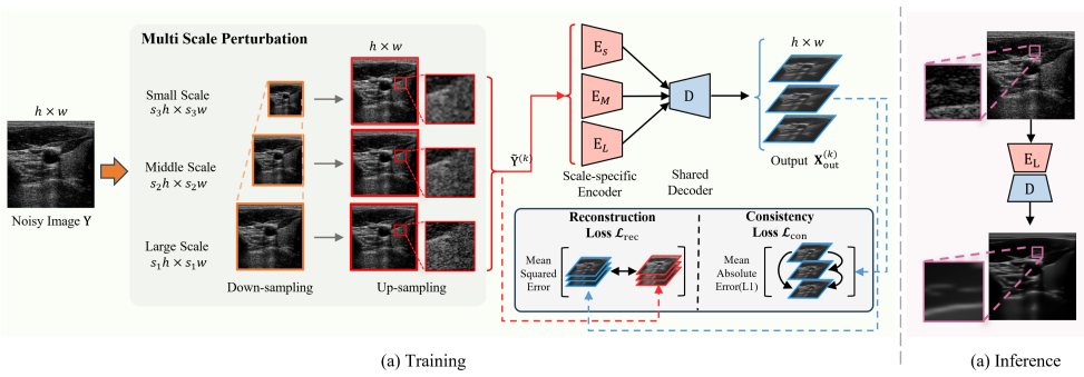
The load-bearing mechanism is the multi-scale perturbation (MSP): controlled downsampling and upsampling of the input at scale factors 1.0, 0.5, and 0.25, which is assumed to leave the clean tissue structure X unchanged while reshuffling the speckle pattern. Around this, the framework is a multi-encoder network, one encoder per scale sharing a single decoder, trained with a reconstruction loss (MSE to each perturbed view) and a consistency loss (L1 between all pairs of reconstructed outputs). The low-rank prior on clean tissue, the assumption that anatomy lies on a low-rank subspace while speckle is sparse or high-rank, is what gives the consistency loss its meaning: the network is rewarded for outputs that are common across scales and penalized for outputs that follow scale-specific speckle.
What would settle it
On a simulated phantom with a known clean image X and sharp point scatterers, run Speckle2Self and compare the output to X and to X passed through the same downsampling-upsampling at scale 0.25; if the output is closer (higher SSIM) to the smoothed version than to X, the assumption that MSP preserves structure fails and the method is doing low-pass estimation rather than true despeckling.
Extended reading notes
Core claim
On the paper's own terms, the discovery is that ultrasound speckle can be removed from a single noisy image by treating the clean anatomy as a low-rank signal and the speckle as sparse, scale-dependent variation. The multi-scale perturbation (MSP) operation, with scale factors 1.0, 0.5, and 0.25, is claimed to preserve the shared anatomical structure X while producing distinct speckle realizations N(k) for each scale (Eq. 7). Three scale-specific encoders and a shared decoder are trained with an MSE reconstruction loss toward each perturbed input plus an L1 consistency loss between all pairs of reconstructions. The consistency loss forces the network to find what is invariant across scales, and the authors report that MSE reconstruction plus L1 consistency is the only loss combination that removes speckle, while symmetric combinations blur structure or retain speckle. The outcome is a despeckled image that, in their experiments, beats established filters (SRAD, NLM, BM3D, OBNLM) and prior self-supervised methods (N2N, N2V, Noise2Self, Neighbor2Neighbor, DIP, ZS-N2N) on SSIM, LPIPS, and homogeneity, with only a supervised method trained on clean targets scoring higher.
Load-bearing premise
The whole training signal rests on the premise that downsampling and upsampling changes only the speckle while leaving the true anatomy untouched, but downsampling also removes fine tissue detail, so the shared structure is really a smoothed image, which likely explains the reported boundary blurring.
Editorial extensions
If this is right
- Speckle reduction no longer requires clean images, paired noisy observations, or plane-wave hardware; one B-mode frame suffices for training, with about 7 ms per image at inference.
- Because no tissue-specific noise model is assumed, the same trained network transfers to unseen central frequencies (3.75 to 9.38 MHz) and to images from two unseen commercial scanners, with zero-shot use and small fine-tuning gains.
- Reducing speckle improves downstream analysis: on carotid artery segmentation with a rule-based flood-fill segmenter, AUC rose from 0.589 to 0.816 and IoU from 0.175 to 0.497 after Speckle2Self denoising.
- The method is architecture-light (about three million parameters) and operates on standard B-mode output, so it can be deployed on portable and point-of-care systems where clean data do not exist.
Reading between the lines
- If cross-scale agreement is the right training signal, the same perturbation idea could be tested on other deterministic, spatially correlated image degradations, such as CT metal artifacts or MRI ghosting, where independent-noise self-supervision fails.
- The reported boundary blurring suggests the method may be estimating a low-pass-filtered version of the anatomy rather than the true X; a direct test would compare denoised output against both clean X and its downsampled-upsampled version on synthetic data.
- Because speckle is itself a deterministic signal used in elastography and motion tracking, the network's ability to separate speckle from tissue could be repurposed to extract speckle maps, not only to discard them.
- A cheap diagnostic for the mechanism is replacing the three-scale perturbation with two scales plus stronger perturbation to see whether performance tracks the degree of speckle decorrelation.
Editorial analysis
A structured set of objections, weighed in public.
Referee Report
Summary. The paper proposes Speckle2Self, a self-supervised method for ultrasound speckle reduction that requires only single noisy B-mode images. The method applies a multi-scale perturbation (MSP) operation, implemented as downsampling-upsampling at scales {1.0, 0.5, 0.25}, to create three variants of the input; it assumes each variant can be written as X + N^(k) with shared clean structure X and scale-specific speckle N^(k), and trains three encoders plus a shared decoder with an MSE reconstruction loss and an L1 cross-scale consistency loss, motivated by low-rank/sparse decomposition. The paper evaluates the method on three synthetic MUST datasets, in-vivo carotid images from a Clarius scanner, cross-configuration and cross-device generalization, a loss/interpolation/scale ablation, and a downstream carotid segmentation experiment.
Significance. The proposed setting is practically important: ultrasound speckle is tissue-dependent and deterministic, so N2N-style paired observations and blind-spot assumptions are often inapplicable, and a method that works from single images would fill a real gap. The paper's strengths are its extensive comparison set, the use of a realistic simulator, the cross-device generalization attempt, the ablation study, and the reported 7 ms inference time. The central weakness is that the MSP assumption has not been separated from low-pass smoothing: the metrics on which Speckle2Self wins are substantially the ones improved by blur, and the in-vivo evidence is a single smoothing-favoring metric. If the authors can provide evidence that the method preserves high-frequency tissue structure while suppressing speckle, this would be a valuable contribution to the field.
major comments (4)
- [Section 3.3, Eq. (7)] The decomposition P_k(Y) = X + N^(k) is the load-bearing assumption, but downsampling-upsampling does not leave X unchanged: rescaling by 0.25 and interpolating back removes high-frequency tissue signal, so the component shared across scales is a low-pass filtered version of X, not X itself. With the loss in Eq. (11), the network can minimize both the MSE reconstruction term and the L1 consistency term by emitting a smoothed image that sits near all three filtered views; MSE is dominated by low-frequency fidelity. The Discussion's admission of 'slight blurring along tissue boundaries' is therefore not a cosmetic limitation but direct evidence for this mechanism, and the pattern in Table 4 (PSNR not best on S-I/S-II while SSIM/LPIPS/homogeneity improve) is consistent with low-pass filtering. Please provide a test that disentangles speckle suppression from smoothing, e.g., quantify P_k(X) - X on the synthetic clean targets, compare with a model trained on LPF perturbations of matched variation, or report a high-frequency/edge-preservation metric.
- [Section 4.4.4, Table 4 (Carotid column)] The in-vivo evaluation rests on a single no-reference metric, GLCM homogeneity, which explicitly rewards local smoothness; no error bars, confidence intervals, or paired statistical tests are reported for the 104 test images. Given the smoothing concern above, this single metric cannot support the statement that Speckle2Self 'consistently outperforms' other methods on real data. Please report per-image distributions or mean +/- std, add a structure-sensitive no-reference metric (e.g., edge/gradient preservation), and ideally a blinded clinician preference study.
- [Sections 4.4.1 and 4.6.2] The baseline set omits the most relevant self-supervised ultrasound methods, including the deep ultrasound denoising approach already cited as Goudarzi and Rivaz (2023) and the AP-BSN/C-BSN variants (Lee et al. 2022b; Jang et al. 2023) designed for spatially correlated noise; the plane-wave-based N2N variants cited in the Introduction are also not compared. The cross-device evaluation in Section 4.6.2 is qualitative only, with no metric reported on the Siemens and Cephasonics frames before or after fine-tuning. Without these quantitative comparisons, the claims of consistent superiority over self-supervised methods and strong cross-device generalization are not fully supported.
- [Section 4.3] The SVD singular-value decay analysis shows that Speckle2Self outputs have faster singular-value decay, but any low-pass filter produces this effect; without a control for detail preservation, the experiment does not demonstrate that the method preserves tissue structure while removing speckle. On the synthetic data where ground truth exists, please complement this analysis with a quantitative measure of how much of the clean image's high-frequency structure is retained.
minor comments (7)
- [Section 2.4] The heading 'Nosie2Void' contains a typo and should read 'Noise2Void'.
- [Section 5] The heading 'Discusssion' contains a typo and should read 'Discussion'.
- [Section 4.7] The text refers to the 'Folld fill algorithm'; this should be 'Flood Fill algorithm'.
- [Section 4.5.2] In the sentence describing Figure 9, 'the proposed MPS' should be 'the proposed MSP'.
- [Section 6] The sentence 'This denied result will enhance image consistency and thereby boost the development of reproducible computer-aided diagnosis' is malformed and unclear; it should be rewritten.
- [Section 4.4.1] The reference for N2S(*) is given as 'Krull et al. (2019)', but Noise2Self should cite Batson and Royer (2019).
- [Table 4] All synthetic and carotid metrics are reported as point estimates without standard deviations or confidence intervals, so the statistical significance of the performance gaps cannot be assessed.
Circularity Check
No circularity: the method is trained from single noisy images and validated on independent simulated ground truth; the low-rank assumption is a stated prior, not a fitted result.
full rationale
The derivation chain is self-contained and non-circular. The training signal is generated entirely from single noisy inputs via the MSP operation (Eq. 7), and the objective (Eq. 11) is a sum of reconstruction and cross-scale consistency losses with no fitted parameter tied to the reported metrics. Synthetic evaluation uses independently simulated clean ground truth (Sec. 4.2.1), and the method is benchmarked against external methods; there is no fitted input renamed as a prediction. The low-rank assumption (Sec. 3.2–3.3) is a stated prior, not a consequence of the data or of a self-citation. The Discussion's admission of 'slight blurring along tissue boundaries' is a correctness and assumption limitation, not circular reasoning. Self-citations to prior robotic-ultrasound work are contextual and not load-bearing; no central claim reduces to an author's prior result. The frequency-domain analysis in Sec. 3.3 is weak supporting evidence because low-frequency content is preserved by construction under downsampling, but this is not the core derivation and does not make the reported results equivalent to the inputs.
Assumptions & free parameters
free parameters (5)
- MSP scale factors =
s_k in {1.0, 0.5, 0.25}
- Number of MSP scales =
K = 3
- Loss weights =
1.0 for both L_rec and L_con
- Interpolation method =
bilinear (default)
- Training hyperparameters =
lr=0.001, batch=16, 3000 epochs
assumptions (5)
- domain assumption RF signal is multiplicative: Y_RF = X_RF * N_RF (Eq. 1).
- domain assumption After log compression, speckle becomes approximately additive in B-mode (Eq. 2).
- ad hoc to paper Clean tissue signal X is low-rank; speckle N is sparse or high-rank.
- ad hoc to paper The MSP operation produces variants with identical X and different N^(k) (Eq. 7).
- domain assumption Speckle in B-mode has non-zero mean and is deterministic, making N2N inapplicable.
Cite this review
Pith. "Pith review of Speckle2Self: Self-Supervised Ultrasound Speckle Reduction Without Clean Data." pith.science (2026). https://pith.science/paper/DPO3X537
@misc{pith2026250706828,
  author       = {Pith},
  title        = {Pith review of: Speckle2Self: Self-Supervised Ultrasound Speckle Reduction Without Clean Data},
  year         = {2026},
  howpublished = {\url{https://pith.science/paper/DPO3X537}},
  note         = {Machine review of arXiv:2507.06828}
}
read the original abstract
Image denoising is a fundamental task in computer vision, particularly in medical ultrasound (US) imaging, where speckle noise significantly degrades image quality. Although recent advancements in deep neural networks have led to substantial improvements in denoising for natural images, these methods cannot be directly applied to US speckle noise, as it is not purely random. Instead, US speckle arises from complex wave interference within the body microstructure, making it tissue-dependent. This dependency means that obtaining two independent noisy observations of the same scene, as required by pioneering Noise2Noise, is not feasible. Additionally, blind-spot networks also cannot handle US speckle noise due to its high spatial dependency. To address this challenge, we introduce Speckle2Self, a novel self-supervised algorithm for speckle reduction using only single noisy observations. The key insight is that applying a multi-scale perturbation (MSP) operation introduces tissue-dependent variations in the speckle pattern across different scales, while preserving the shared anatomical structure. This enables effective speckle suppression by modeling the clean image as a low-rank signal and isolating the sparse noise component. To demonstrate its effectiveness, Speckle2Self is comprehensively compared with conventional filter-based denoising algorithms and SOTA learning-based methods, using both realistic simulated US images and human carotid US images. Additionally, data from multiple US machines are employed to evaluate model generalization and adaptability to images from unseen domains. Project page: https://noseefood.github.io/us-speckle2self/
Figures


Figures from the paper (9 more)


Reference graph
Works this paper leans on
-
[5]
arXiv preprint arXiv:2504.00264
Diffdenoise: self-supervised medical image denoising with condi- tional diffusion models. arXiv preprint arXiv:2504.00264 . Frost, V .S., Stiles, J.A., Shanmugan, K.S., Holtzman, J.C.,
-
[8]
Motion magnification in robotic sonography: Enabling pulsation-aware artery segmentation, in: 2023 IEEE/RSJ International Conference on Intelligent Robots and Systems (IROS), IEEE. pp. 6565–6570. Huang, D., Li, C., Karlas, A., Chu, X., Au, K.S., Navab, N., Jiang, Z.,
work page 2023
-
[10]
Speckle- constrained filtering of ultrasound images, in: 2005 IEEE Computer Society Conference on Computer Vision and Pattern Recognition (CVPR’05), IEEE. pp. 547–552. Krissian, K., Westin, C.F., Kikinis, R., V osburgh, K.G.,
work page 2005
-
[11]
Learning sparse and low-rank priors for image recovery via iterative reweighted least squares minimization. arXiv preprint arXiv:2304.10536 . Lehtinen, J.,
-
[12]
arXiv preprint arXiv:1803.04189
Noise2noise: Learning image restoration without clean data. arXiv preprint arXiv:1803.04189 . Li, J., Su, T., Zhao, B., Lv, F., Wang, Q., Navab, N., Hu, Y ., Jiang, Z.,
-
[15]
Cross-modal attention for mri and ultrasound volume regis- tration, in: Medical Image Computing and Computer Assisted Intervention– MICCAI 2021: 24th International Conference, Strasbourg, France, Septem- ber 27–October 1, 2021, Proceedings, Part IV 24, Springer. pp. 66–75. Stevens, T.S., Meral, F.C., Yu, J., Apostolakis, I.Z., Robert, J.L., Van Sloun, R.J.,
work page 2021
-
[1973]
IEEE Transactions on Systems, Man, and Cybernetics SMC- 3, 610–621
Textural features for image classification. IEEE Transactions on Systems, Man, and Cybernetics SMC- 3, 610–621. doi: 10.1109/TSMC.1973.4309314. He, K., Zhang, X., Ren, S., Sun, J.,
arXiv 1973
-
[2005]
A non-local algorithm for image denoising, in: 2005 IEEE computer society conference on computer vision and pattern recognition (CVPR’05), Ieee. pp. 60–65. Calis, M., Mischi, M., van der Veen, A.J., Hunyadi, B.,
work page 2005
Show all 16 references
-
[2007]
IEEE Transactions on image processing 16, 2080–2095
Image denoising by sparse 3-d transform-domain collaborative filtering. IEEE Transactions on image processing 16, 2080–2095. Dantas, R.G., Costa, E.T., Leeman, S.,
-
[2010]
Signal Processing 90, 1963–1975
Ramp preserving perona– malik model. Signal Processing 90, 1963–1975. Chen, Y ., Fang, B., Li, H., Huang, L., Luo, J.,
1963
-
[2012]
Practical low- rank matrix approximation under robust l 1-norm, in: 2012 IEEE Confer- ence on Computer Vision and Pattern Recognition, IEEE. pp. 1410–1417. Zhou, Y ., Bi, Y ., Tong, W., Wang, W., Navab, N., Jiang, Z.,
2012
-
[2018]
BioMed research international 2018, 5137904
Machine learning in ultrasound computer- aided diagnostic systems: a survey. BioMed research international 2018, 5137904. Huang, T., Li, S., Jia, X., Lu, H., Liu, J.,
2018
-
[2021]
A bias-reducing loss function for ct image denoising, in: ICASSP 2021-2021 IEEE International Conference on Acoustics, Speech and Signal Processing (ICASSP), IEEE. pp. 1175–1179. Nyrnes, S.A., Fadnes, S., Wigen, M.S., Mertens, L., Lovstakken, L.,
2021
-
[2023]
Deep ultrasound denoising without clean data, in: Medical Imaging 2023: Ultrasonic Imaging and Tomography, SPIE. pp. 131–136. Gu, S., Li, Y ., Gool, L.V ., Timofte, R.,
2023
-
[2024]
arXiv preprint arXiv:2408.00714
Sam 2: Segment anything in images and videos. arXiv preprint arXiv:2408.00714 . Ronneberger, O., Fischer, P., Brox, T.,
-
[2025]
arXiv preprint arXiv:2506.21499
Lightweight physics- informed zero-shot ultrasound plane wave denoising. arXiv preprint arXiv:2506.21499 . Batson, J., Royer, L.,
Reviewed August 6, 2026 · model on record in the stance chip above.
Discussion (0). Sign in to comment.