REVIEW 4 major objections 5 minor 49 references
Semi-supervised learning and integration of multi-sequence MR-images for carotid vessel wall and plaque segmentation
T0 review · 4 major / 5 minor · reviewed 2026-08-06 · deepseek-v4-flash
Pith's one-line read This paper claims that the fusion point in a U-Net—bottleneck rather than input—drives multi-sequence carotid MRI segmentation quality, with the best binary Dice of 0.8725 and IoU of 0.7930.
desk verdict Competent empirical study whose headline fusion-point claim is confounded by model capacity; worth reviewing with a parameter-matched ablation required. read the letter →
The pith
A machine-rendered reading of the paper's core claim, the machinery that carries it, and where it could break.
The reading
What carries the argument
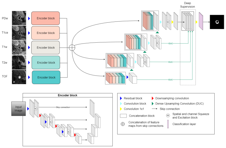
The central mechanism is the fusion point of a multi-sequence U-Net: each of the five sequences (PDw, T1w, T1ce, T2w, TOF) is fed to its own encoder branch, and the per-sequence feature maps are concatenated at the bottleneck and at every decoder level, so high-level modality-specific features are preserved before merging. Around this sit two supporting mechanisms: a one-way consistency semi-supervised scheme, where a clean teacher (exponential moving average of student weights) supervises a perturbed student under geometric and photometric transformations with an uncertainty-weighted MSE loss; and a coarse localization model whose loss includes a prior that penalizes wrong numbers of connected components and excessive asymmetry of the two carotid artery centers.
What would settle it
Deliberately misalign the five sequences by known small translations and rotations and repeat the input-vs-bottleneck fusion comparison: if bottleneck fusion no longer beats input fusion as misalignment grows, the claim that fusion-point selection is the operative factor is refuted; alternatively, a multi-center replication with consensus ground truth that fails to reproduce the reported Dice gap (0.8725 vs 0.8265) would also settle it.
Extended reading notes
Core claim
The paper establishes that fusion-point selection is a first-order design choice in U-Net-based multi-sequence segmentation. Processing each MRI sequence through its own encoder and merging the streams at the bottleneck—with the merged maps also forwarded through the decoder's skip connections—yields binary Dice 0.8725 and IoU 0.7930 for carotid vessel wall and plaque, beating the same architecture with input fusion (0.8549/0.7706) and a basic U-Net with input fusion (0.8265/0.7350). The same ordering holds for a basic U-Net, where bottleneck fusion adds more than four Dice points over input fusion. The paper also develops a semi-supervised training scheme that enforces transformation consistency between a perturbed student and an EMA teacher, filters the consistency signal by Monte-Carlo-dropout uncertainty, and reports expert-rated predicted masks close in quality to ground truth (mean 4.56 vs 4.75 on a 1–5 scale for binary segmentation).
Load-bearing premise
The whole comparison rests on the assumption that the five MRI sequences are accurately registered to the labeled PDw sequence, so that the same anatomical locations line up across modalities; if the registration is imperfect, the measured fusion-point differences could be artifacts of misalignment rather than real architectural effects.
Editorial extensions
If this is right
- A basic U-Net gains more than four Dice points by switching from input fusion to bottleneck fusion, so fusion-point choice should be an explicit design variable in any multi-sequence segmentation pipeline.
- Because the proposed U-Net shrinks the input-vs-bottleneck gap relative to the basic U-Net, architectural upgrades (residual blocks, SE modules, deep supervision) can partly compensate for a poor fusion choice.
- Uncertainty-gated one-way consistency training improves binary Dice from 0.8645 to 0.8773 and is reported to produce visually more stable predictions on slices with artifacts and unusual plaque morphology.
- The geometric prior lifts carotid detection from 95.18% of slices to 99.87%, showing that simple anatomical constraints can replace additional data.
- Because vessel wall and plaque are segmented jointly, downstream plaque-only analysis is possible without a separate network to isolate plaque from wall.
Reading between the lines
- The bottleneck-over-input ordering should transfer to other multi-sequence U-Net applications, such as brain tumor or cardiac MRI, since the mechanism—preserving per-modality high-level features before merging—is not specific to carotid anatomy; this is a testable prediction the paper does not make.
- The small semi-supervised gain may reflect the near-balanced labeled/unlabeled ratio; adding many more unlabeled patients while holding the architecture fixed should make consistency regularization contribute more.
- Because the authors flag registration mismatches as a remaining limitation, an experiment that measures fusion gain under controlled misregistration could separate a true architectural benefit from tolerance to alignment error.
- Re-annotating the test set with consensus ground truth would test the paper's explanation for the multiclass gap—that annotation errors, not model errors, dominate the vessel-wall/plaque confusion.
Editorial analysis
A structured set of objections, weighed in public.
Referee Report
Summary. The paper proposes a two-stage semi-supervised segmentation pipeline for carotid artery vessel wall and plaque in five-sequence MRI data. A coarse localization network, guided by anatomical priors on carotid number and symmetry, identifies a region of interest; a fine segmentation network, built on a multi-level multi-sequence U-Net, then delineates vessel wall and plaque. The authors compare input-level fusion with bottleneck fusion in both a basic U-Net and their proposed architecture, evaluate binary and multiclass segmentation on a 52-patient dataset, and extend a perturbed-student/clean-teacher consistency framework with an uncertainty-weighted loss. Quantitative results are complemented by an expert rating study.
Significance. If the fusion-point claim holds, it is practically valuable: the paper suggests that a simple architectural choice, fusing multi-sequence feature maps at the bottleneck rather than at the input, substantially improves carotid segmentation, and that this effect can be larger than architectural upgrades. The prior-based localization result is also notable, reducing failed detections from 38/789 to 1/789 slices. The semi-supervised framework for multi-sequence carotid MRI, including uncertainty-aware consistency, is a sensible extension of existing self-ensembling methods. However, the quantitative evidence is weakened by a capacity confound in the central fusion comparison and by the absence of variance or significance reporting; the results are promising but not yet conclusive.
major comments (4)
- [Section 3.1, Section 5, Table 2] The central claim that 'bottleneck fusion outperforms early fusion in U-Net-based architectures' rests on Table 2, but that comparison is not capacity-matched. In the bottleneck-fusion condition, each of the five MRI sequences is processed by its own independent encoder path, whereas input fusion concatenates the five channels into a single shared encoder. The bottleneck-fusion models therefore have roughly five times the encoder parameters and compute of the input-fusion models. The observed gains, such as 0.8265 to 0.8702 Dice for basic U-Net, could be explained by increased capacity rather than by fusion point alone. Please provide parameter counts, FLOPs, and an ablation that matches capacity (for example, a wider shared encoder, or proportionally fewer channels in each of the five encoder paths) before attributing the improvement to fusion location. This is load-bearing because the Discussion in Section 5 explicitly frames the key finding as the choice of fusion point.
- [Section 4.2, Tables 2-5] All quantitative segmentation results are reported only as mean metrics across folds, with no standard deviations, confidence intervals, or significance tests. Many comparisons that drive the narrative are small: 0.8725 versus 0.8702 in Table 2, 0.8773 versus 0.8725 in Table 4, and 0.6884 versus 0.6787 in Table 5. With five folds and a limited patient cohort, such differences are plausibly within fold-to-fold or run-to-run variation. Please report per-fold results, standard deviations, and a paired significance test over patient-level predictions, so that the claimed benefits of the proposed architecture and of semi-supervised learning can be assessed.
- [Section 4.2, Table 3 and Figure 9] The multiclass results reverse the binary-case ranking of the two architectures: basic U-Net with bottleneck fusion achieves Dice 0.7010, while the proposed U-Net with bottleneck fusion achieves Dice 0.6787. The paper explains this by arguing that the proposed model's 'wrong' plaque regions reflect ground-truth labeling errors, but this explanation is post hoc and is not backed by a quantitative analysis, such as a re-evaluation on adjudicated or corrected labels, a label-noise model, or an inter-expert consistency measure for the specific test cases. Since one of the contributions is the proposed architecture, this contradiction needs direct supporting evidence rather than visual inspection alone.
- [Section 4.1, Section 5] The multi-sequence fusion comparison assumes that all five sequences are accurately aligned to the annotated PDw space. The paper states only that rigid and scaling transformations were used for registration and that some slices with distortions were manually removed; no registration error is reported. If residual misalignment is non-negligible, it could affect the fusion-point comparison, because input fusion and bottleneck fusion combine misregistered features differently. The manuscript itself acknowledges this by listing 'addressing registration mismatches' as future work. Please quantify registration accuracy (for example, target registration error on anatomical landmarks, or the number of slices rejected for registration failure) or otherwise bound the effect of residual misalignment on the central comparison.
minor comments (5)
- [Title page and Section 5] There are several typos and spacing issues, including 'V ASCage' on the title page and 'medial image analysis' in the Discussion; these should be corrected.
- [Table 2] The precision and recall entries in the second row appear as '0.83390.9200' without a separating space; please fix the formatting.
- [Section 4.1] The T2w sequence description lists 'fat saturated' twice; this duplication should be removed.
- [Section 3.2] The notation for the total loss, L_total_t(Phi_theta(X), Y), is slightly inconsistent because the supervised and consistency terms are written with different arguments in the following line; please unify the notation for clarity.
- [Section 5] The Discussion states that 'the wrong plaque' regions suggest label inaccuracy, but the term is not defined; clarifying the criterion would help readers interpret Figure 9.
Circularity Check
No circularity: all reported gains come from held-out empirical comparisons, and no load-bearing claim reduces to a fitted parameter or self-citation.
full rationale
This paper is an empirical segmentation study. The central claims—that bottleneck fusion improves Dice/IoU over input fusion (Table 2) and that semi-supervised consistency and uncertainty weighting add small gains (Tables 4 and 5)—are evaluated on patient-wise held-out splits with fixed hyperparameters, and the reported Dice/IoU metrics are not used to define or fit any loss term. The consistency loss is a training regularizer, not the evaluation target, so its contribution is an ablation rather than a self-fulfilling prediction. The prior-knowledge localization loss is an independent anatomical constraint (connected components and approximate symmetry), and its effect is measured by a with/without ablation in Table 1 rather than asserted by construction. No equation in the paper defines a prediction in terms of the quantity it is claimed to predict, and no load-bearing argument relies on a self-citation: the cited comparison architecture [35] is an external prior work, not an author self-citation. The capacity-confound concern about Table 2—that bottleneck fusion uses separate encoder paths and therefore more parameters—is a threat to the internal-validity interpretation of the fusion-point comparison, but it is an experimental confound, not a circularity, because the comparison remains an empirical measurement rather than a definitional equivalence. The acknowledged limitations (small dataset, registration mismatches, imperfect ground truth) are validity concerns for generality, not circular steps. No circular step was found.
Assumptions & free parameters
free parameters (12)
- lambda_loc =
0.5
- delta_loc =
0.7
- lambda_seg =
0.5
- delta_seg =
0.6
- theta =
0.25
- omega =
0.1
- alpha =
0.999
- k_seg =
20 (k_loc = 10)
- R =
60 for localization, 40 for fine segmentation
- T_uncertainty =
8
- symmetry_threshold =
20 pixels
- num_connected_components =
1 to 2 per side
assumptions (4)
- domain assumption Each side of the image contains one or two carotid artery vessels, and the two carotid artery centers lie approximately on the same horizontal line (max y-difference <= 20 pixels).
- domain assumption Rigid and scaling registration of the five MRI sequences to the PDw sequence is sufficiently accurate that labels transfer between sequences.
- domain assumption Manual annotations by two radiologists are reliable enough for training and evaluation.
- ad hoc to paper The uncertainty threshold tau(t) = ln(2) * (3/4 + lambda(t)/4) is a reasonable ad hoc schedule.
Cite this review
Pith. "Pith review of Semi-supervised learning and integration of multi-sequence MR-images for carotid vessel wall and plaque segmentation." pith.science (2026). https://pith.science/paper/Y52WYHZL
@misc{pith2026250707496,
  author       = {Pith},
  title        = {Pith review of: Semi-supervised learning and integration of multi-sequence MR-images for carotid vessel wall and plaque segmentation},
  year         = {2026},
  howpublished = {\url{https://pith.science/paper/Y52WYHZL}},
  note         = {Machine review of arXiv:2507.07496}
}
read the original abstract
The analysis of carotid arteries, particularly plaques, in multi-sequence Magnetic Resonance Imaging (MRI) data is crucial for assessing the risk of atherosclerosis and ischemic stroke. In order to evaluate metrics and radiomic features, quantifying the state of atherosclerosis, accurate segmentation is important. However, the complex morphology of plaques and the scarcity of labeled data poses significant challenges. In this work, we address these problems and propose a semi-supervised deep learning-based approach designed to effectively integrate multi-sequence MRI data for the segmentation of carotid artery vessel wall and plaque. The proposed algorithm consists of two networks: a coarse localization model identifies the region of interest guided by some prior knowledge on the position and number of carotid arteries, followed by a fine segmentation model for precise delineation of vessel walls and plaques. To effectively integrate complementary information across different MRI sequences, we investigate different fusion strategies and introduce a multi-level multi-sequence version of U-Net architecture. To address the challenges of limited labeled data and the complexity of carotid artery MRI, we propose a semi-supervised approach that enforces consistency under various input transformations. Our approach is evaluated on 52 patients with arteriosclerosis, each with five MRI sequences. Comprehensive experiments demonstrate the effectiveness of our approach and emphasize the role of fusion point selection in U-Net-based architectures. To validate the accuracy of our results, we also include an expert-based assessment of model performance. Our findings highlight the potential of fusion strategies and semi-supervised learning for improving carotid artery segmentation in data-limited MRI applications.
Figures


Figures from the paper (9 more)


Reference graph
Works this paper leans on
-
[1]
Georgios Athanasiou, Josep Lluis Arcos, and Jesus Cerquides. Enhancing medical image segmentation: Ground truth optimization through evaluating uncertainty in expert annotations.Mathematics, 11(17):3771, 2023
work page 2023
-
[2]
There are many consistent explanations of unlabeled data: Why you should average, 2019
Ben Athiwaratkun, Marc Finzi, Pavel Izmailov, and Andrew Gordon Wilson. There are many consistent explanations of unlabeled data: Why you should average, 2019. URLhttps://arxiv.org/abs/1806.05594
arXiv 2019
-
[3]
Multi Modal Convolutional Neural Networks for Brain Tumor Segmentation
Mehmet Ayg¨ un, Yusuf Huseyin Sahin, and G¨ ozde B.¨Unal. Multi modal convolutional neural networks for brain tumor segmentation.CoRR, abs/1809.06191, 2018. URL http://arxiv.org/abs/1809.06191
work page Pith review arXiv 2018
-
[4]
Matthews, and Daniel Rueck- ert
Wenjia Bai, Ozan Oktay, Matthew Sinclair, Hideaki Suzuki, Martin Rajchl, Gia- como Tarroni, Ben Glocker, Andrew King, Paul M. Matthews, and Daniel Rueck- ert. Semi-supervised learning for network-based cardiac mr image segmentation. In Medical Image Computing and Computer-Assisted Intervention - MICCAI 2017: 20th International Conference, Quebec City, QC,...
2017
-
[5]
Albumentations: fast and flexible image augmen- tations.Information, 11(2):125, 2020
Alexander Buslaev, Vladimir I Iglovikov, Eugene Khvedchenya, Alex Parinov, Mikhail Druzhinin, and Alexandr A Kalinin. Albumentations: fast and flexible image augmen- tations.Information, 11(2):125, 2020
work page 2020
-
[6]
Hippe, Xihai Zhao, Rui Li, Thomas S
Li Chen, Jie Sun, Gador Canton, Niranjan Balu, Daniel S. Hippe, Xihai Zhao, Rui Li, Thomas S. Hatsukami, Jenq-Neng Hwang, and Chun Yuan. Automated artery localiza- tion and vessel wall segmentation using tracklet refinement and polar conversion.IEEE Access, 8:217603–217614, 2020. ISSN 2169-3536. doi: 10.1109/ACCESS.2020.3040616
-
[7]
Xuxin Chen, Ximin Wang, Ke Zhang, Kar-Ming Fung, Theresa C Thai, Kathleen Moore, Robert S Mannel, Hong Liu, Bin Zheng, and Yuchen Qiu. Recent advances and clinical applications of deep learning in medical image analysis.Medical image analysis, 79:102444, 2022
work page 2022
-
[8]
Ya-Fang Chen, Zhen-Jie Chen, You-Yu Lin, Zhi-Qiang Lin, Chun-Nuan Chen, Mei-Li Yang, Jin-Yin Zhang, Yuan-zhe Li, Yi Wang, and Yin-Hui Huang. Stroke risk study based on deep learning-based magnetic resonance imaging carotid plaque automatic segmentation algorithm.Frontiers in Cardiovascular Medicine, 10, 2023
work page 2023
Show all 49 references
-
[9]
Semi-supervised brain lesion segmentation with an adapted mean teacher model.ArXiv, abs/1903.01248, 2019
Wenhui Cui, Yanling Liu, Yuxing Li, Meng-Hao Guo, Yiming Li, Xiuli Li, Tianle Wang, Xiangzhu Zeng, and Chuyang Ye. Semi-supervised brain lesion segmentation with an adapted mean teacher model.ArXiv, abs/1903.01248, 2019. URLhttps: //api.semanticscholar.org/CorpusID:67855384
1903 arXiv
-
[10]
Deep learning technology in vascular image segmentation and disease diagnosis.Journal of Intelligent Medicine, 2024
Chengyang Du, Jie Zhuang, and Xinglu Huang. Deep learning technology in vascular image segmentation and disease diagnosis.Journal of Intelligent Medicine, 2024. 30
2024
-
[11]
Semi-supervised learning for pelvic mr image segmentation based on multi-task residual fully convolutional net- works
Zishun Feng, Dong Nie, Li Wang, and Dinggang Shen. Semi-supervised learning for pelvic mr image segmentation based on multi-task residual fully convolutional net- works. In2018 IEEE 15th International Symposium on Biomedical Imaging (ISBI 2018), pages 885–888, 2018. doi: 10.11...
2018
-
[13]
Semi-supervised learning by entropy minimiza- tion
Yves Grandvalet and Yoshua Bengio. Semi-supervised learning by entropy minimiza- tion. InProceedings of the 18th International Conference on Neural Information Pro- cessing Systems, NIPS’04, page 529–536, Cambridge, MA, USA, 2004. MIT Press
2004
-
[14]
Revisiting consistency for semi- supervised semantic segmentation.Sensors, 23(2):940, 2023
Ivan Grubiˇ si´ c, Marin Orˇ si´ c, and Siniˇ saˇSegvi´ c. Revisiting consistency for semi- supervised semantic segmentation.Sensors, 23(2):940, 2023
2023
-
[15]
Sheng, Yuqing Song, Yi Liu, Chengjian Qiu, Siqi Ma, and Zhe Liu
Kai Han, Victor S. Sheng, Yuqing Song, Yi Liu, Chengjian Qiu, Siqi Ma, and Zhe Liu. Deep semi-supervised learning for medical image segmentation: A review.Ex- pert Systems with Applications, 245:123052, 2024. ISSN 0957-4174. doi: https://doi. org/10.1016/j.eswa.2023.123052. UR...
2024
-
[16]
Howard, Menglong Zhu, Bo Chen, Dmitry Kalenichenko, Weijun Wang, Tobias Weyand, Marco Andreetto, and Hartwig Adam
Andrew G. Howard, Menglong Zhu, Bo Chen, Dmitry Kalenichenko, Weijun Wang, Tobias Weyand, Marco Andreetto, and Hartwig Adam. Mobilenets: Efficient convo- lutional neural networks for mobile vision applications.CoRR, abs/1704.04861, 2017. URLhttp://arxiv.org/abs/1704.04861
2017 arXiv
-
[17]
Squeeze-and-excitation networks.CoRR, abs/1709.01507, 2017
Jie Hu, Li Shen, and Gang Sun. Squeeze-and-excitation networks.CoRR, abs/1709.01507, 2017. URLhttp://arxiv.org/abs/1709.01507
2017 arXiv
-
[18]
J¨ ager, Simon A
Fabian Isensee, Paul F. J¨ ager, Simon A. A. Kohl, Jens Petersen, and Klaus Maier- Hein. nnu-net: a self-configuring method for deep learning-based biomedical image segmentation.Nature Methods, 18:203 – 211, 2021. doi: https://doi.org/10.1038/ s41592-020-01008-z
2021
-
[19]
Averaging weights leads to wider optima and better generalization.arXiv preprint arXiv:1803.05407, 2018
Pavel Izmailov, Dmitrii Podoprikhin, Timur Garipov, Dmitry Vetrov, and Andrew Gor- don Wilson. Averaging weights leads to wider optima and better generalization.arXiv preprint arXiv:1803.05407, 2018
2018 arXiv
-
[20]
Deep learning applications in medical image analysis.Ieee Access, 6:9375–9389, 2017
Justin Ker, Lipo Wang, Jai Rao, and Tchoyoson Lim. Deep learning applications in medical image analysis.Ieee Access, 6:9375–9389, 2017
2017
-
[21]
Comparative review on traditional and deep learning methods for medical image segmentation
Shadi Mahmoodi Khaniabadi, Haidi Ibrahim, Ilyas Ahmad Huqqani, Farzad Mahmoodi Khaniabadi, Harsa Amylia Mat Sakim, and Soo Siang Teoh. Comparative review on traditional and deep learning methods for medical image segmentation. In2023 IEEE 14th control and system graduate resea...
2023
-
[22]
Londhe, S
Anita Khanna, Narendra D. Londhe, S. Gupta, and Ashish Semwal. A deep resid- ual u-net convolutional neural network for automated lung segmentation in com- puted tomography images.Biocybernetics and Biomedical Engineering, 40(3):1314– 1327, 2020. ISSN 0208-5216. doi: https://d...
2020 doi
-
[23]
Ran Li, Jie Zheng, Mohamed A Zayed, Jeffrey E Saffitz, Pamela K Woodard, and Abhinav K Jha. Carotid atherosclerotic plaque segmentation in multi-weighted mri using a two-stage neural network: advantages of training with high-resolution imaging and histology.Frontiers in Cardio...
2023
-
[24]
Transformation-consistent self-ensembling model for semisupervised medical image seg- mentation.IEEE transactions on neural networks and learning systems, 32(2):523–534, 2020
Xiaomeng Li, Lequan Yu, Hao Chen, Chi-Wing Fu, Lei Xing, and Pheng-Ann Heng. Transformation-consistent self-ensembling model for semisupervised medical image seg- mentation.IEEE transactions on neural networks and learning systems, 32(2):523–534, 2020
2020
-
[25]
A comprehensive review of deep neural networks for medical image processing: Recent developments and future opportunities.Healthcare Analytics, page 100216, 2023
Pawan Kumar Mall, Pradeep Kumar Singh, Swapnita Srivastav, Vipul Narayan, Marcin Paprzycki, Tatiana Jaworska, and Maria Ganzha. A comprehensive review of deep neural networks for medical image processing: Recent developments and future opportunities.Healthcare Analytics, page ...
2023
-
[26]
Ozan Oktay, Jo Schlemper, Lo ¨ ıc Le Folgoc, Matthew C. H. Lee, Mattias P. Heinrich, Kazunari Misawa, Kensaku Mori, Steven G. McDonagh, Nils Y. Hammerla, Bernhard Kainz, Ben Glocker, and Daniel Rueckert. Attention u-net: Learning where to look for the pancreas.CoRR, abs/1804.0...
2018 arXiv
-
[27]
Synthetic ground truth for validation of brain tumor mri segmentation
Marcel Prastawa, Elizabeth Bullitt, and Guido Gerig. Synthetic ground truth for validation of brain tumor mri segmentation. InInternational Conference on Medical Image Computing and Computer-Assisted Intervention, pages 26–33. Springer, 2005
2005
-
[28]
Semi-supervised segmentation of retinoblastoma tumors in fundus images
Amir Rahdar, Mohamad Javad Ahmadi, Masood Naseripour, Abtin Akhtari, Ahad Sedaghat, Vahid Zare Hosseinabadi, Parsa Yarmohamadi, Samin Hajihasani, and Reza Mirshahi. Semi-supervised segmentation of retinoblastoma tumors in fundus images. Research Square, 2023. doi: 10.21203/rs....
2023 doi
-
[29]
Girshick, and Ali Farhadi
Joseph Redmon, Santosh Kumar Divvala, Ross B. Girshick, and Ali Farhadi. You only look once: Unified, real-time object detection.CoRR, abs/1506.02640, 2015. URL http://arxiv.org/abs/1506.02640
2015 arXiv
-
[30]
U-net: Convolutional networks for biomedical image segmentation.CoRR, abs/1505.04597, 2015
Olaf Ronneberger, Philipp Fischer, and Thomas Brox. U-net: Convolutional networks for biomedical image segmentation.CoRR, abs/1505.04597, 2015. URLhttp://arxiv. org/abs/1505.04597
2015 arXiv
-
[31]
Automated medical image segmentation techniques.Journal of Medical Physics / Association of Medical Physicists of India, 35:3 – 14, 2010
Neeraj Sharma and Lalit Mohan Aggarwal. Automated medical image segmentation techniques.Journal of Medical Physics / Association of Medical Physicists of India, 35:3 – 14, 2010. URLhttps://api.semanticscholar.org/CorpusID:30824724. 32
2010
-
[32]
Mean teachers are better role models: Weight- averaged consistency targets improve semi-supervised deep learning results.Advances in neural information processing systems, 30, 2017
Antti Tarvainen and Harri Valpola. Mean teachers are better role models: Weight- averaged consistency targets improve semi-supervised deep learning results.Advances in neural information processing systems, 30, 2017
2017
-
[33]
Tsakanikas, Panagiotis K
Vassilis D. Tsakanikas, Panagiotis K. Siogkas, Michalis D. Mantzaris, Vassiliki T. Potsika, Vassiliki I. Kigka, Themis P. Exarchos, Igor B. Koncar, Marija Jovanovic, Aleksandra Vujcic, Stefan Ducic, Jaroslav Pelisek, and Dimitrios I. Fotiadis. A deep learning oriented method f...
-
[34]
Understanding interobserver agreement: The kappa statistic.Family medicine, 37:360–3, 06 2005
Anthony Viera and Joanne Garrett. Understanding interobserver agreement: The kappa statistic.Family medicine, 37:360–3, 06 2005
2005
-
[35]
Jian Wang, Fan Yu, Mengze Zhang, Jie Lu, and Zhen Qian. A 3d framework for segmentation of carotid artery vessel wall and identification of plaque compositions in multi-sequence mr images.Computerized Medical Imaging and Graphics, 116:102402, September 2024. ISSN 08956111. doi...
2024
-
[36]
Cottrell
Panqu Wang, Pengfei Chen, Ye Yuan, Ding Liu, Zehua Huang, Xiaodi Hou, and Garrison W. Cottrell. Understanding convolution for semantic segmentation.CoRR, abs/1702.08502, 2017. URLhttp://arxiv.org/abs/1702.08502
2017 arXiv
-
[37]
Application of artificial intelligence methods in carotid artery segmentation: a review.IEEE Access, 2023
Yu Wang and Yudong Yao. Application of artificial intelligence methods in carotid artery segmentation: a review.IEEE Access, 2023
2023
-
[38]
Simultaneous truth and perfor- mance level estimation (staple): an algorithm for the validation of image segmentation
Simon K Warfield, Kelly H Zou, and William M Wells. Simultaneous truth and perfor- mance level estimation (staple): an algorithm for the validation of image segmentation. IEEE transactions on medical imaging, 23(7):903–921, 2004
2004
-
[39]
Jiayi Wu, Jingmin Xin, Xiaofeng Yang, Jie Sun, Dongxiang Xu, Nanning Zheng, and Chun Yuan. Deep morphology aided diagnosis network for segmentation of carotid artery vessel wall and diagnosis of carotid atherosclerosis on black-blood vessel wall mri.Medical Physics, 46(12):554...
2019
-
[40]
A comprehensive review of deep learning for medical image segmentation
Qingling Xia, Hong Zheng, Haonan Zou, Dinghao Luo, Hongan Tang, Lingxiao Li, and Bin Jiang. A comprehensive review of deep learning for medical image segmentation. Neurocomputing, page 128740, 2024
2024
-
[41]
A semantic segmentation method with emphasis on the edges for automatic vessel wall analysis.Applied Sciences, 12(14), 2022
Wenjing Xu and Qing Zhu. A semantic segmentation method with emphasis on the edges for automatic vessel wall analysis.Applied Sciences, 12(14), 2022. ISSN 2076-
2022
-
[42]
Deep learning- based automated detection of arterial vessel wall and plaque on magnetic resonance vessel wall images.Frontiers in Neuroscience, 16, June 2022
Wenjing Xu, Xiong Yang, Yikang Li, Guihua Jiang, Sen Jia, Zhenhuan Gong, Yufei Mao, Shuheng Zhang, Yanqun Teng, Jiayu Zhu, Qiang He, Liwen Wan, Dong 33 Liang, Ye Li, Zhanli Hu, Hairong Zheng, Xin Liu, and Na Zhang. Deep learning- based automated detection of arterial vessel wa...
2022
-
[43]
Michael Yeung, Evis Sala, Carola-Bibiane Sch¨ onlieb, and Leonardo Rundo. Uni- fied focal loss: Generalising dice and cross entropy-based losses to handle class imbalanced medical image segmentation.Computerized Medical Imaging and Graphics, 95:102026, 2022. ISSN 0895-6111. do...
2022
-
[44]
Uncertainty-aware self-ensembling model for semi-supervised 3d left atrium segmenta- tion
Lequan Yu, Shujun Wang, Xiaomeng Li, Chi-Wing Fu, and Pheng-Ann Heng. Uncertainty-aware self-ensembling model for semi-supervised 3d left atrium segmenta- tion. InMedical image computing and computer assisted intervention–MICCAI 2019: 22nd international conference, Shenzhen, C...
2019
-
[45]
Road extraction by deep residual u-net.CoRR, abs/1711.10684, 2017
Zhengxin Zhang, Qingjie Liu, and Yunhong Wang. Road extraction by deep residual u-net.CoRR, abs/1711.10684, 2017. URLhttp://arxiv.org/abs/1711.10684
2017 arXiv
-
[46]
Chenglu Zhu, Xiaoyan Wang, Zhongzhao Teng, Shengyong Chen, Xiaojie Huang, Ming Xia, Lizhao Mao, and Cong Bai. Cascaded residual u-net for fully automatic segmen- tation of 3d carotid artery in high-resolution multi-contrast mr images.Physics in Medicine and Biology, 66(4):0450...
2021 doi
-
[47]
Chenglu Zhu, Xiaoyan Wang, Shengyong Chen, Zhongzhao Teng, Cong Bai, Xiaojie Huang, Ming Xia, Zhanpeng Shao, Zheng Gu, and Peiliang Sun. Complex carotid artery segmentation in multi-contrast mr sequences by improved optimal surface graph cuts based on flow line learning.Medica...
2022
-
[2013]
URLhttp://arxiv.org/abs/1311.2524
-
[2020]
doi: 10.1109/EMBC44109.2020.9176532
ISBN 978-1-7281-1990-8. doi: 10.1109/EMBC44109.2020.9176532. URL https://ieeexplore.ieee.org/document/9176532/
1990
-
[3417]
URLhttps://www.mdpi.com/2076-3417/12/14/ 7012
doi: 10.3390/app12147012. URLhttps://www.mdpi.com/2076-3417/12/14/ 7012
Reviewed August 6, 2026 · model on record in the stance chip above.
Discussion (0). Sign in to comment.