REVIEW 5 major objections 6 minor 29 references
Graph Convolutional Neural Networks to Model the Brain for Insomnia
T0 review · 5 major / 6 minor · reviewed 2026-08-06 · deepseek-v4-flash
Pith's one-line read A graph convolutional network built from spectral-coherence brain graphs and power-spectral-density node features can classify insomnia from continuous sleep EEG, reaching 70.1% window-level and 66.7% subject-level accuracy, and…
desk verdict Small-sample insomnia EEG-GCNN paper with a broken coherence formula; the channel-ranking result is not trustable as brain connectivity. read the letter →
The pith
A machine-rendered reading of the paper's core claim, the machinery that carries it, and where it could break.
The reading
What carries the argument
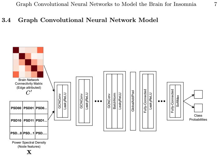
The load-bearing object is the weighted brain graph built per EEG window. Nodes are EEG channels, acting as proxies for cortical regions; edges are spectral coherence values reduced by a random-coherence correction and then linearly combined with geodesic electrode distance, with the result normalized to $[0,1]$. The random-coherence correction, $C^{(random)}_{ij}=\exp(1 - D_{ij}/k)$, is the mechanism intended to remove volume conduction, and the additive distance term is what distinguishes this connectivity estimate from plain coherence. Node features are per-channel power spectral density in six bands computed by Welch's method, and a three-layer graph convolutional network with global add pooling and dense layers maps these graphs to class probabilities.
What would settle it
Record scalp EEG from a known dipole source, or run a realistic head-model simulation with known true connectivity, and check whether the edge weights from this formula recover that ground truth; a miscalibrated $k$ would leave residual distance-dependent coherence. Alternatively, rerun the classification on the same data after replacing the random-coherence correction with a Laplacian spatial filter, and look for a large change in accuracy or in the C4-P4 ranking.
Extended reading notes
Core claim
On its own terms, the paper's central discovery is that a domain-guided graph convolutional neural network, using edge weights $C'_{ij}=C^{(computed)}_{ij}-\exp(1 - D_{ij}/k)+D_{ij}$ normalized to $[0,1]$ and node features from Welch power-spectral-density estimates in six frequency bands, classifies insomnia from continuous sleep EEG at 70.1% window-level and 66.7% subject-level accuracy with a 50-second non-overlapping window. With spectral coherence alone, the same pipeline reaches about 64% window-level and 57% subject-level accuracy, so the additive distance term improves the connectivity estimate. Ablating individual channels shows that removing C4-P4, F4-C4, or C4-A1 degrades accuracy the most, and the paper connects those electrodes to brain regions with previously reported atypical functional connectivity in insomnia. The claim is that a graph representation of functional connectivity, not the raw multichannel time series, is sufficient to carry insomnia-related information in long sleep recordings.
Load-bearing premise
Everything rests on assuming that the random-coherence correction in Eq. (1), with its fixed constant $k$ taken from earlier work, adequately removes volume conduction from the EEG coherence; if that correction is miscalibrated, the brain graphs are artifacts and the accuracy and channel ranking are not measuring brain connectivity.
Editorial extensions
If this is right
- A 50-second window is the best temporal scale in this EEG setting; shorter windows weaken the signal and longer windows likely straddle sleep micro-structure cycle boundaries, reducing accuracy.
- Combining spectral coherence with electrode distance is strictly better than coherence alone, indicating that spatial geometry carries complementary discriminative information for insomnia.
- The channel-importance ranking gives a concrete compression target: a simplified device should keep C4-P4, F4-C4, and C4-A1 before other channels.
- Subject-level accuracy remains above chance but below window-level accuracy, so the learned features are not explained by a single subject, although patient-level variability still matters.
- The same graph-construction and classification pipeline should transfer to other sleep pathologies if the connectivity signature is similarly learnable.
Reading between the lines
- Editorial inference: the channel-importance ranking is a model-internal attribution, not direct evidence of brain-region causality, unless the connectivity estimate is validated against source-localized data.
- A testable extension would redo the classification with a Laplacian spatial filter instead of the Eq. (1) random-coherence correction; if accuracy or the C4-P4 ranking changes sharply, the reported result depends on that correction rather than on the brain signal.
- Another testable extension is sensitivity analysis over the constant $k$ in Eq. (1); if the best window-level accuracy moves by more than a few points when $k$ is re-estimated on this dataset, the edge-weight scale is not robust.
- An inference for future work: feeding the same graphs to a simpler classifier, such as an SVM on graph metrics, would show how much of the 70% comes from the graph features themselves versus the graph convolutional network.
Editorial analysis
A structured set of objections, weighed in public.
Referee Report
Summary. The manuscript proposes a graph convolutional neural network model to classify insomnia from continuous sleep EEG. Brain graphs are built from five channels, using spectral coherence corrected by a random-coherence model and augmented with geodesic distance as edge weights, and power spectral density in six frequency bands as node features. The authors report 5-fold subject-independent cross-validation over 16 subjects (9 insomnia, 7 control), finding that a 50-second window yields 70.1% window-level and 67.7% subject-level accuracy (Table 1), and that combining coherence with distance improves accuracy (Table 2). Channel-ablation experiments identify C4-P4, F4-C4 and C4-A1 as the most important channels (Table 3).
Significance. If the central claims hold, this is an early demonstration of GCNN-based brain modelling for insomnia from long-duration EEG, and the subject-independent cross-validation is a genuine strength. The systematic window-length comparison and channel-ablation protocol are useful, and the use of continuous ~13-hour recordings goes beyond typical short sleep-onset windows in prior work. However, the validity of the connectivity model is not established, and the small sample and lack of statistical inference substantially weaken the quantitative conclusions. The paper currently provides a proof-of-concept rather than a reliable clinical or neurobiological result.
major comments (5)
- [§3.3, Eq. (1)–(2)] The random-coherence model in Eq. (1) is invalid as written: C_random^ij = exp(1 − D_ij/k) exceeds 1 whenever D_ij < k, reaching e at D_ij = 0, which is impossible for a coherence quantity. The constant k in [19] is never specified numerically or fitted, so the reduced coherence in Eq. (2) and every edge weight in Eq. (3) are not reproducible and may take invalid negative values. Since Tables 1–3 depend entirely on these edge weights, the classification and channel-importance results cannot currently be interpreted as brain-connectivity measures. Please correct the formula (or use the actual random-coherence estimator from Nunez et al.), report k and the normalization procedure, and show that the resulting edge weights lie in [0,1].
- [§3.3, Eq. (3)] Eq. (3) adds a physical geodesic distance D_ij, measured in centimetres or similar, to a dimensionless coherence value C_ij without any stated scaling or units. The subsequent normalisation to [0,1] is not described, so it is unclear whether the improvement attributed to the spatial term in Table 2 reflects a genuine connectivity enhancement or just a rescaling of the graph. Provide the normalisation formula and a sensitivity analysis over the relative scaling of coherence and distance.
- [§4.1, Table 1] The 50-second window is selected because it gives the highest accuracy on the same subject-independent 5-fold cross-validation folds that are then used to report the final performance. This makes the reported accuracy optimistic and does not account for model selection. Use nested cross-validation or pre-specify the window length, and report the variance across folds and iterations.
- [§3.1, §4] The study includes only 16 subjects (9 insomnia, 7 control), and no error bars, confidence intervals, or significance tests are reported anywhere. In this setting, the small differences between conditions in Tables 1–3 (e.g., 70.1% vs. 68.5% for Fp2-F4 omission) may easily be within chance. Report per-subject accuracies, exact binomial or permutation tests, and compare against at least one classical baseline (e.g., SVM or logistic regression on the same features) to establish that the GCNN architecture contributes beyond the features.
- [§4.2, Table 3] In the channel-importance experiment, omitting a channel reduces the graph from five to four nodes and changes the model architecture, so the resulting drop in accuracy conflates information loss with a change in model capacity/input size. To support the neurobiological interpretation, the ablation should be compared against removing a randomly chosen channel in a permutation test, or the model should be re-run with a four-node graph using different subsets to separate the effect of losing a particular channel.
minor comments (6)
- [Abstract vs. Table 1] The abstract reports subject-level accuracy as 68%, while Table 1 and the main text report 67.7%; please make these consistent.
- [§3.4 and §4] The GCNN hyperparameters (number of layers, hidden dimensions, dropout, batch size, number of epochs, early stopping) are not reported; include them for reproducibility.
- [§3.3] The exact coherence estimator from [5] and the frequency range or band over which coherence is computed are not specified; the connectivity matrix could differ substantially across bands.
- [§3.2] Clarify whether artefact rejection or sleep-stage selection was performed on the ~13-hour recordings, since the whole recording is used without further description.
- [Figures 3–5] Figures 3–5 show no error bars or spread; given the small sample, add per-fold or per-subject variability.
- [Table 3] Table 3 evaluates only the five channels in the montage; the conclusion about the 'three most important channels' should be explicitly limited to this five-channel setup.
Circularity Check
No circularity: the classification result is an empirical evaluation against external labels; the connectivity construction is a fixed preprocessing choice, not a fitted prediction.
full rationale
The paper makes no first-principles derivation claim that reduces to its own inputs. Its central result (70.1% window-level, 66.7% subject-level accuracy) is an empirical classification accuracy obtained by 5-fold subject-independent cross-validation against external insomnia/control labels from the CAP database; the labels are not constructed from the connectivity features. The connectivity construction in Eqs. (1)-(3) is a fixed preprocessing rule imported from Nunez et al. [19] and Wagh & Varatharajah [24]. Even if Eq. (1) is mis-specified as written or the constant k is unreported (a reproducibility and validity concern, not a circularity), no fitted outcome is fed back into the graph construction or into the accuracy computation. The choice of the 50 s window and the combined connectivity measure is a post-hoc model selection over cross-validated results, which may optimistically bias the reported numbers, but the reported accuracy is a direct measurement under that configuration, not a prediction derived from the selected parameter. The channel-importance ablation is an empirical sensitivity analysis, and its anatomical interpretation is checked against independent clinical studies ([8,14,25]) rather than being used to define the ranking. No self-citation is load-bearing; all cited methods are external. No specific reduction of a result to a fitted input or to a self-citation chain can be exhibited, so the circularity score is 0.
Assumptions & free parameters
free parameters (3)
- k in random coherence model (Eq. 1) =
not specified; cited to Nunez et al. [19]
- window length =
50 s selected by cross-validation
- unreported network hyperparameters =
not specified
assumptions (5)
- domain assumption Spectral coherence between scalp EEG channels reflects true cortical functional connectivity
- domain assumption Random coherence correction of Eq. (1) adequately removes volume conduction
- ad hoc to paper Adding geodesic distance to coherence improves connectivity estimates
- domain assumption EEG is quasi-stationary within segments of 10 to 90 seconds
- domain assumption The CAP database labels (insomnia versus control) are correct
Cite this review
Pith. "Pith review of Graph Convolutional Neural Networks to Model the Brain for Insomnia." pith.science (2026). https://pith.science/paper/7PRUSN6M
@misc{pith2026250714147,
  author       = {Pith},
  title        = {Pith review of: Graph Convolutional Neural Networks to Model the Brain for Insomnia},
  year         = {2026},
  howpublished = {\url{https://pith.science/paper/7PRUSN6M}},
  note         = {Machine review of arXiv:2507.14147}
}
read the original abstract
Insomnia affects a vast population of the world and can have a wide range of causes. Existing treatments for insomnia have been linked with many side effects like headaches, dizziness, etc. As such, there is a clear need for improved insomnia treatment. Brain modelling has helped with assessing the effects of brain pathology on brain network dynamics and with supporting clinical decisions in the treatment of Alzheimer's disease, epilepsy, etc. However, such models have not been developed for insomnia. Therefore, this project attempts to understand the characteristics of the brain of individuals experiencing insomnia using continuous long-duration EEG data. Brain networks are derived based on functional connectivity and spatial distance between EEG channels. The power spectral density of the channels is then computed for the major brain wave frequency bands. A graph convolutional neural network (GCNN) model is then trained to capture the functional characteristics associated with insomnia and configured for the classification task to judge performance. Results indicated a 50-second non-overlapping sliding window was the most suitable choice for EEG segmentation. This approach achieved a classification accuracy of 70% at window level and 68% at subject level. Additionally, the omission of EEG channels C4-P4, F4-C4 and C4-A1 caused higher degradation in model performance than the removal of other channels. These channel electrodes are positioned near brain regions known to exhibit atypical levels of functional connectivity in individuals with insomnia, which can explain such results.
Figures


Figures from the paper (3 more)


Reference graph
Works this paper leans on
-
[19]
Electroencephalography and clinical neurophysiology103(5), 499–515 (1997)
Nunez, P.L., Srinivasan, R., Westdorp, A.F., Wijesinghe, R.S., Tucker, D.M., Sil- berstein, R.B., Cadusch, P.J.: Eeg coherency: I: statistics, reference electrode, vol- ume conduction, laplacians, cortical imaging, and interpretation at multiple scales. Electroencephalography and clinical neurophysiology103(5), 499–515 (1997)
work page 1997
-
[1]
Sleep Medicine82, 186–192 (2021)
Aernout, E., Benradia, I., Hazo, J.B., Sy, A., Askevis-Leherpeux, F., Sebbane, D., Roelandt, J.L.: International study of the prevalence and factors associated with insomnia in the general population. Sleep Medicine82, 186–192 (2021)
work page 2021
-
[2]
Human brain mapping 28(2), 143–157 (2007)
Astolfi, L., Cincotti, F., Mattia, D., Marciani, M.G., Baccala, L.A., de Vico Fallani, F., Salinari, S., Ursino, M., Zavaglia, M., Ding, L., et al.: Comparison of different cortical connectivity estimators for high-resolution eeg recordings. Human brain mapping 28(2), 143–157 (2007)
work page 2007
-
[3]
Journal of medical systems39, 1–10 (2015)
Aydın, S., Tunga, M.A., Yetkin, S.: Mutual information analysis of sleep eeg in de- tecting psycho-physiological insomnia. Journal of medical systems39, 1–10 (2015)
work page 2015
-
[4]
Scientific reports5(1), 9748 (2015)
Bajo, R., Pusil, S., Lopez, M.E., Canuet, L., Pereda, E., Osipova, D., Maestú, F., Pekkonen, E.: Scopolamine effects on functional brain connectivity: a pharmaco- logical model of alzheimer’s disease. Scientific reports5(1), 9748 (2015)
work page 2015
-
[5]
Bendat, J.S., Piersol, A.G.: Random data: analysis and measurement procedures. John Wiley & Sons (2011)
work page 2011
-
[6]
Bertolini, D., Loukianov, A.D., Smith, A.M., Li-Bland, D., Pouliot, Y., Walsh, J.R., Fisher, C.K.: Modeling disease progression in mild cognitive impairment and alzheimer’s disease with digital twins (2020)
work page 2020
-
[7]
Clinical Neurophysiology 130(10), 1937–1944 (2019)
Chaturvedi, M., Bogaarts, J.G., Kozak, V.V., Hatz, F., Gschwandtner, U., Meyer, A., Fuhr, P., Roth, V.: Phase lag index and spectral power as qeeg features for identification of patients with mild cognitive impairment in parkinson’s disease. Clinical Neurophysiology 130(10), 1937–1944 (2019)
work page 2019
Show all 29 references
-
[8]
Biological Psychology97, 1–8 (2014)
Chen, M.C., Chang, C., Glover, G.H., Gotlib, I.H.: Increased insula coactivation with salience networks in insomnia. Biological Psychology97, 1–8 (2014)
2014
-
[9]
Frontiers in human neuroscience 12, 110 (2018)
Chriskos, P., Frantzidis, C.A., Gkivogkli, P.T., Bamidis, P.D., Kourtidou-Papadeli, C.: Achieving accurate automatic sleep staging on manually pre-processed eeg data through synchronization feature extraction and graph metrics. Frontiers in human neuroscience 12, 110 (2018)
2018
-
[10]
circulation101(23), e215–e220 (2000)
Goldberger, A.L., Amaral, L.A., Glass, L., Hausdorff, J.M., Ivanov, P.C., Mark, R.G., Mietus, J.E., Moody, G.B., Peng, C.K., Stanley, H.E.: Physiobank, phys- iotoolkit, and physionet: components of a new research resource for complex phys- iologic signals. circulation101(23), ...
2000
-
[11]
Frontiers in computational neu- roscience 13, 85 (2020)
Harmah, D.J., Li, C., Li, F., Liao, Y., Wang, J., Ayedh, W.M., Bore, J.C., Yao, D., Dong, W., Xu, P.: Measuring the non-linear directed information flow in schizophrenia by multivariate transfer entropy. Frontiers in computational neu- roscience 13, 85 (2020)
2020
-
[12]
Sensors21(6), 1988 (2021)
Huang, H., Zhang, J., Zhu, L., Tang, J., Lin, G., Kong, W., Lei, X., Zhu, L.: Eeg-based sleep staging analysis with functional connectivity. Sensors21(6), 1988 (2021)
2021
-
[13]
In: IJCAI
Jia, Z., Lin, Y., Wang, J., Zhou, R., Ning, X., He, Y., Zhao, Y.: Graphsleepnet: Adaptive spatial-temporal graph convolutional networks for sleep stage classifica- tion. In: IJCAI. vol. 2021, pp. 1324–1330 (2020) 14 K. Monteiro et al
2020
-
[14]
Neuroreport 24(5), 233–240 (2013)
Killgore, W.D., Schwab, Z.J., Kipman, M., DelDonno, S.R., Weber, M.: Insomnia- related complaints correlate with functional connectivity between sensory–motor regions. Neuroreport 24(5), 233–240 (2013)
2013
-
[15]
arXiv preprint arXiv:1609.02907 (2016)
Kipf, T.N., Welling, M.: Semi-supervised classification with graph convolutional networks. arXiv preprint arXiv:1609.02907 (2016)
2016 arXiv
-
[16]
IEEE Transactions on Neural Systems and Rehabilitation Engineering 30, 2651–2660 (2022)
Klepl,D.,He,F.,Wu,M.,Blackburn,D.J.,Sarrigiannis,P.:Eeg-basedgraphneural network classification of alzheimer’s disease: An empirical evaluation of functional connectivity methods. IEEE Transactions on Neural Systems and Rehabilitation Engineering 30, 2651–2660 (2022)
2022
-
[17]
PloS one9(4), e95415 (2014)
Lee, Y.Y., Hsieh, S.: Classifying different emotional states by means of eeg-based functional connectivity patterns. PloS one9(4), e95415 (2014)
2014
-
[18]
Medical engineering & physics37(3), 297–308 (2015)
Melia, U., Guaita, M., Vallverdú, M., Embid, C., Vilaseca, I., Salamero, M., Santa- maria, J.: Mutual information measures applied to eeg signals for sleepiness char- acterization. Medical engineering & physics37(3), 297–308 (2015)
2015
-
[20]
Clinical EEG and neuroscience43(4), 268–278 (2012)
Sankari,Z.,Adeli,H.,Adeli,A.:Waveletcoherencemodelfordiagnosisofalzheimer disease. Clinical EEG and neuroscience43(4), 268–278 (2012)
2012
-
[21]
Journal of neuroscience methods166(1), 41–52 (2007)
Srinivasan, R., Winter, W.R., Ding, J., Nunez, P.L.: Eeg and meg coherence: mea- sures of functional connectivity at distinct spatial scales of neocortical dynamics. Journal of neuroscience methods166(1), 41–52 (2007)
2007
-
[22]
Sleep medicine2(6), 537–554 (2001)
Terzano, M.G., Parrino, L., Sherieri, A., Chervin, R., Chokroverty, S., Guillem- inault, C., Hirshkowitz, M., Mahowald, M., Moldofsky, H., Rosa, A., et al.: Atlas, rules, and recording techniques for the scoring of cyclic alternating pattern (cap) in human sleep. Sleep medicin...
2001
-
[23]
Frontiers in immunology12, 669811 (2021)
Voigt, I., Inojosa, H., Dillenseger, A., Haase, R., Akgün, K., Ziemssen, T.: Digital twins for multiple sclerosis. Frontiers in immunology12, 669811 (2021)
2021
-
[24]
In: Machine Learning for Health
Wagh, N., Varatharajah, Y.: Eeg-gcnn: Augmenting electroencephalogram-based neurological disease diagnosis using a domain-guided graph convolutional neural network. In: Machine Learning for Health. pp. 367–378. PMLR (2020)
2020
-
[25]
European Radiology27, 3703–3709 (2017)
Wang, T., Yan, J., Li, S., Zhan, W., Ma, X., Xia, L., Li, M., Lin, C., Tian, J., Li, C., et al.: Increased insular connectivity with emotional regions in primary insomnia patients: a resting-state fmri study. European Radiology27, 3703–3709 (2017)
2017
-
[26]
IEEE Trans- actions on Audio and Electroacoustics 15(2), 70–73 (1967)
Welch, P.: The use of fast fourier transform for the estimation of power spectra: A method based on time averaging over short, modified periodograms. IEEE Trans- actions on Audio and Electroacoustics 15(2), 70–73 (1967). https://doi.org/10. 1109/TAU.1967.1161901
1967
-
[27]
Journal of neuroscience methods183(1), 9–18 (2009)
Wendling, F., Ansari-Asl, K., Bartolomei, F., Senhadji, L.: From eeg signals to brain connectivity: a model-based evaluation of interdependence measures. Journal of neuroscience methods183(1), 9–18 (2009)
2009
-
[28]
Journal of Psychopharma- cology 33(8), 923–947 (2019)
Wilson,S.,Anderson,K.,Baldwin,D.,Dijk,D.J.,Espie,A.,Espie,C.,Gringras,P., Krystal, A., Nutt, D., Selsick, H., Sharpley, A.: British association for psychophar- macology consensus statement on evidence-based treatment of insomnia, para- somnias and circadian rhythm disorders: A...
2019 doi
-
[29]
Biomedical Signal Processing and Control74, 103498 (2022)
Zhang, C., Sun, L., Ge, S., Chang, Y., Jin, M., Xiao, Y., Gao, H., Wang, L., Cong, F.: Quantitative evaluation of short-term resting-state brain networks for primary insomnia diagnosis. Biomedical Signal Processing and Control74, 103498 (2022)
2022
Reviewed August 6, 2026 · model on record in the stance chip above.
Discussion (0). Sign in to comment.