REVIEW 4 major objections 7 minor 37 references
SFNet: A Spatial-Frequency Domain Deep Learning Network for Efficient Alzheimer's Disease Diagnosis
T0 review · 4 major / 7 minor · reviewed 2026-08-06 · deepseek-v4-flash
Pith's one-line read SFNet combines spatial and frequency features from 3D MRI and reports 95.1% accuracy separating Alzheimer's disease from cognitively normal controls.
desk verdict A sensible 3D spatial-frequency architecture with a legitimate new combination, but the evaluation as written does not support the state-of-the-art claim. read the letter →
The pith
A machine-rendered reading of the paper's core claim, the machinery that carries it, and where it could break.
The reading
What carries the argument
The load-bearing machinery is the paired dual-domain architecture. In the spatial branch, dense blocks with dense connectivity are modified by inserting a multi-scale attention module between two convolutions; the module has a channel-attention branch using 1D convolution after global average pooling (following an adaptive kernel-size rule) and a spatial-attention branch using $3\times3\times3$ dilated convolutions with dilation rates 1, 2, and 3, concatenated and passed through a 3D convolution, with residual connections and learnable weights initially set to 0.33. In the frequency branch, the spatial feature map is split into 3D patches, projected into tokens, transformed by a 3D fast Fourier transform, multiplied element-wise by learnable global filters, transformed back, and refined by a low-rank MLP. The global filter layer replaces quadratic self-attention with $O(n\log n)$ Fourier operations, and the low-rank MLP cuts parameters; the learned filters double as a visualization of which spectral components the network weights at each layer.
What would settle it
Run SFNet and the Table II and Table III baselines on exactly the same ADNI folds, preprocessing pipeline, and test set. If SFNet's AD/CN accuracy does not exceed the best baseline by more than the fold-to-fold variation (the ablations report standard deviations around 0.02), the paper's superiority claim would be refuted.
Extended reading notes
Core claim
The central claim is that SFNet, presented as the first end-to-end 3D MRI model to jointly exploit spatial and frequency information, improves Alzheimer's disease classification across all three pairwise tasks on ADNI. In the fully controlled comparison, it reaches 0.951 accuracy, 0.959 sensitivity, 0.939 specificity, and 0.984 AUC for AD versus CN, and outperforms the strongest baseline, Conv-Swinformer, by 1.6 percentage points in accuracy for AD/CN, 6.9 for CN/MCI, and 2.8 for AD/MCI, with fewer parameters (18.75M versus 24.30M). The paper also claims that the multi-scale attention module raises specificity, particularly in AD/MCI, and that visualizing the learned global filters shows a progression from high-frequency emphasis in early layers to low-frequency emphasis in deeper layers, which it offers as evidence of interpretable, hierarchical spectral feature extraction.
Load-bearing premise
The load-bearing premise is that the performance numbers for nine comparison models, taken directly from their original publications, are comparable to SFNet's numbers even though the data subsets, preprocessing pipelines, and evaluation splits differ; if that comparability fails, the claim that SFNet leads the field loses its footing.
Editorial extensions
If this is right
- A dual-domain 3D architecture is a viable alternative to transformer-based models for small medical imaging datasets, because frequency operations capture long-range dependencies without quadratic self-attention cost.
- The reported margins on ADNI imply that frequency-domain global context adds the most value on subtle MCI classifications (a 6.9-point gain on CN/MCI), not just on the easier AD-versus-CN separation.
- The multi-scale attention module, which the ablation credits with raising AD/MCI specificity from 0.682 to 0.80, suggests that dilated convolutions at multiple rates are useful for picking up the diffuse structural changes of early Alzheimer's disease.
- The learned global filter visualizations provide a concrete, inspectable account of what the network attends to in the spectrum, which could make automated AD screening more acceptable in clinical settings.
Reading between the lines
- A testable extension not explored in the paper is to use the learned global filters as candidate imaging biomarkers, checking whether their high- versus low-frequency emphasis correlates with atrophy severity or with progression from MCI to AD.
- Because the CN/MCI gain is the largest, the design is likely to transfer to early-detection tasks such as predicting conversion from mild cognitive impairment to Alzheimer's disease; a longitudinal ADNI follow-up would settle this.
- The frequency module is domain-general, so the same dual-domain recipe could be tried on other 3D neuroimaging tasks such as tumor segmentation or multiple sclerosis lesion detection, though the paper reports only AD classification.
- Independent validation on a standardized split would clarify how much of the reported edge over the nine external models is due to architecture versus evaluation setup.
Editorial analysis
A structured set of objections, weighed in public.
Referee Report
Summary. The paper proposes SFNet, an end-to-end 3D CNN-Transformer hybrid for Alzheimer's disease (AD) diagnosis from structural MRI. The architecture combines a DenseNet-style spatial feature extractor with a multi-scale attention module (channel attention plus dilated-convolution spatial attention) and a global frequency module based on 3D FFT/IFFT with learnable filters and a low-rank MLP. Experiments on ADNI report ACC/SEN/SPE/AUC for AD/CN, AD/MCI, and CN/MCI, with the headline result being 95.1% accuracy for AD/CN classification using 18.75M parameters. The paper also includes ablation studies and visualizations of the learned frequency filters. The central claim is that SFNet is the first end-to-end framework to jointly exploit spatial and frequency information in 3D MRI for AD diagnosis and that it outperforms existing methods.
Significance. If the performance claims hold, the architectural idea of combining local multi-scale spatial features with global frequency-domain modeling in 3D is a sensible and potentially useful contribution to medical image classification, particularly because the frequency module offers an alternative to quadratic-complexity self-attention. The model is comparatively lightweight (18.75M parameters), and the ablation study (Figure 4) with error bars gives some evidence for the contribution of each component. The frequency-filter visualization in Section IV-E is a positive step toward interpretability. However, the paper's headline 'state-of-the-art' claim rests on uncontrolled comparisons (Table III) and on Table II, which lacks error bars and statistical tests despite the authors' own ablations showing ±0.02–0.03 variability. The evaluation protocol is also described inconsistently. These issues must be resolved before the central outperformance claim can be accepted.
major comments (4)
- [§IV-A] The evaluation protocol is internally inconsistent: the text first states 'A five-fold cross-validation strategy is adopted' and then immediately says '15% of the entire dataset is reserved as the test set, while the remaining 85% is used for training and validation.' These two statements describe different protocols. If a single 15% test split is used, the claim of five-fold cross-validation is misleading and no cross-validated performance estimate is provided. If five-fold CV is performed on the 85% subset, the relationship to the 15% hold-out set is unclear. This ambiguity is load-bearing because Table II reports no variance or test procedure, making it impossible to assess the reliability of the reported metrics.
- [§IV-B, Table II] Table II reports only point estimates for ACC, SEN, SPE, and AUC with no standard deviations, confidence intervals, or statistical significance tests. This is especially problematic because the authors' own ablation experiments (Figure 4) report standard deviations of ±0.02–0.03 in accuracy on the same tasks. The reported margins over CE-AH (0.002 in ACC) and Conv-Swinformer (0.016 in ACC) are within the range of noise observed in the authors' own experiments. The paper should report mean ± std over cross-validation folds and, ideally, paired statistical tests (e.g., McNemar's test or a paired bootstrap) to support the claim of superiority.
- [§IV-C, Table III] The comparison with nine external methods is performed by 'directly extract[ing]' metrics 'from the respective original publications.' These methods use different ADNI subject selections, different preprocessing pipelines (slice-based, patch-based, region-based, 3D), and different evaluation splits. This is not a controlled comparison, and the 0.2–2.6% margins over CE-AH and Conv-Swinformer cannot be attributed to model quality under these conditions. The state-of-the-art claim in the abstract and conclusion is therefore not supported. The authors should either re-implement the baselines under the identical preprocessing, subject selection, and evaluation protocol, or substantially temper the claims to describe the results as 'reported on different protocols.'
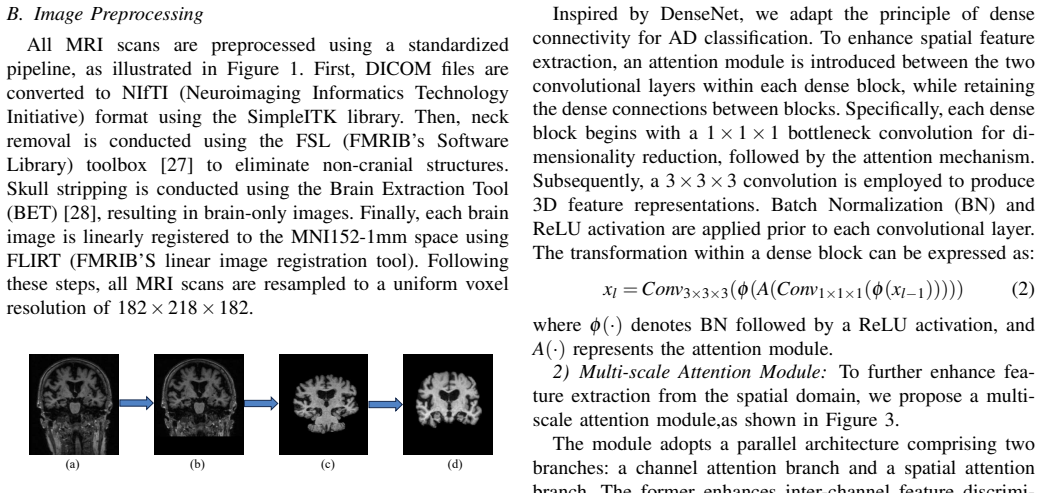
- [§III-B, §IV-A] Several hyperparameters that are central to the architecture are never reported: the patch size P, the low-rank MLP ranks r1 and r2, and the dilation rates used in the multi-scale attention module are mentioned in the text but no numerical values are given in the experimental settings. The model also has learnable fusion weights λ1, λ2, λ3 whose values or training behavior are not discussed. Without these values, the architecture cannot be reproduced, and the sensitivity of the results to them is unknown. Please provide a complete hyperparameter table or state the values in Section IV-A.
minor comments (7)
- [§IV-C] The text states that SFNet achieves 'AUC (0.84)' in the AD/CN comparison, but Table III lists AUC as 0.984. This appears to be a typographical error and should be corrected.
- [§IV-A] The heading 'Model Comparsion On All Three Classification Tasks' contains a typo ('Comparsion' should be 'Comparison'), and the same heading in Section IV-C says 'Comparison' but is inconsistently capitalized.
- [§III-B] Equation (8) uses the notation 'X̃′ ∈ R^{D×L}', whereas the preceding text describes the tokens as X̃ ∈ R^{L×D}. The dimension order is inconsistent and should be unified.
- [§III-A] The definition of the channel attention in Equation (3) references the parameter k, but the text does not define what k represents until the following sentence; please clarify the notation in the equation caption or the surrounding text.
- [§IV-D] The ablation study in Figure 4 reports standard deviations for the three variants, which is good, but it does not report whether the differences between variants are statistically significant. Adding a significance test would strengthen the claim that each component contributes meaningfully.
- [References] References [13] and [14] appear to be unrelated to the topic of AD diagnosis (they concern time-series Granger causality and Kolmogorov-Arnold networks). If they are not used in the introduction, they should be removed or replaced.
- [§IV-D] The hyperparameter selection for the number of dense blocks and frequency module depth (Figure 5) is performed on the same dataset used for evaluation. If the reported test metrics are used to choose these hyperparameters, the final numbers may be optimistically biased. Please clarify whether this selection was performed on a validation split and, if not, state the limitation.
Circularity Check
No significant circularity: SFNet's contribution is an empirical architecture evaluated on held-out data, and the cited prior work is external to the authors' own derivation.
full rationale
The paper does not present a derivation chain in which a predicted quantity is equivalent by construction to an input. The central claim is an empirical performance result: SFNet is trained and evaluated on ADNI, with accuracy, sensitivity, specificity, F1-score, and AUC reported. No parameter is fitted to the test set and then renamed as a prediction; the model is trained with cross-entropy loss and evaluated on a held-out portion, so the classification numbers are genuine empirical outcomes rather than identities. The architectural components—DenseNet, ECA-Net-style channel attention, dilated spatial attention, FFT-based global filtering, and low-rank MLP—are assembled from established, externally published methods, and the paper's Eqs. (1)–(9) describe the forward computation rather than a result that assumes its own conclusion. The self-citations in the reference list ([13], [14]) are not load-bearing: they appear only inside a bracketed citation block in the introduction and are not used to justify the model design, the evaluation protocol, or the claim of being first. The ablation study selecting three dense blocks and frequency depth six is standard model selection on validation performance, not a circular derivation, because the final test evaluation is separate and the paper does not present the selected configuration as an independent prediction derived from first principles. Concerns about Table III, where comparison numbers are 'directly extracted from the respective original publications' and therefore may come from different cohorts, preprocessing, and splits, are legitimate threats to the strength of the state-of-the-art claim, but they are threats to external validity and comparability, not circularity. Similarly, the ambiguity in Section IV-A between 'a five-fold cross-validation strategy is adopted' and '15% of the entire dataset is reserved as the test set' is a reporting inconsistency that affects reproducibility, not a self-referential reduction. Under the stated rules, matters of comparison fairness and evaluation rigor belong to correctness risk rather than circularity, and no specific equation or fitted parameter can be exhibited as circular. Therefore, the appropriate finding is no significant circularity with a score of 0.
Assumptions & free parameters
free parameters (6)
- Number of dense blocks =
3
- Global frequency module depth =
6
- Patch size P =
Not reported
- Low-rank MLP ranks r1 and r2 =
Not reported
- Dilation rates in spatial attention =
1, 2, 3
- Initial fusion weights lambda1, lambda2, lambda3 =
0.33 each
assumptions (4)
- standard math Standard properties of the fast Fourier transform and inverse FFT
- domain assumption ADNI clinical labels are reliable ground truth for CN, MCI, and AD
- domain assumption Preprocessing to MNI152 preserves AD-related structural signal
- ad hoc to paper Non-overlapping patch tokenization is a suitable input representation for the frequency module
Cite this review
Pith. "Pith review of SFNet: A Spatial-Frequency Domain Deep Learning Network for Efficient Alzheimer's Disease Diagnosis." pith.science (2026). https://pith.science/paper/F57PSOCS
@misc{pith2026250716267,
  author       = {Pith},
  title        = {Pith review of: SFNet: A Spatial-Frequency Domain Deep Learning Network for Efficient Alzheimer's Disease Diagnosis},
  year         = {2026},
  howpublished = {\url{https://pith.science/paper/F57PSOCS}},
  note         = {Machine review of arXiv:2507.16267}
}
read the original abstract
Alzheimer's disease (AD) is a progressive neurodegenerative disorder that predominantly affects the elderly population and currently has no cure. Magnetic Resonance Imaging (MRI), as a non-invasive imaging technique, is essential for the early diagnosis of AD. MRI inherently contains both spatial and frequency information, as raw signals are acquired in the frequency domain and reconstructed into spatial images via the Fourier transform. However, most existing AD diagnostic models extract features from a single domain, limiting their capacity to fully capture the complex neuroimaging characteristics of the disease. While some studies have combined spatial and frequency information, they are mostly confined to 2D MRI, leaving the potential of dual-domain analysis in 3D MRI unexplored. To overcome this limitation, we propose Spatio-Frequency Network (SFNet), the first end-to-end deep learning framework that simultaneously leverages spatial and frequency domain information to enhance 3D MRI-based AD diagnosis. SFNet integrates an enhanced dense convolutional network to extract local spatial features and a global frequency module to capture global frequency-domain representations. Additionally, a novel multi-scale attention module is proposed to further refine spatial feature extraction. Experiments on the Alzheimer's Disease Neuroimaging Initiative (ADNI) dataset demonstrate that SFNet outperforms existing baselines and reduces computational overhead in classifying cognitively normal (CN) and AD, achieving an accuracy of 95.1%.
Figures


Figures from the paper (3 more)


Reference graph
Works this paper leans on
-
[1]
”Alzheimer’s disease facts and figures.” Alzheimers Dement 19.4 (2023): 1598-1695
Better, MAPPING A. ”Alzheimer’s disease facts and figures.” Alzheimers Dement 19.4 (2023): 1598-1695
work page 2023
-
[2]
Clinical trials of new drugs for Alzheimer disease[J]
Huang L K, Chao S P, Hu C J. Clinical trials of new drugs for Alzheimer disease[J]. Journal of biomedical science, 2020, 27: 1-13
work page 2020
-
[3]
The clinical use of structural MRI in Alzheimer disease[J]
Frisoni G B, Fox N C, Jack Jr C R, et al. The clinical use of structural MRI in Alzheimer disease[J]. Nature reviews neurology, 2010, 6(2): 67-77
work page 2010
-
[4]
Wen J, Thibeau-Sutre E, Diaz-Melo M, et al. Convolutional neural networks for classification of Alzheimer’s disease: Overview and re- producible evaluation[J]. Medical image analysis, 2020, 63: 101694
work page 2020
-
[5]
Korolev S, Safiullin A, Belyaev M, et al. Residual and plain con- volutional neural networks for 3D brain MRI classification[C]//2017 IEEE 14th international symposium on biomedical imaging (ISBI 2017). IEEE, 2017: 835-838
work page 2017
-
[6]
U-net based analysis of MRI for Alzheimer’s disease diagnosis[J]
Fan Z, Li J, Zhang L, et al. U-net based analysis of MRI for Alzheimer’s disease diagnosis[J]. Neural Computing and Applications, 2021, 33: 13587-13599
work page 2021
-
[7]
Wang H, Shen Y , Wang S, et al. Ensemble of 3D densely connected convolutional network for diagnosis of mild cognitive impairment and Alzheimer’s disease[J]. Neurocomputing, 2019, 333: 145-156. Fig. 6: Learnable global filters visualization. (a): Direction XY; (b): Direction YZ; (c): Direction XZ
work page 2019
-
[9]
Dutta T K, Nayak D R, Zhang Y D. Arm-net: Attention-guided residual multiscale cnn for multiclass brain tumor classification using mr images[J]. Biomedical Signal Processing and Control, 2024, 87: 105421
work page 2024
Show all 37 references
-
[10]
MPS-FFA: A multiplane and multiscale feature fusion attention network for Alzheimer’s disease prediction with structural MRI[J]
Liu F, Wang H, Liang S N, et al. MPS-FFA: A multiplane and multiscale feature fusion attention network for Alzheimer’s disease prediction with structural MRI[J]. Computers in Biology and Medicine, 2023, 157: 106790
2023
-
[11]
MSFNet-2SE: A multi-scale fusion convolutional network for Alzheimer’s disease classification on magnetic resonance images[J]
Zhang L, Xia R, Yang B, et al. MSFNet-2SE: A multi-scale fusion convolutional network for Alzheimer’s disease classification on magnetic resonance images[J]. International Journal of Imaging Systems and Technology, 2024, 34(4): e23112
2024
-
[12]
MACFNet: Detection of Alzheimer’s dis- ease via multiscale attention and cross-enhancement fusion network[J]
Tang C, Xi M, Sun J, et al. MACFNet: Detection of Alzheimer’s dis- ease via multiscale attention and cross-enhancement fusion network[J]. Computer Methods and Programs in Biomedicine, 2024, 254: 108259
2024
-
[13]
Kolmogorov-Arnold Networks for Time Series Granger Causality Inference[J]
Liu M, Xu Y , Li Z, et al. Kolmogorov-Arnold Networks for Time Series Granger Causality Inference[J]. arXiv preprint arXiv:2501.08958, 2025
2025 arXiv
-
[14]
Gradient Regularization-based Neural Granger Causality[J]
Liu M, Dong H, Yang X, et al. Gradient Regularization-based Neural Granger Causality[J]. arXiv preprint arXiv:2507.11178, 2025
2025 arXiv
-
[15]
Attention is all you need[J]
Vaswani A, Shazeer N, Parmar N, et al. Attention is all you need[J]. Advances in neural information processing systems, 2017, 30
2017
-
[16]
”Transformers in vision: A survey.” ACM comput- ing surveys (CSUR) 54.10s (2022): 1-41
Khan, Salman, et al. ”Transformers in vision: A survey.” ACM comput- ing surveys (CSUR) 54.10s (2022): 1-41
2022
-
[17]
”End-to-end object detection with transformers.” European conference on computer vision
Carion, Nicolas, et al. ”End-to-end object detection with transformers.” European conference on computer vision. Cham: Springer International Publishing, 2020
2020
-
[18]
Efficient self-attention mechanism and struc- tural distilling model for Alzheimer’s disease diagnosis[J]
Zhu J, Tan Y , Lin R, et al. Efficient self-attention mechanism and struc- tural distilling model for Alzheimer’s disease diagnosis[J]. Computers in Biology and Medicine, 2022, 147: 105737
2022
-
[19]
Trans-resnet: Integrating transformers and cnns for alzheimer’s disease classification[C]//2022 IEEE 19th International Symposium on Biomedical Imaging (ISBI)
Li C, Cui Y , Luo N, et al. Trans-resnet: Integrating transformers and cnns for alzheimer’s disease classification[C]//2022 IEEE 19th International Symposium on Biomedical Imaging (ISBI). IEEE, 2022: 1-5
2022
-
[20]
M3T: three-dimensional Medical image classifier using Multi-plane and Multi-slice Transformer[C]//Proceedings of the IEEE/CVF conference on computer vision and pattern recognition
Jang J, Hwang D. M3T: three-dimensional Medical image classifier using Multi-plane and Multi-slice Transformer[C]//Proceedings of the IEEE/CVF conference on computer vision and pattern recognition. 2022: 20718-20729
2022
-
[21]
Conv-Swinformer: Integration of CNN and shift window attention for Alzheimer’s disease classification[J]
Hu Z, Li Y , Wang Z, et al. Conv-Swinformer: Integration of CNN and shift window attention for Alzheimer’s disease classification[J]. Computers in Biology and Medicine, 2023, 164: 107304
2023
-
[22]
Diagnosis of Alzheimer’s disease via optimized lightweight convolution-attention and structural MRI[J]
Khatri U, Kwon G R. Diagnosis of Alzheimer’s disease via optimized lightweight convolution-attention and structural MRI[J]. Computers in Biology and Medicine, 2024, 171: 108116
2024
-
[23]
MMTFN: Multi-modal multi-scale trans- former fusion network for Alzheimer’s disease diagnosis[J]
Miao S, Xu Q, Li W, et al. MMTFN: Multi-modal multi-scale trans- former fusion network for Alzheimer’s disease diagnosis[J]. Interna- tional Journal of Imaging Systems and Technology, 2024, 34(1): e22970
2024
-
[24]
Global filter networks for image classification[J]
Rao Y , Zhao W, Zhu Z, et al. Global filter networks for image classification[J]. Advances in neural information processing systems, 2021, 34: 980-993
2021
-
[25]
3d global fourier network for alzheimer’s disease diagnosis using structural mri[C]//International Conference on Medical Image Computing and Computer-Assisted Intervention
Zhang S, Chen X, Ren B, et al. 3d global fourier network for alzheimer’s disease diagnosis using structural mri[C]//International Conference on Medical Image Computing and Computer-Assisted Intervention. Cham: Springer Nature Switzerland, 2022: 34-43
2022
-
[26]
Addformer: Alzheimer’s disease detection from structural mri using fusion transformer[C]//2022 IEEE 19th International Symposium on Biomedical Imaging (ISBI)
Kushol R, Masoumzadeh A, Huo D, et al. Addformer: Alzheimer’s disease detection from structural mri using fusion transformer[C]//2022 IEEE 19th International Symposium on Biomedical Imaging (ISBI). IEEE, 2022: 1-5
2022
-
[27]
Jenkinson M, Beckmann C F, Behrens T E J, et al. Fsl[J]. Neuroimage, 2012, 62(2): 782-790
2012
-
[28]
Automated brain extraction of multisequence MRI using artificial neural networks[J]
Isensee F, Schell M, Pflueger I, et al. Automated brain extraction of multisequence MRI using artificial neural networks[J]. Human brain mapping, 2019, 40(17): 4952-4964
2019
-
[29]
Densely connected convo- lutional networks[C]//Proceedings of the IEEE conference on computer vision and pattern recognition
Huang G, Liu Z, Van Der Maaten L, et al. Densely connected convo- lutional networks[C]//Proceedings of the IEEE conference on computer vision and pattern recognition. 2017: 4700-4708
2017
-
[30]
ECA-Net: Efficient channel attention for deep convolutional neural networks[C]//Proceedings of the IEEE/CVF conference on computer vision and pattern recognition
Wang Q, Wu B, Zhu P, et al. ECA-Net: Efficient channel attention for deep convolutional neural networks[C]//Proceedings of the IEEE/CVF conference on computer vision and pattern recognition. 2020: 11534- 11542
2020
-
[31]
Dual attention multi-instance deep learning for Alzheimer’s disease diagnosis with structural MRI[J]
Zhu W, Sun L, Huang J, et al. Dual attention multi-instance deep learning for Alzheimer’s disease diagnosis with structural MRI[J]. IEEE Transactions on Medical Imaging, 2021, 40(9): 2354-2366
2021
-
[32]
sMRI-PatchNet: A novel efficient explainable patch-based deep learning network for Alzheimer’s disease diagnosis with Structural MRI[J]
Zhang X, Han L, Han L, et al. sMRI-PatchNet: A novel efficient explainable patch-based deep learning network for Alzheimer’s disease diagnosis with Structural MRI[J]. IEEE Access, 2023, 11: 108603- 108616
2023
-
[33]
Multi-relation graph convolutional network for Alzheimer’s disease diagnosis using structural MRI[J]
Zhang J, He X, Qing L, et al. Multi-relation graph convolutional network for Alzheimer’s disease diagnosis using structural MRI[J]. Knowledge- Based Systems, 2023, 270: 110546
2023
-
[34]
A hybrid multi-scale attention convolution and aging transformer network for Alzheimer’s disease diagnosis[J]
Gao X, Cai H, Liu M. A hybrid multi-scale attention convolution and aging transformer network for Alzheimer’s disease diagnosis[J]. IEEE Journal of Biomedical and Health Informatics, 2023, 27(7): 3292-3301
2023
-
[35]
Interpretable medical deep framework by logits-constraint attention guiding graph-based multi-scale fusion for Alzheimer’s disease analysis[J]
Xu J, Yuan C, Ma X, et al. Interpretable medical deep framework by logits-constraint attention guiding graph-based multi-scale fusion for Alzheimer’s disease analysis[J]. Pattern Recognition, 2024, 152: 110450
2024
-
[36]
Chen Q, Fu Q, Bai H, et al. Longformer: longitudinal transformer for Alzheimer’s disease classification with structural MRIs[C]//Proceedings of the IEEE/CVF winter conference on applications of computer vision. 2024: 3575-3584
2024
-
[37]
A quantitatively interpretable model for Alzheimer’s disease prediction using deep counterfactuals[J]
Oh K, Heo D W, Mulyadi A W, et al. A quantitatively interpretable model for Alzheimer’s disease prediction using deep counterfactuals[J]. NeuroImage, 2025: 121077
2025
-
[38]
CE-AH: A Contrast-Enhanced Attention Hierar- chical Network for Alzheimer’s Disease Diagnosis Based on Structural MRI[J]
Wang T, Dai Q, Lu H. CE-AH: A Contrast-Enhanced Attention Hierar- chical Network for Alzheimer’s Disease Diagnosis Based on Structural MRI[J]. Pattern Recognition, 2025: 111986
2025
Reviewed August 6, 2026 · model on record in the stance chip above.
Discussion (0). Sign in to comment.