REVIEW 4 major objections 7 minor 1 cited by
HOG-CNN: Integrating Histogram of Oriented Gradients with Convolutional Neural Networks for Retinal Image Classification
T0 review · 4 major / 7 minor · reviewed 2026-08-06 · deepseek-v4-flash
Pith's one-line read A hybrid HOG-CNN model fuses handcrafted gradient histograms with frozen pretrained CNN features, and the paper claims this raises retinal disease classification accuracy and AUC over pure deep-learning baselines across three public…
desk verdict Useful appendix, but the headline numbers are a per-dataset best-backbone selection, so the claim that a single HOG-CNN model performs consistently well does not hold as written. read the letter →
The pith
A machine-rendered reading of the paper's core claim, the machinery that carries it, and where it could break.
The reading
What carries the argument
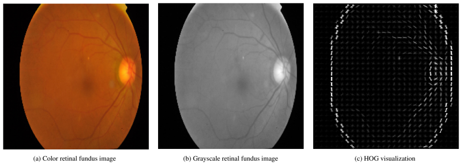
The central mechanism is a two-branch architecture. A grayscale version of each fundus image is converted into a 26,244-dimensional HOG descriptor with 9 orientation bins, 8x8 cells, and 2x2 normalized blocks, then compressed by a three-layer MLP of sizes 800, 256, and 128; in parallel, the RGB image passes through a frozen pretrained CNN with its classification head removed, followed by a 64-filter 3x3 convolution, 2x2 max pooling, flattening, and a 64-unit dense layer. The two embeddings are concatenated into a 192-dimensional vector and fed to two further dense layers with dropout 0.2 before a sigmoid or softmax output. This design lets the model combine local gradient histograms with deep semantic features.
What would settle it
Run the same four tasks with a single pre-registered backbone and without inspecting test labels; if the HOG+CNN variant does not beat the CNN-only variant in accuracy or AUC on held-out folds, the claimed complementarity is refuted.
Extended reading notes
Core claim
The central claim is that adding a handcrafted gradient-histogram branch to a deep CNN improves retinal disease classification beyond either feature type alone. In the paper's experiments the fused model beats the CNN-only version on nearly every backbone and dataset combination reported in the appendix, and the headline configuration outperforms published baselines: 98.5% accuracy and 99.2 AUC for binary diabetic retinopathy, 83.2% accuracy with 94.2 AUC for five-class grading, 92.8% accuracy and 94.5 AUC for AMD, and 83.9% accuracy and 87.2 AUC for glaucoma. The author treats this as evidence that local texture information captured by HOG is complementary to the semantic features learned by CNNs in ophthalmic imaging.
Load-bearing premise
The headline results use a different pretrained backbone for each dataset, and the paper never states that those backbone choices were made before seeing test performance, so the claimed consistent advantage could be an artifact of picking the best of several configurations rather than a property of the HOG+CNN fusion.
Editorial extensions
If this is right
- If the reported results hold under fair comparison, HOG-CNN shows that handcrafted descriptors still add value on top of modern CNN features in medical imaging.
- The method reaches near-state-of-the-art accuracy without data augmentation, segmentation, or fine-tuning of the backbone, which shortens training and lowers compute requirements.
- Because the HOG branch is computed in grayscale and the CNN branch is frozen, the model can be trained on CPU and deployed in low-resource clinics.
- The consistent gains of HOG+CNN over CNN-only in the appendix support the general claim that gradient and texture cues are complementary to deep features across three diseases.
- The four-task results establish a single fusion recipe that transfers across DR, AMD, and glaucoma without task-specific engineering.
Reading between the lines
- The appendix tests ten backbones with and without HOG, and the headline numbers are the best per dataset; the one-model framing is strongest if each backbone choice was fixed before test performance was inspected.
- A testable extension is to check whether the HOG gain persists on external or out-of-distribution fundus datasets where illumination and resolution vary; if it does, the method's value is robustness rather than only benchmark accuracy.
- The reported precision and recall asymmetry on IC-AMD, 94.8% precision versus 72.0% recall, suggests the model is conservative in flagging AMD, so future work could tune the decision threshold to meet clinical recall requirements.
- The same dual-branch fusion could be combined with other handcrafted descriptors, such as color histograms or topological features, which the paper leaves to future work and which might improve recall on the minority AMD and glaucoma classes.
Editorial analysis
A structured set of objections, weighed in public.
Referee Report
Summary. The manuscript proposes HOG-CNN, a hybrid architecture that concatenates Histogram of Oriented Gradients (HOG) descriptors with features from a frozen pretrained CNN backbone, and evaluates it on three retinal fundus benchmarks: binary and five-class diabetic retinopathy on APTOS 2019, age-related macular degeneration on IChallenge-AMD, and glaucoma on ORIGA. The main text reports state-of-the-art or near-state-of-the-art accuracy, precision, recall, and AUC numbers for each task, and an appendix tabulates results for ten different CNN backbones with and without HOG fusion. The paper argues that the fusion of handcrafted gradient features with deep features yields consistently high performance while remaining lightweight and interpretable.
Significance. If the reported results were produced by a single, well-specified model under controlled comparison, the paper would provide a useful data point on combining handcrafted HOG features with transfer-learned CNN features for retinal screening, supported by public code and a broad set of per-backbone experiments. The appendix's systematic reporting of ten backbones is a strength. However, the central claim of a single HOG-CNN architecture achieving consistent state-of-the-art performance is not currently supported, because the headline numbers are drawn from different backbones across datasets and the comparisons to prior work are made under incompatible evaluation protocols. The empirical contribution is therefore real but overstated, and the paper in its present form does not support the generalization claims in the abstract and conclusion.
major comments (4)
- [Section 5 and Appendix Tables .6-.9] The headline result for each dataset is not produced by a single HOG-CNN configuration. The binary APTOS result in Table 3 corresponds to HOG+EfficientNetB3 in Table .6; the five-class APTOS and IC-AMD results correspond to HOG+EfficientNetB2 in Tables .7 and .8; and the ORIGA result corresponds to HOG+EfficientNetB0 in Table .9. The manuscript does not specify a validation-based rule for selecting these backbones, so the reported numbers are consistent with selecting the best of the ten configurations after observing test performance. This is not a cosmetic issue: in Table .7, plain VGG19 achieves a five-class DR AUC of 95.65, above the reported HOG-CNN AUC of 94.2, and in Table .8, HOG+EfficientNetB3 has an IC-AMD AUC of 93.61 while HOG+EfficientNetB2 has 94.49. The text's claim that a single 'HOG-CNN' model performs consistently at the state of the art is therefore not established by the present experiments.
- [Tables 2-5] The comparisons against prior work use different train/test splits or cross-validation schemes across methods: HOG-CNN uses 80:20 for APTOS and ORIGA while many baselines use 85:15 or 10-fold CV; Table 4 lists no split for MobileNetV2-SVM; Table 5 mixes 70:30, 10-fold, and pretrained setups; and the CycleGAN row in Table 2 uses a different class balance (933:267). No confidence intervals or repeated-run variability are reported. Under these conditions, differences of a fraction of a percentage point, which the text interprets as 'surpassing' (e.g., 98.5 vs 98.4 accuracy in Table 3), cannot be distinguished from split-induced variation. The superiority claims in Section 5 require evaluation on identical splits with confidence intervals or significance tests.
- [Section 6, first paragraph] The sentence 'the proposed model achieved an accuracy, precision, and recall of 92.8%, along with an AUC of 94.5%' is contradicted by Table 2, which lists precision 94.8 and recall 72.0 for HOG-CNN on the IC-AMD dataset. This factual error also undermines the subsequent argument that HOG-CNN offers 'a more balanced performance across all metrics' than S2C2L, since the reported recall is ten points below S2C2L's 82.4%.
- [Section 5, DR Detection Results] In the five-class DR setting, the paper reports precision 86.0, recall 78.6, accuracy 83.2, and AUC 94.2 as the HOG-CNN result. Appendix Table .7 shows that no single backbone simultaneously attains these values: HOG+EfficientNetB0 reaches 83.49 accuracy, while HOG+EfficientNetB2 reaches 94.15 AUC, and the two configurations differ in precision and recall. Reporting a composite of two different configurations as one model's result reinforces the selection-bias concern raised above and should be clarified or corrected.
minor comments (7)
- [Highlights and Section 4.1] The dataset is referred to inconsistently as 'IC-Dataset', 'IChallenge-AMD', and 'IC-AMD'; one name should be used throughout.
- [Section 3.2] The text describing the HOG pathway reads 'the first dense layer contains 800 ReLU-activated units, the second dense layer contains 256 ReLU-activated units, and the second contains 128 ReLU-activated units'; the last occurrence should be 'the third'.
- [Algorithm 1 reference] The phrase 'summarized in here 1' should be 'summarized in Algorithm 1'.
- [Table .8] In the InceptionResNetV2 row, the precision value '2152' appears to be a typo for 21.52.
- [Table 5] The CNN-SVM row lists a class balance of 660:453 for ORIGA, although the dataset is described as containing 650 images; this discrepancy needs clarification.
- [General reporting] AUC values are reported with percent signs (e.g., 99.2%), which is nonstandard for a unitless quantity; either report AUC as a decimal or state the convention explicitly.
- [Section 2] The related-work section contains an extended discussion of topological data analysis that is not connected to the proposed method or experiments, making the section harder to follow.
Circularity Check
No circularity: empirical benchmark evaluation; headline-backbone variation is a selection/reporting concern, not a derivation that reduces to its inputs.
full rationale
This is an empirical benchmark paper rather than a theoretical derivation, so the circularity burden is low. HOG features are computed by the standard Dalal-Triggs descriptor, CNN embeddings come from frozen pretrained backbones, and the fusion is a fixed concatenation followed by dense layers. The reported accuracies and AUCs are measured on external public benchmarks (APTOS, ORIGA, IC-AMD) under stated train/test protocols; no equation defines the model in terms of the target result, and no parameter is fitted to a headline metric and then renamed as a prediction. The author self-citations in the Related Work (Refs. [12,13,14]) are background TDA references and are not load-bearing for the HOG-CNN claims. The appendix shows that the headline results use different pretrained backbones across datasets and that some plain backbones outperform the reported HOG-CNN figures on individual metrics; this is a possible model-selection and reporting concern, but it is not circularity, because the reported numbers are not forced to equal their inputs by construction. No uniqueness theorem, ansatz-smuggling citation, or renamed known result carries the argument. The derivation chain, such as it is, is self-contained against external benchmarks, so the appropriate circularity finding is no significant circularity.
Assumptions & free parameters
free parameters (6)
- Backbone selection per dataset =
EfficientNetB3 (binary APTOS), EfficientNetB2 (5-class APTOS and IC-AMD), EfficientNetB0 (ORIGA)
- HOG cell size =
8x8 pixels
- HOG block size =
2x2 cells
- HOG orientation bins =
9
- Dense layer sizes and dropout =
800, 256, 128; 256, 128; dropout 0.2
- Batch size and training epochs =
32; 50
assumptions (4)
- domain assumption Pretrained ImageNet CNN features transfer to retinal fundus images.
- domain assumption HOG descriptors carry discriminative information for retinal disease classification.
- domain assumption No pixel normalization is needed for the pretrained CNN.
- domain assumption Comparing models across different train/test splits is fair.
Cite this review
Pith. "Pith review of HOG-CNN: Integrating Histogram of Oriented Gradients with Convolutional Neural Networks for Retinal Image Classification." pith.science (2026). https://pith.science/paper/5ACAMZFG
@misc{pith2026250722274,
  author       = {Pith},
  title        = {Pith review of: HOG-CNN: Integrating Histogram of Oriented Gradients with Convolutional Neural Networks for Retinal Image Classification},
  year         = {2026},
  howpublished = {\url{https://pith.science/paper/5ACAMZFG}},
  note         = {Machine review of arXiv:2507.22274}
}
read the original abstract
The analysis of fundus images is critical for the early detection and diagnosis of retinal diseases such as Diabetic Retinopathy (DR), Glaucoma, and Age-related Macular Degeneration (AMD). Traditional diagnostic workflows, however, often depend on manual interpretation and are both time- and resource-intensive. To address these limitations, we propose an automated and interpretable clinical decision support framework based on a hybrid feature extraction model called HOG-CNN. Our key contribution lies in the integration of handcrafted Histogram of Oriented Gradients (HOG) features with deep convolutional neural network (CNN) representations. This fusion enables our model to capture both local texture patterns and high-level semantic features from retinal fundus images. We evaluated our model on three public benchmark datasets: APTOS 2019 (for binary and multiclass DR classification), ORIGA (for Glaucoma detection), and IC-AMD (for AMD diagnosis); HOG-CNN demonstrates consistently high performance. It achieves 98.5\% accuracy and 99.2 AUC for binary DR classification, and 94.2 AUC for five-class DR classification. On the IC-AMD dataset, it attains 92.8\% accuracy, 94.8\% precision, and 94.5 AUC, outperforming several state-of-the-art models. For Glaucoma detection on ORIGA, our model achieves 83.9\% accuracy and 87.2 AUC, showing competitive performance despite dataset limitations. We show, through comprehensive appendix studies, the complementary strength of combining HOG and CNN features. The model's lightweight and interpretable design makes it particularly suitable for deployment in resource-constrained clinical environments. These results position HOG-CNN as a robust and scalable tool for automated retinal disease screening.
Figures


Figures from the paper (1 more)

Forward citations
Cited by 1 Pith paper
-
RepViT-CXR: A Channel Replication Strategy for Vision Transformers in Chest X-ray Tuberculosis and Pneumonia Classification
Applying a standard channel-replication trick to a pretrained Vision Transformer yields very high reported accuracy for TB and pneumonia classification on three chest X-ray datasets.
Reference graph
Works this paper leans on
-
[1]
W. H. Organization, Blindness and vision im- pairment, 2023. URL: https://www.who. int/news-room/fact-sheets/detail/ blindness-and-visual-impairment , accessed: 2025-05-02
work page 2023
-
[2]
P. S. Silva, D. Zhang, C. M. P. Jacoba, W. Fickweiler, D. Lewis, J. Leitmeyer, K. Curran, R. P. Salongcay, D. Doan, M. Ashraf, et al., Automated machine learning for predicting diabetic retinopathy progression from ultra-widefield retinal images, JAMA ophthalmology 142 (2024) 171–178
work page 2024
-
[3]
E. R. Dow, H. K. Jeong, E. A. Katz, C. A. Toth, D. Wang, T. Lee, D. Kuo, M. J. Allingham, M. Hadziahmetovic, P. S. Mettu, et al., A deep-learning algorithm to predict short-term progression to geographic atrophy on spectral-domain optical coherence to- mography, JAMA ophthalmology 141 (2023) 1052–1061
work page 2023
-
[4]
T. Li, W. Bo, C. Hu, H. Kang, H. Liu, K. Wang, H. Fu, Appli- cations of deep learning in fundus images: A review, Medical Image Analysis 69 (2021) 101971
work page 2021
-
[5]
D. S. Ting, et al., Deep learning in ophthalmology: the technical and clinical considerations, Progress in retinal and eye research 72 (2019) 100759
work page 2019
-
[6]
M. H. Sarhan, et al., Machine learning techniques for oph- thalmic data processing: a review, IEEE Journal of Biomedical and Health Informatics 24 (2020) 3338–3350
work page 2020
-
[7]
D. S. W. Ting, et al., Artificial intelligence and deep learning in ophthalmology, British Journal of Ophthalmology 103 (2019) 167–175
work page 2019
- [8]
Show all 61 references
-
[9]
Singh, S
A. Singh, S. Sengupta, V . Lakshminarayanan, Explainable deep learning models in medical image analysis, Journal of imaging 6 (2020) 52
2020
-
[10]
Fourcade, R
A. Fourcade, R. H. Khonsari, Deep learning in medical image analysis: A third eye for doctors, Journal of stomatology, oral and maxillofacial surgery 120 (2019) 279–288
2019
-
[11]
Pratt, F
H. Pratt, F. Coenen, D. M. Broadbent, S. P. Harding, Y . Zheng, Convolutional neural networks for diabetic retinopathy, Proce- dia computer science 90 (2016) 200–205
2016
-
[12]
Ahmed, M
F. Ahmed, M. A. N. Bhuiyan, B. Coskunuzer, Topo-cnn: Reti- nal image analysis with topological deep learning, Journal of Imaging Informatics in Medicine (2025) 1–17
2025
-
[13]
Ahmed, B
F. Ahmed, B. Coskunuzer, Tofi-ml: Retinal image screening with topological machine learning, in: Annual Conference on Medical Image Understanding and Analysis, Springer, 2023, pp. 281–297
2023
-
[14]
Ahmed, Topological Machine Learning in Medical Image Analysis, Ph.D
F. Ahmed, Topological Machine Learning in Medical Image Analysis, Ph.D. thesis, The University of Texas at Dallas, 2023
2023
-
[15]
Qaiser, Y .-W
T. Qaiser, Y .-W. Tsang, D. Taniyama, N. Sakamoto, K. Nakane, D. Epstein, N. Rajpoot, Fast and accurate tumor segmentation of histology images using persistent homology and deep convo- lutional features, Medical image analysis 55 (2019) 1–14
2019
-
[16]
Lawson, A
P. Lawson, A. B. Sholl, J. Q. Brown, B. T. Fasy, C. Wenk, Per- sistent homology for the quantitative evaluation of architectural features in prostate cancer histology, Scientific reports 9 (2019) 1139
2019
-
[17]
Yadav, F
A. Yadav, F. Ahmed, O. Daescu, R. Gedik, B. Coskunuzer, Histopathological cancer detection with topological signatures, in: 2023 IEEE International Conference on Bioinformatics and Biomedicine (BIBM), 2023, pp. 1610–1619. doi: 10.1109/ BIBM58861.2023.10385822
2023
-
[18]
Berry, Y .-C
E. Berry, Y .-C. Chen, J. Cisewski-Kehe, B. T. Fasy, Functional summaries of persistence diagrams, Journal of Applied and Computational Topology 4 (2020) 211–262
2020
-
[19]
Crawford, et
L. Crawford, et. al., Predicting clinical outcomes in glioblas- toma: an application of topological and functional data analysis, J.Amer.Stat.Assoc. 115 (2020) 1139–1150
2020
-
[20]
Yadav, F
A. Yadav, F. Ahmed, O. Daescu, R. Gedik, B. Coskunuzer, Histopathological cancer detection with topological signatures, in: 2023 IEEE International Conference on Bioinformatics and Biomedicine (BIBM), IEEE, 2023, pp. 1610–1619
2023
-
[21]
Ahmed, B
F. Ahmed, B. Nuwagira, F. Torlak, B. Coskunuzer, Topo-CXR: Chest X-ray TB and Pneumonia Screening with Topological Machine Learning, in: Proceedings of the IEEE/CVF Interna- tional Conference on Computer Vision, 2023, pp. 2326–2336
2023
-
[22]
Kanari, et al., A topological representation of branching neu- ronal morphologies, Neuroinformatics 16 (2018) 3–13
L. Kanari, et al., A topological representation of branching neu- ronal morphologies, Neuroinformatics 16 (2018) 3–13
2018
-
[23]
Bendich, J
P. Bendich, J. S. Marron, E. Miller, A. Pieloch, S. Skwerer, Per- sistent homology analysis of brain artery trees, The annals of applied statistics 10 (2016) 198
2016
-
[24]
Rieck, et al., Uncovering the topology of time-varying fmri data using cubical persistence, NeurIPS 33 (2020) 6900–6912
B. Rieck, et al., Uncovering the topology of time-varying fmri data using cubical persistence, NeurIPS 33 (2020) 6900–6912
2020
-
[25]
B. J. Stolz, et al., Topological data analysis of task-based fmri data from experiments on schizophrenia, Journal of Physics: Complexity 2 (2021) 035006
2021
-
[26]
P. G. C ´amara, A. J. Levine, R. Rabadan, Inference of ancestral recombination graphs through topological data analysis, PLoS computational biology 12 (2016) e1005071
2016
-
[27]
J. I. Orlando, et al., An ensemble deep learning based approach for red lesion detection in fundus images, Computer methods and programs in biomedicine 153 (2018) 115–127
2018
-
[28]
A. You, J. K. Kim, I. H. Ryu, T. K. Yoo, Application of gen- erative adversarial networks (gan) for ophthalmology image do- mains: a survey, Eye and Vision 9 (2022) 1–19
2022
-
[29]
Srivastava, et.al., Artificial intelligence and machine learning in ophthalmology: A review, Indian Journal of Ophthalmology 71 (2023) 11–17
O. Srivastava, et.al., Artificial intelligence and machine learning in ophthalmology: A review, Indian Journal of Ophthalmology 71 (2023) 11–17
2023
-
[30]
N. D. Koseoglu, T. Liu, Predictive deep learning applications in ophthalmology, Rev. Ophthalmol 17 (2023)
2023
-
[31]
Ashtari-Majlan, M
M. Ashtari-Majlan, M. M. Dehshibi, D. Masip, Glaucoma di- agnosis in the era of deep learning: A survey, Expert Systems with Applications 256 (2024) 124888
2024
-
[32]
D. M. Nguyen, H. M. T. Alam, T. Nguyen, D. Srivastav, H.- J. Profitlich, N. Le, D. Sonntag, Deep learning for ophthal- mology: The state-of-the-art and future trends, arXiv preprint arXiv:2501.04073 (2025)
2025 arXiv
-
[33]
Fang, et al., ADAM challenge: Detecting age-related mac- ular degeneration from fundus images, IEEE Transactions on Medical Imaging (2022)
H. Fang, et al., ADAM challenge: Detecting age-related mac- ular degeneration from fundus images, IEEE Transactions on Medical Imaging (2022)
2022
-
[34]
Fu, et al., ADAM: Automatic detection challenge on AMD,
H. Fu, et al., ADAM: Automatic detection challenge on AMD,
-
[35]
Z. Zhang, et al., Origa-light: An online retinal fundus image database for glaucoma analysis and research, in: 2010 Annual international conference of the IEEE engineering in medicine and biology, IEEE, 2010, pp. 3065–3068
2010
-
[36]
https://www.kaggle.com/c/ aptos2019-blindness-detection
APTOS, Asia Pacific Tele-Ophthalmology So- ciety (APTOS) 2019 Blindness Detection Dataset, 2019. https://www.kaggle.com/c/ aptos2019-blindness-detection
2019
-
[37]
Goutam, M
B. Goutam, M. F. Hashmi, Z. W. Geem, N. D. Bokde, A com- prehensive review of deep learning strategies in retinal disease diagnosis using fundus images, IEEE Access (2022)
2022
-
[38]
M. R. Islam, et al., Applying supervised contrastive learning for the detection of DR and its severity levels from fundus images, Computers in Biology and Medicine 146 (2022) 105602
2022
-
[39]
J. D. Bodapati, et. al., Composite deep neural network with gated-attention mechanism for DR severity classification, J. 14 Amb. Int. Hum. Compt. 12 (2021) 9825–9839
2021
-
[40]
Macsik, J
P. Macsik, J. Pavlovicova, J. Goga, S. Kajan, Local binary cnn for diabetic retinopathy classification on fundus images, Acta Polytech. Hung. 19 (2022) 27–45
2022
-
[41]
A. R. Singh, G. Singh, N. Saluja, L. Garg, Mobilenetv2-svm hybrid model classification of diabetic retinopathy effected hu- man eye, in: 2024 Global Conference on Communications and Information Technologies (GCCIT), IEEE, 2024, pp. 1–5
2024
-
[42]
L. K. Singh, M. Khanna, H. Garg, R. Singh, M. Iqbal, A three- stage novel framework for efficient and automatic glaucoma classification from retinal fundus images, Multimedia Tools and Applications 83 (2024) 85421–85481
2024
-
[43]
Latif, S
J. Latif, S. Tu, C. Xiao, S. Ur Rehman, A. Imran, Y . Latif, Odgnet: a deep learning model for automated optic disc local- ization and glaucoma classification using fundus images, SN Applied Sciences 4 (2022) 1–11
2022
-
[44]
Liao, et al., Clinical interpretable deep learning model for glaucoma diagnosis, IEEE journal of biomedical and health in- formatics 24 (2019) 1405–1412
W. Liao, et al., Clinical interpretable deep learning model for glaucoma diagnosis, IEEE journal of biomedical and health in- formatics 24 (2019) 1405–1412
2019
-
[45]
Elangovan, M
P. Elangovan, M. K. Nath, Glaucoma assessment from color fundus images using convolutional neural network, Int. J. of Imaging Systems and Technology 31 (2021) 955–971
2021
-
[46]
X. Zhao, F. Guo, Y . Mai, J. Tang, X. Duan, B. Zou, L. Jiang, Glaucoma screening pipeline based on clinical measurements and hidden features, IET Image Processing 13 (2019) 2213– 2223
2019
-
[47]
Ajitha, J
S. Ajitha, J. D. Akkara, M. Judy, Identification of glaucoma from fundus images using deep learning techniques, Indian Journal of Ophthalmology 69 (2021) 2702
2021
-
[48]
Z. Wu, Y . Xiong, S. X. Yu, D. Lin, Unsupervised feature learning via non-parametric instance discrimination, in: CVPR, 2018, pp. 3733–3742
2018
-
[49]
M. Ye, X. Zhang, P. C. Yuen, S.-F. Chang, Unsupervised em- bedding learning via invariant and spreading instance feature, in: CVPR, 2019, pp. 6210–6219
2019
-
[50]
Z. Feng, C. Xu, D. Tao, Self-supervised representation learning by rotation feature decoupling, in: CVPR, 2019, pp. 10364– 10374
2019
-
[51]
T. Chen, S. Kornblith, M. Norouzi, G. Hinton, A simple frame- work for contrastive learning of visual representations, in: ICML, PMLR, 2020, pp. 1597–1607
2020
-
[52]
X. Li, X. Hu, X. Qi, L. Yu, W. Zhao, P.-A. Heng, L. Xing, Rotation-oriented collaborative self-supervised learning for reti- nal disease diagnosis, IEEE Transactions on Medical Imaging 40 (2021) 2284–2294
2021
-
[53]
Zhang, Z
Z. Zhang, Z. Ji, Q. Chen, S. Yuan, W. Fan, Joint optimization of cyclegan and cnn classifier for detection and localization of retinal pathologies on color fundus photographs, IEEE Journal of Biomedical and Health Informatics 26 (2021) 115–126
2021
-
[54]
Chakraborty, ., A
R. Chakraborty, ., A. Pramanik, Dcnn-based prediction model for detection of AMD from color fundus images, Medical & Bio. Eng. & Comp. 60 (2022) 1431–1448
2022
-
[55]
Q. Bi, H. Zheng, X. Sun, J. Yi, W. Zhang, Y . Huang, Y . Li, Y . Zheng, Self-supervised cross-level consistency learning for fundus image classification, in: ICASSP 2024-2024 IEEE Inter- national Conference on Acoustics, Speech and Signal Process- ing (ICASSP), IEEE, 2024, pp....
2024
-
[56]
S. S. Chaturvedi, K. Gupta, V . Ninawe, P. S. Prasad, Automated diabetic retinopathy grading using deep convolutional neural network, arXiv preprint arXiv:2004.06334 (2020)
2020 arXiv
-
[57]
J. D. Bodapati, et. al., Blended multi-modal deep convnet fea- tures for diabetic retinopathy severity prediction, Electronics 9 (2020) 914
2020
-
[58]
Kumar, S
G. Kumar, S. Chatterjee, C. Chattopadhyay, Dristi: a hybrid deep neural network for diabetic retinopathy diagnosis, Signal, Image and Video Processing 15 (2021) 1679–1686
2021
-
[59]
F. Long, H. Xiong, J. Sang, A classification method for diabetic retinopathy based on self-supervised learning, in: International Conference on Intelligent Computing, Springer, 2024, pp. 347– 357
2024
-
[60]
I. A. Taj, et al., An ensemble framework based on deep CNNs for glaucoma classification., Mathematical Biosciences and En- gineering 18 (2021) 5321–5347. 15
2021
-
[2020]
doi:10.21227/dt4f-rt59
Reviewed August 6, 2026 · model on record in the stance chip above.
Discussion (0). Sign in to comment.