REVIEW 3 major objections 3 minor 73 references
Correlated cell movements drive epithelial finger formation
T0 review · 3 major / 3 minor · reviewed 2026-08-06 · deepseek-v4-flash
Pith's one-line read Correlated active cell motion alone, without leader cells or signalling, is sufficient to produce the finger-like protrusions at the front of spreading epithelial sheets.
desk verdict Strong sufficiency result: fingers emerge from correlated cell noise plus a soft contractile edge, but the continuum theory's 25x stiffness rescaling and the abstract's causal claims need fixing. read the letter →
The pith
A machine-rendered reading of the paper's core claim, the machinery that carries it, and where it could break.
The reading
What carries the argument
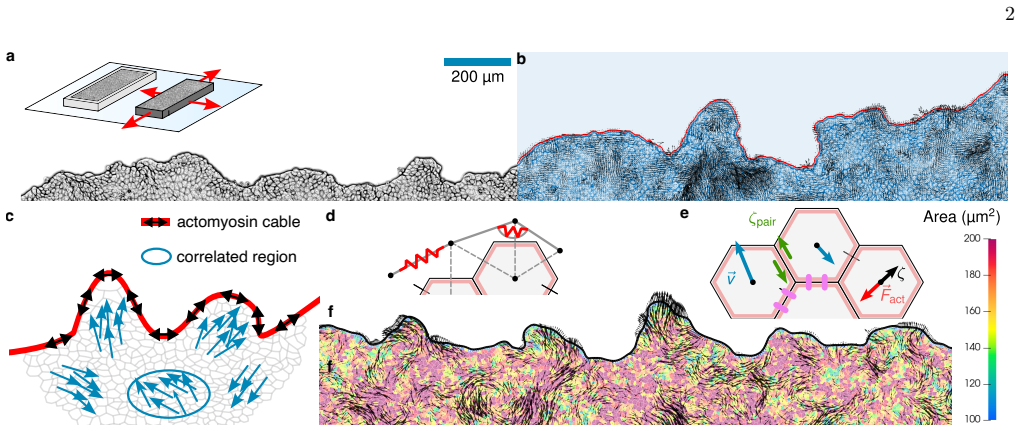
The load-bearing object is the advancing edge as a stretched, contractile semiflexible polymer, an effective worm-like chain driven by a correlated active noise field that represents interior cell motion. In the continuum description this becomes a linear height equation for the boundary height $h(x,t)$, whose relaxation is controlled by line tension $\lambda$, bending stiffness $\kappa$, and an internal pair-dissipation coefficient $\eta_h$, and whose driving is the velocity field of a viscoelastic model of the monolayer interior. The analysis shows that the dominant finger length scale is the shear length scale of the interior velocity correlations, $\xi_{\perp,p}=\sqrt{(\mu\tau+\eta)/\zeta_c}$, and that finger lifetimes are set by the slow $q^{-2}$ relaxation of the height modes at that scale. Alongside it, the active vertex model supplies the agent-based testbed: cells crawl as active Brownian particles with persistence time $\tau$, interactions include substrate friction and pair friction, and the boundary is implemented as coupled linear and angular springs that can grow and shrink.
What would settle it
Measure whether the advancing edge is actually passive: if inhibiting actomyosin contractility at the leading edge abolishes fingers while the interior velocity correlations (length scale of about 100 micrometres, persistence of about 1 hour) remain unchanged, the passive-cable picture fails; if fingers persist, the claim that interior correlations alone drive them survives.
Extended reading notes
Core claim
On its own terms, the paper's central claim is: correlated active cell motion alone suffices to produce fingers; leader cells, signalling, and proliferation modulate, but do not trigger, this pattern. The authors present fingers as long-lived active fluctuations of the boundary rather than the product of a finite-wavelength instability. They demonstrate this by combining in vitro imaging of spreading epithelial monolayers with simulations of an active vertex model whose edge is a contractile semiflexible polymer, and with a linear viscoelastic height-equation theory for the edge driven by correlated noise from the interior. The quantitative match between theory, simulation, and experiment on tangent-tangent correlations, roughness correlations, height spectra, and long finger lifetimes is presented as evidence that no feedback instability is needed.
Load-bearing premise
The load-bearing premise is that the actomyosin cable at the edge can be treated as a passive, very soft elastic string whose line tension and bending stiffness are about 25 times smaller than the values obtained by directly mapping the boundary springs of the simulation.
Editorial extensions
If this is right
- Leader cells appear at the tips of fingers as a response to the local mechanical environment of a forming protrusion, so they should amplify rather than initiate the pattern.
- Proliferation and cell flattening contribute to the average border speed but leave the finger statistics unchanged, so division is not the trigger.
- Finger length and lifetime are predicted to be set by the interior correlation length and cable parameters: changing cell-cell adhesion, friction, crawling speed, or cable line tension should shift both interior and boundary scales in a predictable way.
- The absence of a peak in the boundary height spectrum over time implies that no finite-wavelength instability is at work; the low-$q$ fluctuations saturate to a steady state.
- Initial roughness of the border explains the early-time differences between experiment and simulation, and the nonlinear short-scale behaviour shifts the roughness exponent from about 2 to 1.
Reading between the lines
- Inference: the same null model could be tested in other epithelial systems whose interior correlation lengths differ; the theory predicts their finger length scales should track the interior shear length scale rather than any cell-autonomous fingerprint.
- Inference: if the edge is passive, then transiently suppressing the actomyosin cable should make fingers wider and shorter-lived, while suppressing interior correlations, for example by raising density toward a jammed state, should suppress fingers even with a normal cable; both experiments are directly doable.
- Inference: because the height correlator contains a leading $1/q^2$ term that dominates at long times and large system sizes, very long experiments should show slowly coarsening roughness even though tangent-tangent correlations saturate; this coarsening is an untested corollary.
Signed reviews
Editorial analysis
A structured set of objections, weighed in public.
Referee Report
Summary. The paper combines MDCK wound-healing experiments, self-propelled Voronoi/active vertex model (AVM) simulations, and a continuum viscoelastic theory with a boundary height equation and active polymer model to argue that fingers at the edge of expanding epithelial monolayers arise as long-lived active fluctuations driven by correlated interior cell motion, without requiring a finite-wavelength instability or leader-cell feedback. The interior velocity statistics are matched by the AVM and by an active viscoelastic continuum model, while the boundary is modelled as a semiflexible polymer driven by interior noise; the height equation reproduces tangent-tangent, roughness, and temporal correlation functions. The paper concludes that leader cells, signalling, and proliferation modulate but do not trigger finger formation.
Significance. If the mechanism is correct, the paper is significant: it provides a quantitative null model for epithelial fingering, connects finger length and time scales to bulk active-matter correlation lengths, and makes falsifiable predictions about how perturbations to adhesion, friction, crawling speed, and cable properties affect fingers. Strengths include the direct experimental measurement of velocity and boundary correlations, the explicit AVM with a dynamic boundary, the detailed analytical derivations in the SI, and the clear statement of the model as a fluctuation-driven null model. The claim that fingers do not require an instability is supported by the absence of peaks in the Fourier spectra and by the AVM's spontaneous finger formation without division or leader cells. However, the quantitative predictive power of the continuum theory is weakened by an unexplained rescaling of the boundary stiffness, and the dominant-length-scale prediction is partly built into the model construction.
major comments (3)
- [SI Appendix II.D and main text Eq. (8)] The continuum theory and the 2D polymer model are not actually obtained from the AVM boundary parameters by dimensional mapping: SI Appendix II.D states that the estimated line tension and bending stiffness on the boundary of the AVM should be taken roughly a factor of 25 smaller to match theory, and the main text admits that a very small value of the spring constant ks had to be fitted. Because finger width, roughness, and lifetime in the height equation depend directly on lambda and kappa, the quantitative agreement shown in Fig. 4c, f, and i is partly a consequence of this tuning rather than an independent validation. As written, the abstract's claim that the model 'quantitatively predicts' the edge statistics is overstated; the authors should either derive the reduction factor from the boundary point addition/removal relaxation process or explicitly treat lambda and kappa as fitted parameters and state how many free parameters the comparison uses.
- [SI Appendix II.D] The noise-generating chain modulus for the 2D polymer is set to mu_h/zeta_h = 600 um^2/h 'by hand from fitting the real space spatial correlation of the 2d polymer noise generating chain velocity to the real space velocity correlations of the AVM'. This is an additional free parameter that is not listed in Table I and is calibrated against the same AVM velocity field that was already matched to experiments. This further weakens the claim that the polymer and height-equation simulations are parameter-free descendants of the AVM; mu_h should be listed with its uncertainty, and the sensitivity of the boundary correlation functions to its value should be reported.
- [Main text, Eq. (S86) and Discussion] The statement that finger length scales are dominated by the interior shear correlation length q1 = sqrt(zeta/(mu*tau + eta)) is close to a restatement of the model construction: the boundary noise vf in Eq. (S41) is derived from the same viscoelastic interior field with the same mu, eta, zeta, and tau, so the edge inheriting that scale is a consistency check rather than an independent prediction. A stronger, falsifiable test would be a perturbation experiment or simulation, for example varying KP, zeta_pair, or v0 and showing that the finger length and lifetime scales track the predicted q1; the Discussion proposes such dependencies but does not report them. The authors should either perform such a perturbation test or clearly label the dominant-scale result as a self-consistency statement.
minor comments (3)
- [Table I] The fitted interval for ks/zeta is reported as 78 +/- 50 h^-1, which is a very large range; the authors should show the sensitivity of the tangent-tangent correlation to ks within this range, for example as a supplemental figure, so that the constraint is visually verifiable.
- [Main text, Fig. 2 caption and text near Eq. (3)] The phrase 'again computed using dimensional scaling only' is misleading because the AVM parameters themselves were fitted to the experimental velocity statistics; the continuum theory is parameterized by mapping those fitted AVM parameters, so it is not an independent calculation.
- [SI Appendix II.D, Fig. S10] The text states that bond lengths can be approximated by a constant a(t) about a(0) = 5 um, but Fig. S10 shows a(t) growing from about 5 um to about 10 um over 15 h; the approximation should be justified more carefully or its effect on the mapping estimated.
Circularity Check
Quantitative finger-statistics 'predictions' are partly fitted (AVM ks from tangent-tangent; height-equation lambda/kappa rescaled 25x) and the dominant finger scale q1 is built into the 1D noise ansatz.
-
fitted input called prediction
[Results - finger formation (first paragraph) and Table I]
"We now compare simulated and measured advancing edges quantitatively by defining three boundary correlation functions, which allow us to constrain the remaining parameters of the A VM, the boundary spring constant ks and refine the crawling speed v0. ... After matching the edge stretching stiffness ks and refining v0 (constrained by vrms) to match the amplitude of the temporal decay, it reaches the same steady state value, although not as quickly as in the experiments (Fig. 4b)."
Table I states that ks is constrained by the 'shape of tangent-tangent correlation'. The AVM tangent-tangent curve in Fig. 4b is therefore the calibration target, not an independent prediction. The abstract's claim that the model 'quantitatively predicts tangent-tangent ... correlation functions' overstates this: for the AVM the match is obtained by fitting ks to that very observable. The qualitative emergence of fingers is independent, but this specific quantitative agreement is forced by construction.
-
fitted input called prediction
[SI Appendix II.D; main text 'Polymer and height equation theory for finger formation']
"However, we found that the estimated line tension and bending stiffness on the boundary of the A VM should be taken roughly a factor of 25 smaller, to match with theory, likely stemming from the relaxation effects when new points are added to the A VM boundary, see [42] for details. ... Note, however, that we had to fit a very small value of spring constant ks, likely reflecting the effectively viscoelastic relaxation mechanism of the cable in both experiment and A VM (SI Appendix, section II.D)."
The height equation (Eq. 8) and 2D polymer model (Eq. 7) are presented as derived from the AVM boundary springs by dimensional mapping (SI II.D, Eq. S51: ks/(zeta a^2) = lambda/zeta_h, kb/(zeta a^4) = kappa/zeta_h). The derivation then rescales both lambda and kappa by an undocumented factor of 25 'to match with theory'. Since tangent-tangent, roughness and spectrum predictions all depend on lambda and kappa, the quantitative agreement in Figs. 4c,f,i is obtained by tuning these effective parameters rather than by a parameter-free first-principles mapping; the factor is justified only by a stated but unquantified 'relaxation effects' mechanism.
1 more flagged steps
-
self definitional
[SI Appendix II.C (Eq. S41); main text analysis after Eq. (8)]
"Therefore, we cannot directly use the fluctuations of the 2d bulk velocity field to inform 1d boundary fluctuations. Therefore, as a matter of simplicity, we will consider that the driving fluctuating velocity field vf is generated by the following chain model ... The largest and dominant of these length scales is for q1 = ..., the same as the shear length scale of the interior velocity correlations."
The 1D noise chain (Eq. S41) is not derived from the 2D interior theory but posited as the same viscoelastic equation in 1D, carrying the same mu, eta, tau, zeta. Its correlation length xi_perp,p = sqrt((mu tau + eta)/zeta) (Eq. S20) is thus input by ansatz. The subsequent statement that the dominant finger length scale q1 equals 1/xi_perp,p and that finger length scales are 'directly set by the spatiotemporal correlations of the interior' is a restatement of this construction, not an independent prediction. The paper is transparent about the 'matter of simplicity' choice, but the central quantitative scale-selection claim reduces to that input.
full rationale
The paper contains an independent, non-circular core: the AVM is calibrated to bulk velocity correlations (tau, v0, zeta_pair, KP), and fingers emerge spontaneously in that model; the boundary observables are then compared, with ks fitted to the tangent-tangent shape, and roughness/spectrum following. This supports a qualitative sufficiency claim. However, several quantitative 'predictions' in the chain reduce to fitted inputs. (1) The AVM tangent-tangent match is the calibration target for ks (Table I), so the abstract's 'quantitatively predicts tangent-tangent' is an overstatement for the AVM. (2) The height-equation/polymer theory is advertised as dimensionally mapped from AVM boundary springs, but SI II.D rescales lambda and kappa by an unexplained factor of 25 'to match with theory'; the main text admits 'we had to fit a very small value of spring constant ks'. Finger width, roughness, and lifetime scale with lambda and kappa, so the agreement in Figs. 4c,f,i is partly a fit. (3) The dominant finger length scale q1 is built into the model: the 1D noise chain driving the edge is chosen 'as a matter of simplicity' to be the 1D version of the same viscoelastic interior equation, so q1 = sqrt(zeta/(mu tau + eta)) equals the interior shear correlation length by construction; the paper's claim that finger scales are 'directly set by' interior correlations is therefore a model input, not an independent derivation. Self-citation of Ref. [36] is not load-bearing here because the model is re-validated against new experimental velocity-correlation data in this paper. Overall: partial circularity, with the central sufficiency claim retaining independent AVM support, so score 6 rather than 8-10.
Assumptions & free parameters
free parameters (8)
- crawling persistence time tau =
1.0 +/- 0.5 h
- crawling speed v0 =
255 +/- 20 µm/h
- relative pair friction zeta_pair/zeta =
2.0 +/- 0.5
- perimeter modulus KP/zeta =
14 +/- 2 h^-1
- edge stretching stiffness ks/zeta =
78 +/- 50 h^-1
- effective line tension lambda in height equation =
roughly 25x smaller than AVM-mapped value
- noise-generating chain modulus mu_h/zeta_h =
600 µm^2/h
- target shape index p0 =
3.72
assumptions (6)
- domain assumption The monolayer interior is a linear, isotropic active viscoelastic solid with bulk and shear elasticity B, mu and bulk and shear viscosities K, eta (Eq. 2).
- domain assumption Active crawling is spatially uncorrelated on the cell scale and temporally persistent with timescale tau (ABP noise); there is no explicit cell-cell alignment or feedback in the driving.
- domain assumption The advancing edge is a passive semiflexible polymer under line tension, with overdamped dynamics and pair dissipation (Eqs. 7 and 8).
- domain assumption Boundary fluctuations are small enough that the height function is single-valued and the small-fluctuation expansion of the worm-like chain applies (SI II.B).
- domain assumption The boundary is driven passively by the interior velocity field and does not feed back onto the interior dynamics.
- domain assumption The velocity correlation functions have reached steady state before barrier removal (SI III.B).
Cite this review
Pith. "Pith review of Correlated cell movements drive epithelial finger formation." pith.science (2026). https://pith.science/paper/ILHRKZUH
@misc{pith2026250801046,
  author       = {Pith},
  title        = {Pith review of: Correlated cell movements drive epithelial finger formation},
  year         = {2026},
  howpublished = {\url{https://pith.science/paper/ILHRKZUH}},
  note         = {Machine review of arXiv:2508.01046}
}
read the original abstract
Epithelia form protective barriers in multicellular organisms. To maintain homeostasis, they must be able to regenerate and heal damaged areas. This occurs through collective cell migration, during which finger-like protrusions commonly appear. Whether these protrusions are driven by specialised leader cells, biochemical cues, or generic physical interactions remains unclear. Integrating in vitro imaging, agent-based simulations, and continuum modelling, we show that correlated active cell motion alone suffices to produce fingers. Leader cells, signalling, and proliferation modulate, but do not trigger, this pattern. Our results show that the key mechanism underlying a complex biological process can be understood using a general framework of the physics of dense active matter.
Figures


Figures from the paper (2 more)


Reference graph
Works this paper leans on
-
[1]
Honda, The world of epithelial sheets, Development, Growth & Differentiation 59, 306 (2017)
H. Honda, The world of epithelial sheets, Development, Growth & Differentiation 59, 306 (2017)
work page 2017
-
[2]
D. L. Nikoli´ c, A. N. Boettiger, D. Bar-Sagi, J. D. Car- beck, and S. Y. Shvartsman, Role of boundary conditions in an experimental model of epithelial wound healing, American Journal of Physiology-Cell Physiology 291, C68 (2006)
work page 2006
-
[3]
M. Poujade, E. Grasland-Mongrain, A. Hertzog, J. Jouanneau, P. Chavrier, B. Ladoux, A. Buguin, and P. Silberzan, Collective migration of an epithelial mono- layer in response to a model wound, Proceedings of the National Academy of Sciences 104, 15988 (2007)
work page 2007
-
[4]
L. Petitjean, M. Reffay, E. Grasland-Mongrain, M. Pou- jade, B. Ladoux, A. Buguin, and P. Silberzan, Velocity Fields in a Collectively Migrating Epithelium, Biophysi- cal Journal 98, 1790 (2010)
work page 2010
- [5]
-
[6]
G. Y. Ouaknin and P. Z. Bar-Yoseph, Stochastic Collec- tive Movement of Cells and Fingering Morphology: No Maverick Cells, Biophysical Journal 97, 1811 (2009)
work page 2009
-
[7]
S. Mark, R. Shlomovitz, N. S. Gov, M. Poujade, E. Grasland-Mongrain, and P. Silberzan, Physical Model of the Dynamic Instability in an Expanding Cell Culture, Biophysical Journal 98, 361 (2010)
work page 2010
- [8]
Show all 73 references
-
[9]
M. H. K¨ opf and L. M. Pismen, A continuum model of epithelial spreading, Soft Matter 9, 3727 (2013)
2013
-
[10]
Sep´ ulveda, L
N. Sep´ ulveda, L. Petitjean, O. Cochet, E. Grasland- Mongrain, P. Silberzan, and V. Hakim, Collective Cell Motion in an Epithelial Sheet Can Be Quantitatively De- scribed by a Stochastic Interacting Particle Model, PLOS Computational Biology 9, e1002944 (2013)
2013
-
[11]
Reffay, M
M. Reffay, M. C. Parrini, O. Cochet-Escartin, B. Ladoux, A. Buguin, S. Coscoy, F. Amblard, J. Camonis, and P. Silberzan, Interplay of RhoA and mechanical forces in collective cell migration driven by leader cells, Nature Cell Biology 16, 217 (2014)
2014
-
[12]
Zimmermann, M
J. Zimmermann, M. Basan, and H. Levine, An instability at the edge of a tissue of collectively migrating cells can lead to finger formation during wound healing, The Eu- ropean Physical Journal Special Topics 223, 1259 (2014)
2014
-
[13]
Tarle, A
V. Tarle, A. Ravasio, V. Hakim, and N. S. Gov, Model- ing the finger instability in an expanding cell monolayer, Integrative Biology 7, 1218 (2015)
2015
-
[14]
Vishwakarma, J
M. Vishwakarma, J. Di Russo, D. Probst, U. S. Schwarz, T. Das, and J. P. Spatz, Mechanical interactions among followers determine the emergence of leaders in migrat- ing epithelial cell collectives, Nature Communications 9, 3469 (2018)
2018
-
[15]
Alert, C
R. Alert, C. Blanch-Mercader, and J. Casademunt, Ac- tive Fingering Instability in Tissue Spreading, Physical Review Letters 122, 088104 (2019)
2019
-
[16]
Yang and H
Y. Yang and H. Levine, Leader-cell-driven epithelial sheet fingering, Physical Biology 17, 046003 (2020)
2020
-
[17]
Oguma, H
T. Oguma, H. Takigawa-Imamura, and T. Miura, Mech- anism underlying dynamic scaling properties observed in the contour of spreading epithelial monolayer, Physical Review E 102, 062408 (2020)
2020
-
[18]
Vishwakarma, J
M. Vishwakarma, J. P. Spatz, and T. Das, Mechanobiol- ogy of leader–follower dynamics in epithelial cell migra- tion, Current Opinion in Cell Biology Cell Dynamics, 66, 97 (2020)
2020
-
[19]
B¨ uscher, A
T. B¨ uscher, A. L. Diez, G. Gompper, and J. Elgeti, Insta- bility and fingering of interfaces in growing tissue, New Journal of Physics 22, 083005 (2020). 12
2020
-
[20]
Trenado, L
C. Trenado, L. L. Bonilla, and A. Mart ´ ınez-Calvo, Fin- gering instability in spreading epithelial monolayers: Roles of cell polarisation, substrate friction and contrac- tile stresses, Soft Matter 17, 8276 (2021)
2021
-
[21]
Mukhtar, E
N. Mukhtar, E. N. Cytrynbaum, and L. Edelstein- Keshet, A multiscale computational model of YAP sig- naling in epithelial fingering behavior, Biophysical Jour- nal 121, 1940 (2022)
2022
-
[22]
Killeen, B
A. Killeen, B. Partridge, T. Bertrand, and C. F. Lee, Modeling growing confluent tissues using a lattice Boltz- mann method: Interface stability and fluctuations, Phys- ical Review Research 5, 043096 (2023)
2023
-
[23]
Jeong, J
H. Jeong, J. Yeo, S. Ryu, and J. H. Shin, The fingering patterns in the epithelial layer control the gap closure rate via curvature-mediated force (2023)
2023
-
[24]
Ye and J
Y. Ye and J. Lin, Fingering Instability Accelerates Popu- lation Growth of a Proliferating Cell Collective, Physical Review Letters 132, 018402 (2024)
2024
-
[25]
L. Qin, D. Yang, W. Yi, H. Cao, and G. Xiao, Roles of leader and follower cells in collective cell migration, Molecular Biology of the Cell 32, 1267 (2021)
2021
-
[26]
Rapin, N
G. Rapin, N. Caballero, I. Gaponenko, B. Ziegler, A. Rawleigh, E. Moriggi, T. Giamarchi, S. A. Brown, and P. Paruch, Roughness and dynamics of proliferating cell fronts as a probe of cell–cell interactions, Scientific Reports 11, 8869 (2021)
2021
-
[27]
Ladoux and R.-M
B. Ladoux and R.-M. M` ege, Mechanobiology of collective cell behaviours, Nature Reviews Molecular Cell Biology 18, 743 (2017)
2017
-
[28]
Rossetti, S
L. Rossetti, S. Grosser, J. F. Abenza, L. Valon, P. Roca- Cusachs, R. Alert, and X. Trepat, Optogenetic genera- tion of leader cells reveals a force–velocity relation for collective cell migration, Nature Physics , 1 (2024)
2024
-
[29]
C. F. Lee, It takes more than forceful leaders, Nature Physics , 1 (2024)
2024
-
[30]
Vazquez, A
K. Vazquez, A. Saraswathibhatla, and J. Notbohm, Ef- fect of substrate stiffness on friction in collective cell mi- gration, Scientific Reports 12, 2474 (2022)
2022
-
[31]
Khataee, A
H. Khataee, A. Czirok, and Z. Neufeld, Multiscale mod- elling of motility wave propagation in cell migration, Sci- entific Reports 10, 8128 (2020)
2020
-
[32]
L. L. Bonilla, A. Carpio, and C. Trenado, Tracking col- lective cell motion by topological data analysis, PLOS Computational Biology 16, e1008407 (2020)
2020
-
[33]
Lee and C
P. Lee and C. W. Wolgemuth, Crawling Cells Can Close Wounds without Purse Strings or Signaling, PLOS Com- putational Biology 7, e1002007 (2011)
2011
-
[34]
Nesbitt, G
D. Nesbitt, G. Pruessner, and C. F. Lee, Edge instability in incompressible planar active fluids, Physical Review E 96, 062615 (2017)
2017
-
[35]
Garcia, E
S. Garcia, E. Hannezo, J. Elgeti, J.-F. Joanny, P. Sil- berzan, and N. S. Gov, Physics of active jamming during collective cellular motion in a monolayer, Proceedings of the National Academy of Sciences 112, 15314 (2015)
2015
-
[36]
Henkes, K
S. Henkes, K. Kostanjevec, J. M. Collinson, R. Sknep- nek, and E. Bertin, Dense active matter model of motion patterns in confluent cell monolayers, Nature Communi- cations 11, 1405 (2020)
2020
-
[37]
Nagai and H
T. Nagai and H. Honda, A dynamic cell model for the formation of epithelial tissues, Philosophical Magazine B 81, 699 (2001)
2001
-
[38]
Farhadifar, J.-C
R. Farhadifar, J.-C. R¨ oper, B. Aigouy, S. Eaton, and F. J¨ ulicher, The Influence of Cell Mechanics, Cell-Cell In- teractions, and Proliferation on Epithelial Packing, Cur- rent Biology 17, 2095 (2007)
2007
-
[39]
A. G. Fletcher, M. Osterfield, R. E. Baker, and S. Y. Shvartsman, Vertex Models of Epithelial Morphogenesis, Biophysical Journal 106, 2291 (2014)
2014
-
[40]
Li and S
B. Li and S. X. Sun, Coherent Motions in Confluent Cell Monolayer Sheets, Biophysical Journal 107, 1532 (2014)
2014
-
[41]
D. Bi, X. Yang, M. C. Marchetti, and M. L. Manning, Motility-Driven Glass and Jamming Transitions in Bio- logical Tissues, Physical Review X 6, 021011 (2016)
2016
-
[42]
D. L. Barton, S. Henkes, C. J. Weijer, and R. Sknepnek, Active Vertex Model for cell-resolution description of ep- ithelial tissue mechanics, PLOS Computational Biology 13, e1005569 (2017)
2017
-
[43]
Farooqui and G
R. Farooqui and G. Fenteany, Multiple rows of cells be- hind an epithelial wound edge extend cryptic lamellipo- dia to collectively drive cell-sheet movement, Journal of Cell Science 118, 51 (2005)
2005
-
[44]
S. Tong, R. Sknepnek, and A. Koˇ smrlj, Linear viscoelas- tic response of the vertex model with internal and exter- nal dissipation: Normal modes analysis, Physical Review Research 5, 013143 (2023)
2023
-
[45]
Rozman, C
J. Rozman, C. K. V. S, J. M. Yeomans, and R. Sknep- nek, From Substrate Dissipation to Internal Dissipation Vertex Model Dynamics: Generating Sustained Flows (2024), arXiv:2312.11756
2024 arXiv
-
[46]
Caprini, U
L. Caprini, U. M. B. Marconi, C. Maggi, M. Paoluzzi, and A. Puglisi, Hidden velocity ordering in dense suspen- sions of self-propelled disks, Physical Review Research 2, 023321 (2020)
2020
-
[47]
Mandal, P
R. Mandal, P. J. Bhuyan, P. Chaudhuri, C. Dasgupta, and M. Rao, Extreme active matter at high densities, Nature Communications 11, 2581 (2020)
2020
-
[48]
Y.-E. Keta, R. L. Jack, and L. Berthier, Disordered Col- lective Motion in Dense Assemblies of Persistent Parti- cles, Physical Review Letters 129, 048002 (2022)
2022
-
[49]
Szamel and E
G. Szamel and E. Flenner, Long-ranged velocity correla- tions in dense systems of self-propelled particles, Euro- physics Letters 133, 60002 (2021)
2021
-
[50]
Serra-Picamal, V
X. Serra-Picamal, V. Conte, R. Vincent, E. Anon, D. T. Tambe, E. Bazellieres, J. P. Butler, J. J. Fredberg, and X. Trepat, Mechanical waves during tissue expansion, Nature Physics 8, 628 (2012)
2012
-
[51]
Boocock, N
D. Boocock, N. Hino, N. Ruzickova, T. Hirashima, and E. Hannezo, Theory of mechanochemical patterning and optimal migration in cell monolayers, Nature physics 17, 267 (2021)
2021
-
[52]
M. Doi, S. Edwards, and S. Edwards, The Theory of Polymer Dynamics, International series of monographs on physics (Clarendon Press, 1988)
1988
-
[53]
Golestanian and T
R. Golestanian and T. B. Liverpool, Statistical mechanics of semiflexible ribbon polymers, Physical Review E 62, 5488 (2000)
2000
-
[54]
T. B. Liverpool and A. C. Maggs, Dynamic scatter- ing from semiflexible polymers, Macromolecules 34, 6064 (2001), https://doi.org/10.1021/ma001468p
2001 doi
-
[55]
T. B. Liverpool, Anomalous fluctuations of active polar filaments, Physical Review E 67, 031909 (2003)
2003
-
[56]
Agoritsas, V
E. Agoritsas, V. Lecomte, and T. Giamarchi, Disordered elastic systems and one-dimensional interfaces, Physica B: Condensed Matter 407, 1725 (2012)
2012
-
[57]
J. F. Marko and E. D. Siggia, Stretching dna, Macro- molecules 28, 8759 (1995)
1995
-
[58]
van Saarloos, V
W. van Saarloos, V. Vitelli, and Z. Zeravcic, Soft Mat- ter: Concepts, Phenomena, and Applications(Princeton 13 University Press, 2024)
2024
-
[59]
B. S. Khatri and T. C. McLeish, Rouse model with in- ternal friction: A coarse grained framework for single biopolymer dynamics, Macromolecules 40, 6770 (2007)
2007
-
[60]
Kardar, G
M. Kardar, G. Parisi, and Y.-C. Zhang, Dynamic scaling of growing interfaces, Phys. Rev. Lett. 56, 889 (1986)
1986
-
[61]
Caballero, T
N. Caballero, T. Giamarchi, V. Lecomte, and E. Agorit- sas, Microscopic interplay of temperature and disorder of a one-dimensional elastic interface, Physical Review E 105, 044138 (2022)
2022
-
[62]
G. I. Bell and E. C. Anderson, Cell Growth and Division: I. A Mathematical Model with Applications to Cell Vol- ume Distributions in Mammalian Suspension Cultures, Biophysical Journal 7, 329 (1967)
1967
-
[63]
G. T. Eisenhoffer and J. Rosenblatt, Bringing balance by force: live cell extrusion controls epithelial cell numbers, Trends in cell biology 23, 185 (2013)
2013
-
[64]
Zulueta-Coarasa and J
T. Zulueta-Coarasa and J. Rosenblatt, The role of tissue maturity and mechanical state in controlling cell extru- sion, Current opinion in genetics & development 72, 1 (2022)
2022
-
[65]
Adkins, I
R. Adkins, I. Kolvin, Z. You, S. Witthaus, M. C. Marchetti, and Z. Dogic, Dynamics of active liquid in- terfaces, Science 377, 768 (2022)
2022
-
[66]
L. Zhao, P. Gulati, F. Caballero, I. Kolvin, R. Adkins, M. C. Marchetti, and Z. Dogic, Asymmetric fluctuations and self-folding of active interfaces, Proceedings of the National Academy of Sciences 121, e2410345121 (2024)
2024
-
[67]
M. E. Cates and C. Nardini, Active phase separation: new phenomenology from non-equilibrium physics, Re- ports on Progress in Physics 88, 056601 (2025)
2025
-
[68]
Culture-Insert 2 Well in µ-Dish 35 mm — Wound Heal- ing Assays — ibidi — ibidi.com, https://ibidi.com/ culture-inserts/24-culture-insert-2-well.html (n.y.)
-
[69]
Guennebaud, B
G. Guennebaud, B. Jacob, et al. , Eigen v3, http://eigen.tuxfamily.org (2010)
2010
-
[70]
F. W. Byron and R. W. Fuller, Mathematics of Classical and Quantum Physics(Courier Corporation, 2012)
2012
-
[71]
Thielicke and R
W. Thielicke and R. Sonntag, Particle Image Velocime- try for MATLAB: Accuracy and enhanced algorithms in PIVlab, Journal of Open Research Software 9, 10.5334/jors.334 (2021). 14 APPENDIX T ABLE OF CONTENTS I Supporting figures 15 I. Interior 15 I.A. Velocity statistics furthe...
2021 doi
-
[72]
Shading of experiments is the standard deviation of experimental histograms and the shading of the A VM is the standard error of the mean. 18 II. BOUNDAR Y II.A. Polymer simulations starting with initial roughness a b c FIG. S6. Tangent-tangent correlation ( a), spatial Fourie...
-
[73]
µq2/ζq [λq2 + κq4]2 /ζ 2q − (µq2/ζq)2 # (S65) − 1 + e−2[λq2+κq4]t/ζq − 2e−[λq2+κq4]t/ζq−t/τ
and add pair dissipation. The equation of motion for the displacement field u within the cell sheet reads ζ ˙u(r, t) = ζv 0 ˆn(r, t) + ∇ ·← →σ , (S1) 20 where ζ is a friction coefficient, which we write without the c subscript of the main text for ease of notation, ζv 0 ˆn is ...
Reviewed August 6, 2026 · model on record in the stance chip above.
Discussion (0). Continue with ORCID to comment.