REVIEW 4 major objections 5 minor 52 references
DS$^2$Net: Detail-Semantic Deep Supervision Network for Medical Image Segmentation
T0 review · 4 major / 5 minor · reviewed 2026-08-06 · deepseek-v4-flash
Pith's one-line read Detail + semantic supervision wins six medical segmentation benchmarks
desk verdict Solid architecture paper with an overclaimed adaptive loss: DEM/SEM supervision is a real combination, but Table V shows raw uncertainty weights hurt and the softmax/max-scaling are empirical repairs, not a non-heuristic principle. read the letter →
The pith
A machine-rendered reading of the paper's core claim, the machinery that carries it, and where it could break.
The reading
What carries the argument
The Detail Enhancement Module (DEM) and Semantic Enhancement Module (SEM), plus the uncertainty-based adaptive supervision loss. DEM uses a max-pooling, double-convolution, sigmoid path on low-level features to create a detail mask $M_d$, then outputs $S_d = M_d \odot f_{\mathrm{fusion}} + f_l$. SEM uses spatial attention on high-level features to create a semantic mask $M_s$, then outputs $S_s = \mathrm{CA}(M_s \odot f_l) + \mathrm{up}(f_h)$. The uncertainty loss computes each side output's mean deviation from 0.5, converts it via softmax and max-scaling into a stage weight, and sums weighted IoU and BCE losses. Together these carry the claim: the masks inject complementary supervision, and
What would settle it
Compare DS²Net on BUSI and 2018-DSB with equal stage weights, with the paper's uncertainty weights, and with an entropy-based uncertainty. If entropy-based or equal weights match or beat the proposed weights, the specific proxy is not carrying the gain. Also, log per-stage validation Dice against per-stage adaptive weights: the claim predicts high-weight stages are the low-Dice, high-uncertainty stages.
Extended reading notes
Core claim
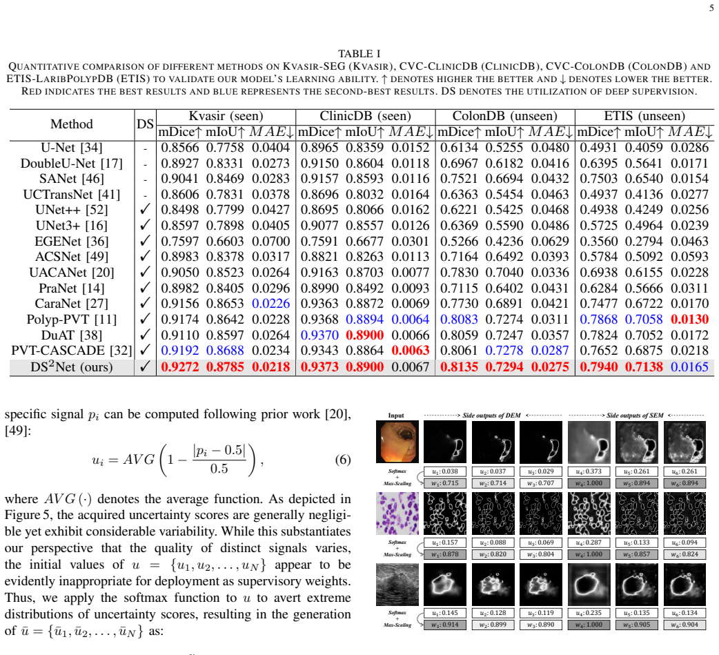
The paper argues that simultaneously supervising low-level detail features and high-level semantic features improves medical image segmentation. DEM derives a detail mask from max-pooled low-level features and multiplies it into a fused low/high-level feature map; SEM derives a semantic mask by spatial attention on high-level features and applies it to low-level features with channel attention. The six resulting side outputs are weighted by $\lambda_i = \bar{u}_i / \max(\bar{u})$, where $\bar{u}$ is the softmax of $u_i = \mathrm{AVG}(1 - |p_i - 0.5|/0.5)$. The paper reports 92.72% mDice on Kvasir-SEG, 79.40% mDice on ETIS-LaribPolypDB, 81.60% mDice on BUSI, and 90.97% mDice on 2018-DSB with
Load-bearing premise
The load-bearing premise is that $u_i = \mathrm{AVG}(1 - |p_i - 0.5|/0.5)$ measures how strongly a side output needs supervision, so that scaling each stage's loss by $\lambda_i$ helps; the paper's own Table V shows the raw uncertainty weights hurt and only the hand-added softmax-plus-max-scaling steps produce small gains.
Editorial extensions
If this is right
- Deep supervision networks no longer need to choose a single supervision view; fusing detail and semantic side outputs is the better default.
- The adaptive supervision loss is a drop-in replacement for fixed stage weights: the paper shows it improves six existing deep supervision networks.
- The largest gains appear on high-resolution and boundary-difficult data, so the method is most valuable for small or pathological structures such as polyps and breast lesions.
- The multi-scale side outputs are summed at inference, giving ensemble-like robustness with no extra test-time forward passes.
Reading between the lines
- The paper's Table V shows the raw uncertainty score hurts on its own, so a meaningful test is substituting an entropy- or Monte-Carlo-dropout-based uncertainty and checking whether the reported gains persist.
- The ablation hints that DEM and SEM matter differently by modality — SEM more on high-resolution polyps, DEM more on nuclei — so a natural extension is data-adaptive gating between detail and semantic enhancement.
- The adaptive weighting principle should transfer to other dense prediction tasks such as salient object detection or instance segmentation, but the paper only validates it on medical segmentation.
Editorial analysis
A structured set of objections, weighed in public.
Referee Report
Summary. The manuscript proposes DS^2Net, a deep-supervision segmentation network that supervises both low-level detail features and high-level semantic features through a Detail Enhancement Module (DEM) and a Semantic Enhancement Module (SEM), and an uncertainty-based adaptive supervision loss that weights each of the six auxiliary losses according to a per-signal uncertainty score. The method is evaluated on six medical segmentation benchmarks (polyp, breast ultrasound, and nuclei), with the strongest reported results being 92.72% mDice on Kvasir-SEG, 79.40% mDice on ETIS-LaribPolypDB, and 81.60% mDice on BUSI. The paper also reports ablations of the modules and of the adaptive loss, and applies the adaptive loss to other deep-supervision baselines.
Significance. The DEM/SEM idea is a plausible extension of deep supervision: it makes the supervision signals deliberately detail-oriented and semantic-oriented rather than merely multi-scale. The ablation results in Table III show large gains over the PVT baseline, and the code is promised for release, which would aid reproducibility. However, the paper's stronger claim—the 'first non-heuristic' uncertainty-based adaptive loss—is not supported by the evidence. The equations are not circular, but the proposed uncertainty proxy and its direction of effect are heuristic and are directly contradicted by the raw-weight ablation in Table V. The paper also lacks multiple-seed statistics, so the small final gains in several comparisons are hard to interpret. If the architectural result holds and the loss claim is substantially toned down or properly validated, the paper would be a useful incremental contribution; in its current form the central novelty claim is overstated.
major comments (4)
- [Section III-C, Eqs. (6)-(8), Table V] The paper's central methodological claim is that the uncertainty-based adaptive supervision loss is 'the first non-heuristic loss' and that it 'autonomously' assigns supervision strength. The evidence in Table V contradicts this. Using raw uncertainty scores as loss weights degrades performance on BUSI (mDice 81.40→80.82, mIoU 72.72→72.03) and on 2018-DSB (mDice 90.70→89.25, mIoU 83.65→81.24). Only after adding softmax and then max-scaling do small gains appear (+0.20 mDice on BUSI, +0.27 mDice on 2018-DSB). These two extra transformations are not derived from any uncertainty principle; the text merely reports that they were found necessary. Thus Eq. (6) is not a validated proxy for supervision-signal quality, and the 'non-heuristic' claim is unsupported. The authors should either supply independent validation of u_i as a quality measure, compare against alternative uncertainty metrics (
- [Section IV-A and Tables I, II, VI] All reported results appear to be single training runs, with no number of random seeds, standard deviations, or statistical significance tests. This is especially important because the headline gains over prior state-of-the-art are small: e.g., 92.72% vs. 91.92% mDice on Kvasir-SEG in Table I, and 81.60% vs. 81.18% on BUSI in Table II. The generality study in Table VI includes differences as small as +0.02 mDice and +0.04 mIoU (PVT-CASCADE on 2018-DSB), which are within run-to-run noise for a single seed. Without at least mean±std over multiple seeds and preferably a paired significance test, the claim that DS^2Net 'consistently outperforms' state-of-the-art methods is not established.
- [Figure 2 and Section I] Figure 2 is the empirical motivation for abandoning fixed loss-weight heuristics, but the manuscript does not define what 'quality' of a supervision signal means, how the quality ranking was computed, or whether the shown values are from the training or test set. The y-axis is labeled DSC, but the caption says 'quality rankings' and the red dashed box is claimed to underscore the 'non-heuristic nature' of signal quality. A ranking alone cannot establish that an uncertainty-based weight is non-heuristic; it only shows that side-output performance changes over training. The paper should specify the exact metric, the evaluation protocol, and ideally provide uncertainty estimates for the curves. This is a load-bearing motivation for the adaptive loss and should be made rigorous.
- [Section III-C, Eq. (6)] The uncertainty score is defined as the average over the entire prediction map of 1 - |p - 0.5|/0.5. This is an average closeness to the decision boundary. The paper asserts, citing prior work, that such uncertainty 'mirrors feature quality,' but no direct link is established between this aggregate quantity and the quality of a multi-stage feature map. In fact, a perfectly calibrated but uncertain prediction (e.g., a low-confidence region near a boundary) would be downweighted, while an overconfident erroneous prediction would be upweighted. This may explain the Table V degradation. The authors should either provide a theoretical or empirical justification for why higher average proximity to 0.5 should receive higher loss weight, or present this as an experimental design choice rather than a principled uncertainty estimate.
minor comments (5)
- [Throughout] The manuscript contains several typos and grammatical errors: 'To present' should be 'To date,' 'hightlight' should be 'highlight,' 'furnishing' is awkward, and 'the performances of the comparative models... are derived' is unclear. A careful proofread is needed.
- [Section IV-A] The paper states 'without any data augmentation' and lists dataset-specific multi-scale scaling ratios. These ratios (0.5/1/1.5 vs. 0.75/1/1.25) are additional hand-set design choices that are not ablated; they should be mentioned in the limitations or ablations.
- [Figure 3] The framework figure is overloaded and hard to read, especially the uncertainty/weight pipeline. Enlarging the relevant panels and explicitly labeling the N=6 signals would improve clarity.
- [Section IV-C, Table III] The ablation table does not include the adaptive supervision loss, even though the full method in Tables I-II uses it. This is fine methodologically, but should be stated in the text more prominently; currently the reader may assume Table III is the full DS^2Net.
- [Section V] The conclusion says the experimental results substantiate 'profound improvement' over state-of-the-art. Given the small margins in several datasets and the lack of error bars, this wording is too strong and should be moderated.
Circularity Check
No significant circularity; the uncertainty-loss design is under-validated but not a circular derivation.
full rationale
The paper's claims are empirical: DS^2Net's DEM/SEM modules and the adaptive loss are tested against held-out test sets and external baselines. The derivation chain in Sec. III is self-contained. Eq. (6) defines an uncertainty score from the model's own predicted probabilities (following prior work [20], [49]), Eqs. (7)-(8) normalize it, and Eq. (9) uses the result as loss weights. Nothing in this chain fits a parameter to the reported benchmark numbers, and the final segmentation is not used to define the loss weights. The softmax and MAX-Scaling steps were added after the ablations in Table V, where raw uncertainty weights actually hurt (BUSI mDice 81.40->80.82; 2018-DSB 90.70->89.25), so the 'first non-heuristic loss' claim is overstated and the proxy remains weakly validated. That is a correctness/validation risk, not circularity: the paper does not rename an input as a prediction nor invoke a self-citation to force the design. Self-citations exist ([14], [48] include author T. Zhou) but only as baselines/related work, not as load-bearing evidence. The method is self-contained against independent benchmarks, so no circular step is present.
Assumptions & free parameters
free parameters (3)
- softmax + max-scaling loss weighting scheme =
softmax then max-scaling (Eqs 7-8)
- multi-scale training scaling ratios =
0.5, 1, 1.5 for colonoscopy; 0.75, 1, 1.25 for ultrasound and microscopy
- DEM convolution kernel sizes per stage =
7x7 for the highest-resolution feature map; other sizes unspecified
assumptions (4)
- domain assumption The uncertainty metric u_i = AVG(1 - |p_i - 0.5| / 0.5) is a valid proxy for supervision signal quality.
- domain assumption Supervising both low-level detail and high-level semantic features simultaneously improves medical segmentation.
- ad hoc to paper The quality ranking in Figure 2 is representative and correctly measured.
- domain assumption PVT pretrained on ImageNet is a suitable backbone for these medical segmentation tasks.
Cite this review
Pith. "Pith review of DS$^2$Net: Detail-Semantic Deep Supervision Network for Medical Image Segmentation." pith.science (2026). https://pith.science/paper/GEDR77QZ
@misc{pith2026250804131,
  author       = {Pith},
  title        = {Pith review of: DS$^2$Net: Detail-Semantic Deep Supervision Network for Medical Image Segmentation},
  year         = {2026},
  howpublished = {\url{https://pith.science/paper/GEDR77QZ}},
  note         = {Machine review of arXiv:2508.04131}
}
abstract
Deep Supervision Networks exhibit significant efficacy for the medical imaging community. Nevertheless, existing work merely supervises either the coarse-grained semantic features or fine-grained detailed features in isolation, which compromises the fact that these two types of features hold vital relationships in medical image analysis. We advocate the powers of complementary feature supervision for medical image segmentation, by proposing a Detail-Semantic Deep Supervision Network (DS$^2$Net). DS$^2$Net navigates both low-level detailed and high-level semantic feature supervision through Detail Enhance Module (DEM) and Semantic Enhance Module (SEM). DEM and SEM respectively harness low-level and high-level feature maps to create detail and semantic masks for enhancing feature supervision. This is a novel shift from single-view deep supervision to multi-view deep supervision. DS$^2$Net is also equipped with a novel uncertainty-based supervision loss that adaptively assigns the supervision strength of features within distinct scales based on their uncertainty, thus circumventing the sub-optimal heuristic design that typifies previous works. Through extensive experiments on six benchmarks captured under either colonoscopy, ultrasound and microscope, we demonstrate that DS$^2$Net consistently outperforms state-of-the-art methods for medical image analysis.
Figures


Figures from the paper (6 more)




Reference graph
Works this paper leans on
-
[1]
Dataset of breast ultrasound images,
W. Al-Dhabyani, M. Gomaa, H. Khaled, and A. Fahmy, “Dataset of breast ultrasound images,” Data in brief , 2020
work page 2020
-
[2]
Phiseg: Capturing uncertainty in medical image segmentation,
C. F. Baumgartner, K. C. Tezcan, K. Chaitanya, A. M. H ¨otker, U. J. Muehlematter, K. Schawkat, A. S. Becker, O. Donati, and E. Konukoglu, “Phiseg: Capturing uncertainty in medical image segmentation,” in International Conference on Medical Image Computing and Computer- Assisted Intervention (MICCAI) , 2019
work page 2019
-
[3]
J. Bernal, F. J. S ´anchez, G. Fern ´andez-Esparrach, D. Gil, C. Rodr ´ıguez, and F. Vilari ˜no, “Wm-dova maps for accurate polyp highlighting in colonoscopy: Validation vs. saliency maps from physicians,” Comput- erized medical imaging and graphics , 2015
work page 2015
-
[4]
Nucleus segmentation across imaging experiments: the 2018 data science bowl,
J. C. Caicedo, A. Goodman, K. W. Karhohs, B. A. Cimini, J. Ackerman, M. Haghighi, C. Heng, T. Becker, M. Doan, C. McQuin, et al., “Nucleus segmentation across imaging experiments: the 2018 data science bowl,” Nature methods, 2019
work page 2018
-
[5]
Swin-unet: Unet-like pure transformer for medical image segmenta- tion,
H. Cao, Y . Wang, J. Chen, D. Jiang, X. Zhang, Q. Tian, and M. Wang, “Swin-unet: Unet-like pure transformer for medical image segmenta- tion,” in European Conference on Computer Vision (ECCV) , 2022
work page 2022
-
[6]
Aau-net: an adaptive attention u-net for breast lesions segmentation in ultrasound images,
G. Chen, L. Li, Y . Dai, J. Zhang, and M. H. Yap, “Aau-net: an adaptive attention u-net for breast lesions segmentation in ultrasound images,” IEEE Transactions on Medical Imaging (IEEE TMI) , 2022
work page 2022
-
[7]
Transunet: Transformers make strong encoders for medical image segmentation,
J. Chen, Y . Lu, Q. Yu, X. Luo, E. Adeli, Y . Wang, L. Lu, A. L. Yuille, and Y . Zhou, “Transunet: Transformers make strong encoders for medical image segmentation,” arXiv preprint arXiv:2102.04306 , 2021
arXiv 2021
-
[8]
A tutorial on the cross-entropy method,
P.-T. De Boer, D. P. Kroese, S. Mannor, and R. Y . Rubinstein, “A tutorial on the cross-entropy method,” Annals of operations research , 2005
work page 2005
Show all 52 references
-
[9]
Imagenet: A large-scale hierarchical image database,
J. Deng, W. Dong, R. Socher, L.-J. Li, K. Li, and L. Fei-Fei, “Imagenet: A large-scale hierarchical image database,” in IEEE/CVF Conference on Computer Vision and Pattern Recognition (CVPR) , 2009
2009
-
[10]
Stamp: Simultane- ous training and model pruning for low data regimes in medical image segmentation,
N. K. Dinsdale, M. Jenkinson, and A. I. Namburete, “Stamp: Simultane- ous training and model pruning for low data regimes in medical image segmentation,” Medical Image Analysis , 2022
2022
-
[11]
Polyp-pvt: Polyp segmentation with pyramid vision transformers,
B. Dong, W. Wang, D.-P. Fan, J. Li, H. Fu, and L. Shao, “Polyp-pvt: Polyp segmentation with pyramid vision transformers,” arXiv preprint arXiv:2108.06932, 2021
2021 arXiv
-
[12]
An image is worth 16x16 words: Transformers for image recognition at scale,
A. Dosovitskiy, L. Beyer, A. Kolesnikov, D. Weissenborn, X. Zhai, T. Unterthiner, M. Dehghani, M. Minderer, G. Heigold, S. Gelly, et al., “An image is worth 16x16 words: Transformers for image recognition at scale,” arXiv preprint arXiv:2010.11929 , 2020
2010 arXiv
-
[13]
Evaluating the clinical acceptability of deep learning contours of prostate and organs-at-risk in an automated prostate treatment planning process,
J. Duan, M. Bernard, L. Downes, B. Willows, X. Feng, W. F. Mourad, W. St Clair, and Q. Chen, “Evaluating the clinical acceptability of deep learning contours of prostate and organs-at-risk in an automated prostate treatment planning process,” Medical Physics, 2022
2022
-
[14]
Pranet: Parallel reverse attention network for polyp segmentation,
D.-P. Fan, G.-P. Ji, T. Zhou, G. Chen, H. Fu, J. Shen, and L. Shao, “Pranet: Parallel reverse attention network for polyp segmentation,” in International Conference on Medical Image Computing and Computer- Assisted Intervention (MICCAI) , 2020
2020
-
[15]
Joint optic disc and cup segmentation based on multi-label deep network and polar transformation,
H. Fu, J. Cheng, Y . Xu, D. W. K. Wong, J. Liu, and X. Cao, “Joint optic disc and cup segmentation based on multi-label deep network and polar transformation,” IEEE Transactions on Medical Imaging (IEEE TMI) , 2018
2018
-
[16]
Unet 3+: A full-scale connected unet for medical image segmentation,
H. Huang, L. Lin, R. Tong, H. Hu, Q. Zhang, Y . Iwamoto, X. Han, Y .- W. Chen, and J. Wu, “Unet 3+: A full-scale connected unet for medical image segmentation,” in IEEE International Conference on Acoustics, Speech and Signal Processing (ICASSP) , 2020
2020
-
[17]
Doubleu-net: A deep convolutional neural network for medical image segmentation,
D. Jha, M. A. Riegler, D. Johansen, P. Halvorsen, and H. D. Johansen, “Doubleu-net: A deep convolutional neural network for medical image segmentation,” in IEEE International Symposium on Computer-Based Medical Systems, 2020
2020
-
[18]
Kvasir-seg: A segmented polyp dataset,
D. Jha, P. H. Smedsrud, M. A. Riegler, P. Halvorsen, T. de Lange, D. Johansen, and H. D. Johansen, “Kvasir-seg: A segmented polyp dataset,” in International Conference on Multimedia Modeling (MMM) , 2020
2020
-
[19]
Crisp-reliable uncertainty estimation for medical image seg- mentation,
T. Judge, O. Bernard, M. Porumb, A. Chartsias, A. Beqiri, and P.-M. Jodoin, “Crisp-reliable uncertainty estimation for medical image seg- mentation,” in International Conference on Medical Image Computing and Computer-Assisted Intervention (MICCAI) , 2022
2022
-
[20]
Uacanet: Uncertainty augmented context attention for polyp segmentation,
T. Kim, H. Lee, and D. Kim, “Uacanet: Uncertainty augmented context attention for polyp segmentation,” in ACM International Conference on Multimedia (ACM MM) , 2021
2021
-
[21]
Deeply-supervised nets,
C.-Y . Lee, S. Xie, P. Gallagher, Z. Zhang, and Z. Tu, “Deeply-supervised nets,” in Artificial intelligence and statistics , 2015
2015
-
[22]
H-denseunet: hybrid densely connected unet for liver and tumor segmentation from ct volumes,
X. Li, H. Chen, X. Qi, Q. Dou, C.-W. Fu, and P.-A. Heng, “H-denseunet: hybrid densely connected unet for liver and tumor segmentation from ct volumes,” IEEE Transactions on Medical Imaging (IEEE TMI) , 2018
2018
-
[23]
Hierarchical fully convolutional network for joint atrophy localization and alzheimer’s disease diagnosis using structural mri,
C. Lian, M. Liu, J. Zhang, and D. Shen, “Hierarchical fully convolutional network for joint atrophy localization and alzheimer’s disease diagnosis using structural mri,” IEEE Transactions on Pattern Analysis and Machine Intelligence (IEEE TPAMI) , 2018
2018
-
[24]
Dna: Deeply supervised nonlinear aggregation for salient object detection,
Y . Liu, M.-M. Cheng, X.-Y . Zhang, G.-Y . Nie, and M. Wang, “Dna: Deeply supervised nonlinear aggregation for salient object detection,” IEEE Transactions on Cybernetics , 2021
2021
-
[25]
Fully convolutional networks for semantic segmentation,
J. Long, E. Shelhamer, and T. Darrell, “Fully convolutional networks for semantic segmentation,” in IEEE/CVF Conference on Computer Vision and Pattern Recognition (CVPR) , 2015
2015
-
[26]
Decoupled weight decay regularization,
I. Loshchilov and F. Hutter, “Decoupled weight decay regularization,” arXiv preprint arXiv:1711.05101 , 2017
2017 arXiv
-
[27]
Caranet: Context axial reverse attention network for segmentation of small medical objects,
A. Lou, S. Guan, H. Ko, and M. H. Loew, “Caranet: Context axial reverse attention network for segmentation of small medical objects,” in Medical Imaging 2022: Image Processing , 2022
2022
-
[28]
Deeproadmapper: Extracting road topology from aerial images,
G. M ´attyus, W. Luo, and R. Urtasun, “Deeproadmapper: Extracting road topology from aerial images,” in International Conference on Computer Vision (ICCV), 2017
2017
-
[29]
A shallow convolu- tional neural network predicts prognosis of lung cancer patients in multi- institutional computed tomography image datasets,
P. Mukherjee, M. Zhou, E. Lee, A. Schicht, Y . Balagurunathan, S. Napel, R. Gillies, S. Wong, A. Thieme, A. Leung, et al., “A shallow convolu- tional neural network predicts prognosis of lung cancer patients in multi- institutional computed tomography image datasets,” Nature m...
2020
-
[30]
Pytorch: An imperative style, high-performance deep learning library,
A. Paszke, S. Gross, F. Massa, A. Lerer, J. Bradbury, G. Chanan, T. Killeen, Z. Lin, N. Gimelshein, L. Antiga, et al. , “Pytorch: An imperative style, high-performance deep learning library,” Conference on Neural Information Processing Systems (NeurIPS) , 2019
2019
-
[31]
Mdf-net: A multi-scale dynamic fusion network for breast tumor segmentation of ultrasound images,
W. Qi, H. Wu, and S. Chan, “Mdf-net: A multi-scale dynamic fusion network for breast tumor segmentation of ultrasound images,” IEEE Transactions on Image Processing (IEEE TIP) , 2023
2023
-
[32]
Medical image segmentation via cascaded attention decoding,
M. M. Rahman and R. Marculescu, “Medical image segmentation via cascaded attention decoding,” in IEEE/CVF Winter Conference on Applications of Computer Vision (WACV) , 2023
2023
-
[33]
Every annotation counts: Multi-label deep supervision for medical image segmentation,
S. Reiss, C. Seibold, A. Freytag, E. Rodner, and R. Stiefelhagen, “Every annotation counts: Multi-label deep supervision for medical image segmentation,” in IEEE/CVF Conference on Computer Vision and Pattern Recognition (CVPR), 2021
2021
-
[34]
U-net: Convolutional networks for biomedical image segmentation,
O. Ronneberger, P. Fischer, and T. Brox, “U-net: Convolutional networks for biomedical image segmentation,” in International Conference on Medical Image Computing and Computer-Assisted Intervention (MIC- CAI), 2015
2015
-
[35]
Malunet: A multi- attention and light-weight unet for skin lesion segmentation,
J. Ruan, S. Xiang, M. Xie, T. Liu, and Y . Fu, “Malunet: A multi- attention and light-weight unet for skin lesion segmentation,” in IEEE International Conference on Bioinformatics and Biomedicine (BIBM) , 2022
2022
-
[36]
Ege-unet: an efficient group enhanced unet for skin lesion segmentation,
J. Ruan, M. Xie, J. Gao, T. Liu, and Y . Fu, “Ege-unet: an efficient group enhanced unet for skin lesion segmentation,” in International Confer- ence on Medical Image Computing and Computer-Assisted Intervention (MICCAI), 2023
2023
-
[37]
Toward embedded detection of polyps in wce images for early diagnosis of colorectal cancer,
J. Silva, A. Histace, O. Romain, X. Dray, and B. Granado, “Toward embedded detection of polyps in wce images for early diagnosis of colorectal cancer,” International journal of computer assisted radiology and surgery, 2014
2014
-
[38]
Duat: Dual- aggregation transformer network for medical image segmentation,
F. Tang, Q. Huang, J. Wang, X. Hou, J. Su, and J. Liu, “Duat: Dual- aggregation transformer network for medical image segmentation,”arXiv preprint arXiv:2212.11677, 2022
2022 arXiv
-
[39]
Kiu-net: Overcomplete convolutional architectures for biomedical image and volumetric segmentation,
J. M. J. Valanarasu, V . A. Sindagi, I. Hacihaliloglu, and V . M. Pa- tel, “Kiu-net: Overcomplete convolutional architectures for biomedical image and volumetric segmentation,” IEEE Transactions on Medical Imaging (IEEE TMI) , 2021
2021
-
[40]
A benchmark for endoluminal scene segmentation of colonoscopy images,
D. V ´azquez, J. Bernal, F. J. S ´anchez, G. Fern ´andez-Esparrach, A. M. L´opez, A. Romero, M. Drozdzal, A. Courville, et al. , “A benchmark for endoluminal scene segmentation of colonoscopy images,” Journal of healthcare engineering , 2017
2017
-
[41]
Uctransnet: rethinking the skip connections in u-net from a channel-wise perspective with transformer,
H. Wang, P. Cao, J. Wang, and O. R. Zaiane, “Uctransnet: rethinking the skip connections in u-net from a channel-wise perspective with transformer,” inAAAI Conference on Artificial Intelligence (AAAI), 2022
2022
-
[42]
Xbound-former: Toward cross-scale boundary modeling in transformers,
J. Wang, F. Chen, Y . Ma, L. Wang, Z. Fei, J. Shuai, X. Tang, Q. Zhou, and J. Qin, “Xbound-former: Toward cross-scale boundary modeling in transformers,” IEEE Transactions on Medical Imaging (IEEE TMI) , 2023
2023
-
[43]
Eca-net: Efficient channel attention for deep convolutional neural networks,
Q. Wang, B. Wu, P. Zhu, P. Li, W. Zuo, and Q. Hu, “Eca-net: Efficient channel attention for deep convolutional neural networks,” in IEEE/CVF Conference on Computer Vision and Pattern Recognition (CVPR), 2020
2020
-
[44]
Pyramid vision transformer: A versatile backbone for 10 dense prediction without convolutions,
W. Wang, E. Xie, X. Li, D.-P. Fan, K. Song, D. Liang, T. Lu, P. Luo, and L. Shao, “Pyramid vision transformer: A versatile backbone for 10 dense prediction without convolutions,” in International Conference on Computer Vision (ICCV) , 2021
2021
-
[45]
Non-local u-nets for biomedical image segmentation,
Z. Wang, N. Zou, D. Shen, and S. Ji, “Non-local u-nets for biomedical image segmentation,” in AAAI Conference on Artificial Intelligence (AAAI), 2020
2020
-
[46]
Shallow attention network for polyp segmentation,
J. Wei, Y . Hu, R. Zhang, Z. Li, S. K. Zhou, and S. Cui, “Shallow attention network for polyp segmentation,” in International Confer- ence on Medical Image Computing and Computer-Assisted Intervention (MICCAI), 2021
2021
-
[47]
Cbam: Convolutional block attention module,
S. Woo, J. Park, J.-Y . Lee, and I. S. Kweon, “Cbam: Convolutional block attention module,” in European Conference on Computer Vision (ECCV), 2018
2018
-
[48]
Attention- guided pyramid context network for polyp segmentation in colonoscopy images,
G. Yue, S. Li, R. Cong, T. Zhou, B. Lei, and T. Wang, “Attention- guided pyramid context network for polyp segmentation in colonoscopy images,” IEEE Transactions on Instrumentation and Measurement, 2023
2023
-
[49]
Adaptive context selection for polyp segmentation,
R. Zhang, G. Li, Z. Li, S. Cui, D. Qian, and Y . Yu, “Adaptive context selection for polyp segmentation,” in International Conference on Med- ical Image Computing and Computer-Assisted Intervention (MICCAI) , 2020
2020
-
[50]
Transfuse: Fusing transformers and cnns for medical image segmentation,
Y . Zhang, H. Liu, and Q. Hu, “Transfuse: Fusing transformers and cnns for medical image segmentation,” in International Conference on Med- ical Image Computing and Computer-Assisted Intervention (MICCAI) , 2021
2021
-
[51]
M2det: A single-shot object detector based on multi-level feature pyramid network,
Q. Zhao, T. Sheng, Y . Wang, Z. Tang, Y . Chen, L. Cai, and H. Ling, “M2det: A single-shot object detector based on multi-level feature pyramid network,” in AAAI Conference on Artificial Intelligence (AAAI), 2019
2019
-
[52]
Unet++: A nested u-net architecture for medical image segmentation,
Z. Zhou, M. M. Rahman Siddiquee, N. Tajbakhsh, and J. Liang, “Unet++: A nested u-net architecture for medical image segmentation,” in Deep Learning in Medical Image Analysis and Multimodal Learning for Clinical Decision Support , 2018
2018
Reviewed August 6, 2026 · model on record in the stance chip above.
Discussion (0). Sign in to comment.