REVIEW 4 major objections 6 minor 59 references
MedVQA-TREE: A Multimodal Reasoning and Retrieval Framework for Sarcopenia Prediction
T0 review · 4 major / 6 minor · reviewed 2026-08-05 · deepseek-v4-flash
Pith's one-line read A three-level visual tree plus retrieval-augmented clinical knowledge pushes sarcopenia ultrasound diagnosis to 99% accuracy.
desk verdict Plausible architecture but the 99% sarcopenia claim rests on image-level leakage and inconsistent dataset counts; needs major revision with patient-grouped evaluation. read the letter →
The pith
A machine-rendered reading of the paper's core claim, the machinery that carries it, and where it could break.
The reading
What carries the argument
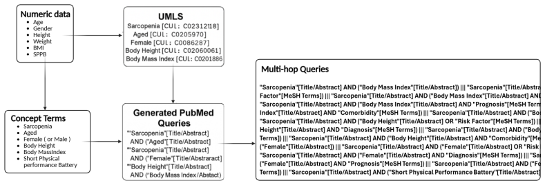
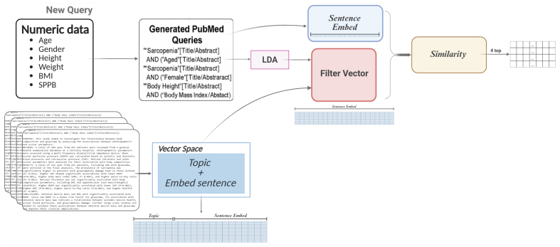
The load-bearing object is the three-level 'tree' of visual representations coupled to a question-guided gate and a retrieval-augmented fusion block. Level 1 is a global feature vector; Level 2 uses a pretrained segmentation model to isolate anatomically relevant muscle regions and extracts regional features; Level 3 builds a spatial graph whose nodes are centroids of superpixel-derived regions and whose edges encode proximity and anatomical adjacency. The gate is a softmax over the projected question text that selects exactly one level, and a low-rank adaptation module transforms the selected vector before it is combined with text and numeric features for classification. The retrieval side
What would settle it
Re-run the full pipeline with patient-disjoint cross-validation—train on a subset of the 24 patients and test on the remaining patients, or leave one patient out at a time—and compare accuracy to the reported 0.99. If accuracy drops substantially, the near-perfect figure is explained by image-level leakage rather than by the hierarchical architecture or retrieval pipeline.
Extended reading notes
Core claim
The paper's central claim is that sarcopenia can be classified from rectus femoris ultrasound with near-perfect accuracy when image interpretation is structured hierarchically and augmented by retrieved clinical knowledge. The vision module extracts three complementary representations: coarse global features, region features from a segmentation model, and node embeddings from a superpixel-based spatial graph that encodes anatomical adjacency. A soft gating function, computed from the projected question text, selects the most relevant level; the chosen features pass through a low-rank adapter and are added to the projected text and numeric representations before classification. In parallel, t
Load-bearing premise
The reported accuracy treats each ultrasound image as an independent sample even though the 3,474 images come from 24 patients; if the cross-validation folds are not grouped by patient, images from the same person can appear in both training and test data, and the model can memorize patient appearance instead of learning generalizable signs of sarcopenia.
Editorial extensions
If this is right
- The reported 99% on the custom ultrasound dataset points toward bedside screening: a portable acquisition device plus this lightweight model could reduce reliance on costly CT or MRI for sarcopenia assessment.
- The ablation progression (coarse ~0.70, fine ~0.68, fused hierarchy ~0.90, fused with clinical data ~0.93, with retrieval ~0.99) implies that no single visual level is sufficient and that external knowledge retrieval is a central ingredient, not a marginal add-on.
- Because the gate processes only the selected visual level, the framework could adapt its computation to the question, an efficiency property that matters for community clinics with limited hardware.
- On VQA-RAD the model reaches 0.88, beating the listed baselines by a small margin; the same hierarchy may transfer to other low-data medical VQA tasks, though the PathVQA result of 0.84 shows the transfer is not uniform.
Reading between the lines
- Editorial extension: the sharp jump from 0.93 to 0.99 when retrieval is added should be tested for leakage—since queries are generated from each patient's own clinical variables, matching could occur between a test patient's profile and similar text seen in training, independent of the image.
- Editorial extension: the paper never states whether its k-fold splits group images by patient; with 24 patients and 3,474 images, patient-disjoint cross-validation is the single most informative next experiment for separating memorization from generalization.
- Editorial extension: the coarse-to-fine visual tree plus ontology-guided retrieval could transfer to other under-labeled musculoskeletal ultrasound tasks, such as frailty or muscle-quality assessment, where cues are subtle and clinical context is essential.
- Editorial extension: a reader should read the 99% as domain-specific; the same architecture reports 0.84 on PathVQA, below several listed baselines, so the contribution is best understood as a low-data sarcopenia-ultrasound method rather than a universal medical-VQA winner.
Editorial analysis
A structured set of objections, weighed in public.
Referee Report
Summary. The paper proposes MedVQA-TREE, a multimodal framework for sarcopenia diagnosis from ultrasound images combined with structured clinical data and retrieved medical text. The architecture combines a three-level hierarchical visual encoder (global features, SAM-based regional features, superpixel spatial-graph features), a question-guided gated fusion mechanism with LoRA, and a UMLS-guided multi-query/multi-hop retrieval pipeline. The authors report experiments on VQA-RAD, PathVQA, and a proprietary sarcopenia ultrasound dataset, claiming up to 99% diagnostic accuracy and improvements over state-of-the-art medical VQA methods by more than 10%. Ablation tables show a progression from individual visual levels (accuracies roughly 0.52–0.70) to combined hierarchical features (0.90) and finally to RAG-enhanced fusion (0.99).
Significance. If the 99% accuracy result were valid, the paper would be a meaningful contribution to low-resource sarcopenia screening and to medical VQA in small-data regimes. The hierarchical visual reasoning, SAM-based region selection, and lightweight gated fusion are reasonable and potentially useful ideas. However, the central quantitative claim is not supported by the evaluation as presented: the k-fold cross-validation is performed on 3,474 images from only 24 patients with no described patient-level split, the dataset description is internally inconsistent, and the RAG module retrieves from the training set at test time. These issues mean the main empirical result cannot currently be distinguished from leakage or circularity, and the comparison claims are contradicted by the paper's own Table 2.
major comments (4)
- [§2.1, §3.2.1, Tables 3–11] The cross-validation is applied to 3,474 images from only 24 patients, and the paper never states that folds are grouped by patient. With an average of roughly 145 images per patient, an image-level split places images from the same patient in both training and test folds, allowing the model to memorize patient-specific appearance and patient-level clinical variables (age, BMI, SPPB). Table 11's 0.99 accuracy is therefore indistinguishable from patient-identity leakage. The authors must report patient-exclusive folds, per-patient predictions, and fold-wise patient counts; all k-fold results in Tables 3–11 need to be re-run under that protocol or explicitly justified.
- [§2.1] The dataset description is numerically inconsistent. Transverse (1,350) and longitudinal (962) image counts sum to 2,312, not 3,474. The sex-stratified counts sum to 3,472 (male: 763+1,240=2,003; female: 297+1,172=1,469). The plane-level sarcopenic/non-sarcopenic counts total 776/1,536=2,312, whereas the stated 30.53%/69.47% proportions applied to 3,474 images correspond to approximately 1,060/2,414 labels. Please correct these counts; the class-imbalance description and all reported metrics depend on them.
- [§3.2.1, Fusion Strategies with RAG] The test-time RAG procedure is described as generating a query from the patient record and matching it with chunks 'aligned with the training set.' This is not external knowledge; it is retrieval from the training data. When combined with image-level splitting, the retrieved chunks can include the exact same patients' clinical variables. Even under patient-level splitting, matching test queries to training-derived topic categories creates a circularity in which predictions are informed by the training distribution. The near-perfect 0.99 result is a fitted outcome under this protocol. The authors should replace this with a genuinely external, label-independent knowledge base, or demonstrate that no information about training labels flows through the retrieval step.
- [Abstract, Table 2] The claim of outperforming previous state-of-the-art methods by over 10% is not supported by the reported numbers. On PathVQA, MedVQA-TREE achieves 0.84, below all listed baselines (0.90–0.91). On VQA-RAD it achieves 0.88 versus 0.87 for PeFoMed, a difference of about one point. The only >10% margin is on the sarcopenia dataset, whose validity is undermined by the issues above. Please restrict the claims to actual comparisons and provide confidence intervals or proper statistical tests for each dataset.
minor comments (6)
- [§3.2.1] The row labels '1-fold' through '5-fold' are ambiguous. Clarify whether these are the five folds of a 5-fold cross-validation and report the number of patients in each fold.
- [Tables 12–15] These tables show high accuracy with very low F1/precision/recall (e.g., Table 12: accuracy 0.70–0.75, F1 0.12–0.19). Explain how this pattern arises and why accuracy alone is a meaningful headline metric in this setting.
- [§2.4.6] The text mentions T5 as the underlying language model, but the formal model in Eqs. (1)–(8) does not include a T5 component. Clarify where T5 enters the architecture.
- [Table 1, References] The citation for PubMedBERT appears to point to a NEJM AI paper on BiomedCLIP rather than the original PubMedBERT publication. Please check the reference list and correct it.
- [Figures 2–3] Figure 2's caption says 'image selected of VQA-RAD dataset', while the surrounding text describes the sarcopenia ultrasound pipeline. Clarify which dataset is actually used for the segmentation illustrations.
- [§2.1, §3.1] The paper refers to the sarcopenia task as 'VQA', but the sarcopenia experiments are binary image classification with clinical features and no natural-language question. Clarify how the gate uses the 'textual query' and whether any free-form question is posed to the model.
Circularity Check
No significant circularity: the framework's modules and evaluation are independently specified; flagged leakage and benchmark issues are correctness risks, not circular reductions.
full rationale
I examined the paper's derivation chain. The visual feature extraction (Sec. 2.3.1), numeric embedding (Sec. 2.3.2), text retrieval (Sec. 2.3.3), LoRA fusion (Sec. 2.4), and loss (Eq. 8) are defined by explicit equations that do not reference the target labels or the reported evaluation results. The VQA-RAD and PathVQA benchmarks are external, and the model's reported accuracy there (0.88 and 0.84) is an independent empirical outcome, not a quantity forced by the method's definitions. The sarcopenia dataset is proprietary and small, and the RAG module builds its retrieval index from training data and matches test queries to 'training-aligned categories' (Sec. 3.2.1); while this is a legitimate methodological concern about generalization and possible overfitting, it is not a case where a predicted quantity is definitionally equal to an input or where a fitted parameter is renamed as a prediction. The absence of an explicit patient-grouped split in the k-fold evaluation (24 patients, 3,474 images) is a serious validity risk, but it concerns data leakage rather than circularity. No load-bearing self-citation or imported uniqueness theorem appears. I therefore find no significant circularity in the manuscript's derivations.
Assumptions & free parameters
free parameters (5)
- LDA topic count T =
not reported
- Number of retrieved sentences C =
C = 10
- Number of SAM regions S =
not reported
- LoRA rank r =
not reported
- LDA similarity threshold =
not reported
assumptions (5)
- domain assumption Images from the same patient can be treated as independent samples in cross-validation.
- domain assumption SAM zero-shot segmentation produces clinically meaningful muscle regions for sarcopenia.
- domain assumption UMLS-expanded PubMed queries retrieve patient-specific evidence that improves diagnosis.
- standard math Plain cross-entropy loss is appropriate for the imbalanced sarcopenia task.
- domain assumption The proprietary dataset labels according to EWGSOP2 criteria are correct.
Cite this review
Pith. "Pith review of MedVQA-TREE: A Multimodal Reasoning and Retrieval Framework for Sarcopenia Prediction." pith.science (2026). https://pith.science/paper/GEZ34EYC
@misc{pith2026250819319,
  author       = {Pith},
  title        = {Pith review of: MedVQA-TREE: A Multimodal Reasoning and Retrieval Framework for Sarcopenia Prediction},
  year         = {2026},
  howpublished = {\url{https://pith.science/paper/GEZ34EYC}},
  note         = {Machine review of arXiv:2508.19319}
}
read the original abstract
Accurate sarcopenia diagnosis via ultrasound remains challenging due to subtle imaging cues, limited labeled data, and the absence of clinical context in most models. We propose MedVQA-TREE, a multimodal framework that integrates a hierarchical image interpretation module, a gated feature-level fusion mechanism, and a novel multi-hop, multi-query retrieval strategy. The vision module includes anatomical classification, region segmentation, and graph-based spatial reasoning to capture coarse, mid-level, and fine-grained structures. A gated fusion mechanism selectively integrates visual features with textual queries, while clinical knowledge is retrieved through a UMLS-guided pipeline accessing PubMed and a sarcopenia-specific external knowledge base. MedVQA-TREE was trained and evaluated on two public MedVQA datasets (VQA-RAD and PathVQA) and a custom sarcopenia ultrasound dataset. The model achieved up to 99% diagnostic accuracy and outperformed previous state-of-the-art methods by over 10%. These results underscore the benefit of combining structured visual understanding with guided knowledge retrieval for effective AI-assisted diagnosis in sarcopenia.
Figures


Figures from the paper (4 more)


Reference graph
Works this paper leans on
-
[1]
J. Dupont, et al., Revised sarcopenia consensus: are we missing the preclinical stage? online comment on, Age And Ageing 48 (1) (2018) 1–1
work page 2018
-
[2]
M.-R. Cho, S. Lee, S.-K. Song, A review of sarcopenia pathophysiology, diagnosis, treatment and future direction, Journal of Korean medical science 37 (18) (2022)
work page 2022
-
[3]
V. Soria-Utrilla, F. J. S´ anchez-Torralvo, F. X. Palmas-Candia, R. Fern´ andez-Jim´ enez, F. Mucarzel-Suarez-Arana, P. Guirado-Pel´ aez, G. Olveira, J. M. Garc ´ ıa-Almeida, R. Burgos-Pel´ aez, Ai-assisted body composition assessment using ct imaging in colorectal cancer patients: Predictive capacity for sarcopenia and malnutrition diagnosis, Nutrients 1...
work page 2024
- [4]
-
[5]
X. Zuo, X. Li, K. Tang, R. Zhao, M. Wu, Y. Wang, T. Li, Sarcopenia and cardiovascular diseases: a systematic review and meta-analysis, Journal of Cachexia, Sarcopenia and Muscle 14 (3) (2023) 1183–1198
work page 2023
-
[6]
Y. Kim, J. Beom, S. Y. Lee, H. C. Jang, K. Kim, M. Kim, G. Y. Shim, C. W. Won, J.-Y. Lim, Comparison of bioelectrical impedance analysis and dual-energy x-ray absorptiometry for the diagnosis of sarcopenia in the older adults with metabolic syndrome: equipment-specific equation development, Aging clinical and experimental research 37 (1) (2025) 1–9. 40
work page 2025
-
[7]
X. Wu, Z. Chen, Y. Zhao, H. Ren, Correlation and predictive value of novel anthropometric indicators with adult sarcopenia and sarcopenia obesity, Scientific Reports 14 (1) (2024) 31776
work page 2024
-
[8]
M. R. Tomaszewski, R. J. Gillies, The biological meaning of radiomic features, Radiology 298 (3) (2021) 505–516
work page 2021
Show all 59 references
-
[9]
Kocak, E
B. Kocak, E. S. Durmaz, C. Erdim, E. Ates, O. K. Kaya, O. Kilickesmez, Radiomics of renal masses: systematic review of reproducibility and validation strategies, American Journal of Roentgenology 214 (1) (2020) 129–136
2020
-
[10]
S. Kim, S. Park, S. Lee, S. H. Seo, H. S. Kim, Y. Cha, J.-T. Kim, J.- W. Kim, Y.-C. Ha, J.-I. Yoo, Assessing physical abilities of sarcopenia patients using gait analysis and smart insole for development of digital biomarker, Scientific Reports 13 (1) (2023) 10602
2023
-
[11]
S. Kim, H. S. Kim, J.-I. Yoo, Sarcopenia classification model for mus- culoskeletal patients using smart insole and artificial intelligence gait analysis, Journal of Cachexia, Sarcopenia and Muscle 14 (6) (2023) 2793–2803
2023
-
[12]
Chen, J.-W
Y.-C. Chen, J.-W. Hsieh, Y.-H. Yang, C.-H. Lee, P.-Y. Yu, P.-Y. Chen, A. S. Santa, Towards deep learning-based sarcopenia screening with body joint composition analysis, in: 2021 IEEE International Confer- ence on Image Processing (ICIP), IEEE, 2021, pp. 3807–3811
2021
-
[13]
Islam, F
S. Islam, F. Kanavati, Z. Arain, O. F. Da Costa, W. Crum, E. Aboagye, A. Rockall, Fully automated deep-learning section-based muscle seg- mentation from ct images for sarcopenia assessment, Clinical Radiology 77 (5) (2022) e363–e371
2022
-
[14]
Sakai, S
K. Sakai, S. Gilmour, E. Hoshino, E. Nakayama, R. Momosaki, N. Sakata, D. Yoneoka, A machine learning-based screening test for sarcopenic dysphagia using image recognition, Nutrients 13 (11) (2021) 4009
2021
-
[15]
Caputo, I
V. Caputo, I. Letteri, S. J. Santini, G. Sinatti, C. Balsano, Towards precision in sarcopenia assessment: the challenges of multimodal data analysis in the era of ai, International Journal of Molecular Sciences 26 (9) (2025) 4428. 41
2025
-
[16]
Aznar-Gimeno, J
R. Aznar-Gimeno, J. L. Perez-Lasierra, P. P´ erez-L´ azaro, I. Bosque- L´ opez, M. Azp ´ ıroz-Puente, P. Salvo-Ib´ a˜ nez, M. Morita-Hernandez, A. C. Hern´ andez-Ruiz, A. G´ omez-Bernal, M. de la Vega Rodrigalvarez- Chamarro, et al., Gait-based ai models for detecting sarcopen...
2024
-
[17]
A. K. Smorchkova, A. V. Petraikin, D. S. Semenov, D. E. Sharova, Sarcopenia: modern approaches to solving diagnosis problems, Digital Diagnostics 3 (3) (2022) 196–211
2022
-
[18]
Perkisas, S
S. Perkisas, S. Baudry, J. Bauer, D. Beckw´ ee, A.-M. De Cock, H. Hobbe- len, H. Jager-Wittenaar, A. Kasiukiewicz, F. Landi, E. Marco, et al., The sarcus project: evidence-based muscle assessment through ultra- sound, European Geriatric Medicine 10 (1) (2019) 157–158
2019
-
[19]
Behboodi, J
B. Behboodi, J. Obrand, J. Afilalo, H. Rivaz, Deepsarc-us: A deep learning framework for assessing sarcopenia using ultrasound images, Applied Sciences 14 (15) (2024) 6726
2024
-
[20]
Z. Ye, A. Saraf, Y. Ravipati, F. Hoebers, P. J. Catalano, Y. Zha, A. Za- paishchykova, J. Likitlersuang, C. Guthier, R. B. Tishler, et al., De- velopment and validation of an automated image-based deep learning platform for sarcopenia assessment in head and neck cancer, JAMA n...
2023
-
[21]
D. Lu, Z. Lin, R. Wang, Z. Chen, J. Zhuo, L. Xu, L. Pan, H. Li, X. Yang, C. He, et al., Multi-omics profiling reveals chitinase-3-like protein 1 as a key mediator in the crosstalk between sarcopenia and liver cancer, Redox biology 58 (2022) 102538
2022
-
[22]
S. Lee, R. M. Summers, Clinical artificial intelligence applications in radiology: chest and abdomen, Radiologic Clinics 59 (6) (2021) 987– 1002
2021
-
[23]
W. Fan, M. N. Suvon, S. Zhou, X. Liu, S. Alabed, V. Osmani, A. J. Swift, C. Chen, H. Lu, Medslip: Medical dual-stream language- image pre-training with pathology-anatomy semantic alignment, arXiv preprint arXiv:2403.10635 (2024)
2024 arXiv
-
[24]
Maani, N
F. Maani, N. Saeed, T. Saleem, Z. Farooq, H. Alasmawi, W. Diehl, A. Mohammad, G. Waring, S. Valappi, L. Bricker, et al., Fetalclip: A 42 visual-language foundation model for fetal ultrasound image analysis, arXiv preprint arXiv:2502.14807 (2025)
2025
-
[25]
P. Ge, T. Su, F. Lv, B. Zhao, P. Zhang, C. H. Wong, L. Yao, Y. Sun, Z. Wang, P. K. Wong, et al., Ultrasound report generation with mul- timodal large language models for standardized texts, arXiv preprint arXiv:2505.08838 (2025)
2025 arXiv
-
[26]
P. H. Yi, H. W. Garner, A. Hirschmann, J. A. Jacobson, P. Omoumi, K. Oh, J. R. Zech, Y. H. Lee, Clinical applications, challenges, and recommendations for artificial intelligence in musculoskeletal and soft- tissue ultrasound: Ajr expert panel narrative review, American Journa...
2024
-
[27]
Y.-W. Chu, K. Zhang, C. Malon, M. R. Min, Reducing hallucina- tions of medical multimodal large language models with visual retrieval- augmented generation, arXiv preprint arXiv:2502.15040 (2025)
2025 arXiv
-
[28]
D. M. Nguyen, N. T. Diep, T. Q. Nguyen, H.-B. Le, T. Nguyen, T. Nguyen, T. Nguyen, N. Ho, P. Xie, R. Wattenhofer, et al., En- riched instruction-following graph alignment for efficient medical vision- language models
-
[29]
S. Toro, A. V. Anagnostopoulos, S. M. Bello, K. Blumberg, R. Cameron, L. Carmody, A. D. Diehl, D. M. Dooley, W. D. Duncan, P. Fey, et al., Dynamic retrieval augmented generation of ontologies using artificial intelligence (dragon-ai), Journal of Biomedical Semantics 15 (1) (2024) 19
2024
-
[30]
D. M. H. Nguyen, N. T. Diep, T. Q. Nguyen, B. H. Le, T. Nguyen, A.-T. Nguyen, T. Nguyen, N. Ho, P. Xie, R. Wattenhofer, et al., Logra- med: Long-context multi-graph alignment for medical visual-language models
-
[31]
J. J. Lau, S. Gayen, A. Ben Abacha, D. Demner-Fushman, A dataset of clinically generated visual questions and answers about radiology im- ages, Scientific data 5 (1) (2018) 1–10
2018
-
[32]
X. He, Y. Zhang, L. Mou, E. Xing, P. Xie, Pathvqa: 30000+ questions for medical visual question answering, arXiv preprint arXiv:2003.10286 (2020). 43
2003 arXiv
-
[33]
National Center for Biotechnology Informa- tion (NCBI), Entrez programming utilities help, https://www.ncbi.nlm.nih.gov/books/NBK25501/, bethesda (MD): National Library of Medicine (US) (2009)
2009
-
[34]
Q. Yang, H. Zuo, R. Su, H. Su, T. Zeng, H. Zhou, R. Wang, J. Chen, Y. Lin, Z. Chen, et al., Dual retrieving and ranking medical large lan- guage model with retrieval augmented generation, Scientific Reports 15 (1) (2025) 18062
2025
-
[35]
Xiong, Q
G. Xiong, Q. Jin, X. Wang, M. Zhang, Z. Lu, A. Zhang, Improv- ing retrieval-augmented generation in medicine with iterative follow-up questions, in: Biocomputing 2025: Proceedings of the Pacific Sympo- sium, World Scientific, 2024, pp. 199–214
2025
-
[36]
L. M. Amugongo, P. Mascheroni, S. Brooks, S. Doering, J. Seidel, Re- trieval augmented generation for large language models in healthcare: A systematic review, PLOS Digital Health 4 (6) (2025) e0000877
2025
-
[37]
Zhang, Y
S. Zhang, Y. Xu, N. Usuyama, H. Xu, J. Bagga, R. Tinn, S. Preston, R. Rao, M. Wei, N. Valluri, et al., Biomedclip: a multimodal biomedical foundation model pretrained from fifteen million scientific image-text pairs, arXiv preprint arXiv:2303.00915 (2023)
2023 arXiv
-
[38]
Kirillov, E
A. Kirillov, E. Mintun, N. Ravi, H. Mao, C. Rolland, L. Gustafson, T. Xiao, S. Whitehead, A. C. Berg, W.-Y. Lo, et al., Segment anything, in: Proceedings of the IEEE/CVF international conference on computer vision, 2023, pp. 4015–4026
2023
-
[39]
Alsentzer, J
E. Alsentzer, J. R. Murphy, W. Boag, W.-H. Weng, D. Jin, T. Nau- mann, M. McDermott, Publicly available clinical bert embeddings, arXiv preprint arXiv:1904.03323 (2019)
1904 arXiv
-
[40]
Zhang, Y
S. Zhang, Y. Xu, N. Usuyama, H. Xu, J. Bagga, R. Tinn, S. Preston, R. Rao, M. Wei, N. Valluri, et al., A multimodal biomedical foundation model trained from fifteen million image–text pairs, NEJM AI 2 (1) (2025) AIoa2400640
2025
-
[41]
P. Deka, A. Jurek-Loughrey, D. P, Evidence extraction to validate med- ical claims in fake news detection, in: International conference on health information science, Springer, 2022, pp. 3–15. 44
2022
-
[42]
Yasunaga, J
M. Yasunaga, J. Leskovec, P. Liang, Linkbert: Pretraining language models with document links, arXiv preprint arXiv:2203.15827 (2022)
2022 arXiv
-
[43]
K. R. Kanakarajan, B. Kundumani, M. Sankarasubbu, Bioelectra: pre- trained biomedical text encoder using discriminators, in: Proceedings of the 20th workshop on biomedical language processing, 2021, pp. 143– 154
2021
-
[44]
Q. Jin, W. Kim, Q. Chen, D. C. Comeau, L. Yeganova, W. J. Wilbur, Z. Lu, Medcpt: Contrastive pre-trained transformers with large-scale pubmed search logs for zero-shot biomedical information retrieval, Bioin- formatics 39 (11) (2023) btad651
2023
-
[45]
Bannur, S
S. Bannur, S. Hyland, Q. Liu, F. Perez-Garcia, M. Ilse, D. C. Castro, B. Boecking, H. Sharma, K. Bouzid, A. Thieme, et al., Learning to exploit temporal structure for biomedical vision-language processing, in: Proceedings of the IEEE/CVF Conference on Computer Vision and Patte...
2023
-
[46]
Zhang, Z
Y. Zhang, Z. Shen, R. Jiao, Segment anything model for medical image segmentation: Current applications and future directions, Computers in Biology and Medicine 171 (2024) 108238
2024
-
[47]
Rohanian, M
O. Rohanian, M. Nouriborji, H. Jauncey, S. Kouchaki, F. Nooralahzadeh, L. Clifton, L. Merson, D. A. Clifton, I. C. C. Group, et al., Lightweight transformers for clinical natural language processing, Natural language engineering 30 (5) (2024) 887–914
2024
-
[48]
Q. Lu, D. Dou, T. Nguyen, Clinicalt5: A generative language model for clinical text, in: Findings of the Association for Computational Linguis- tics: EMNLP 2022, 2022, pp. 5436–5443
2022
-
[49]
G. Ao, M. Chen, J. Li, H. Nie, L. Zhang, Z. Chen, Comparative analysis of large language models on rare disease identification, Orphanet Journal of Rare Diseases 20 (1) (2025) 150
2025
-
[50]
Y. Li, Q. Yang, F. L. Wang, L.-K. Lee, Y. Qu, T. Hao, Asymmetric cross-modal attention network with multimodal augmented mixup for medical visual question answering, Artificial Intelligence in Medicine 144 (2023) 102667. 45
2023
-
[51]
Z. Deng, W. Gao, C. Chen, Z. Niu, Z. Gong, R. Zhang, Z. Cao, F. Li, Z. Ma, W. Wei, et al., Ophglm: An ophthalmology large language-and- vision assistant, Artificial Intelligence in Medicine 157 (2024) 103001
2024
-
[52]
Huang, X
J. Huang, X. Qi, X. Cheng, M. Wang, H. Ju, W. Ding, D. Zhang, Mmf- nns: Multi-modal multi-granularity fusion neural networks for brain net- works and its application to epilepsy identification, Artificial Intelligence in Medicine 157 (2024) 102990
2024
-
[53]
Liu, L.-M
B. Liu, L.-M. Zhan, X.-M. Wu, Contrastive pre-training and representa- tion distillation for medical visual question answering based on radiology images, in: International conference on medical image computing and computer-assisted intervention, Springer, 2021, pp. 210–220
2021
-
[54]
Y. Zhou, J. Mei, Y. Yu, T. Syeda-Mahmood, Medical visual ques- tion answering using joint self-supervised learning, arXiv preprint arXiv:2302.13069 (2023)
2023 arXiv
-
[55]
M. Moor, Q. Huang, S. Wu, M. Yasunaga, Y. Dalmia, J. Leskovec, C. Zakka, E. P. Reis, P. Rajpurkar, Med-flamingo: a multimodal medi- cal few-shot learner, in: Machine Learning for Health (ML4H), PMLR, 2023, pp. 353–367
2023
-
[56]
Z. Wang, Z. Wu, D. Agarwal, J. Sun, Medclip: Contrastive learning from unpaired medical images and text, in: Proceedings of the Conference on Empirical Methods in Natural Language Processing. Conference on Empirical Methods in Natural Language Processing, Vol. 2022, 2022, p. 3876
2022
-
[57]
O. C. Thawakar, A. M. Shaker, S. S. Mullappilly, H. Cholakkal, R. M. Anwer, S. Khan, J. Laaksonen, F. Khan, Xraygpt: Chest radiographs summarization using large medical vision-language models, in: Proceed- ings of the 23rd workshop on biomedical natural language processing, 20...
2024
-
[58]
J. Zhou, X. He, L. Sun, J. Xu, X. Chen, Y. Chu, L. Zhou, X. Liao, B. Zhang, S. Afvari, et al., Pre-trained multimodal large language model enhances dermatological diagnosis using skingpt-4, Nature Communica- tions 15 (1) (2024) 5649. 46
2024
-
[59]
G. Liu, J. He, P. Li, Z. Zhao, S. Zhong, Cross-modal self-supervised vision language pre-training with multiple objectives for medical vi- sual question answering, Journal of Biomedical Informatics 160 (2024) 104748. 47
2024
Reviewed August 5, 2026 · model on record in the stance chip above.
Discussion (0). Sign in to comment.