REVIEW 4 major objections 6 minor 36 references
DNA Dynamics in Dual Nanopore Tug-of-War
T0 review · 4 major / 6 minor · reviewed 2026-08-05 · deepseek-v4-flash
Pith's one-line read A DNA strand escaping a dual-pore tug-of-war crosses the inter-pore gap with a mobility near 30 µm/ms per mV—about 1,000 times its tug-of-war sliding mobility—and a second folded strand slows it.
desk verdict Asymmetric TOW partitioning is real and worth a look, but the free-end mobility claim is off by orders of magnitude and needs to be fixed or removed. read the letter →
The pith
A machine-rendered reading of the paper's core claim, the machinery that carries it, and where it could break.
The reading
What carries the argument
The load-bearing object is the first-passage model of tug-of-war as 1D biased diffusion. The DNA strand stretched between the pores is assumed to be held at roughly 90% extension, which fixes the contour length between the pores; the only dynamical variable is x, the amount of DNA contour in the reservoir adjoining pore 1. The probability density P(x,t) obeys a Smoluchowski equation ∂P/∂t = D ∂²P/∂x² − v ∂P/∂x with absorbing boundaries at x=0 and x=L, so the dwell time is the first-passage time to empty either reservoir. Fitting the cumulative dwell-time distribution fixes the diffusion coefficient D, drift velocity v, and initial contour fraction α; P1-exit probability is then obtained from
What would settle it
Measure the inter-pore strand's extension directly while the tug-of-war is held, for example by attaching fluorescent labels at two points on the linking segment and imaging them, or vary the pore spacing: if the strand is not near full extension, or if the free-end mobility does not fall when the pore spacing (and hence inter-pore contour) increases, the fixed-stretch, one-variable model is not the right description.
Extended reading notes
Core claim
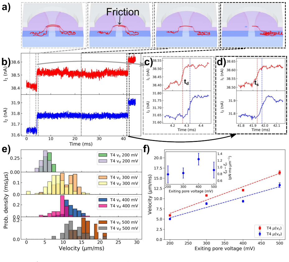
Using a dual-nanopore chip with pores about 600 nm apart and feedback-based control to form and hold tug-of-war states, the authors measured dwell-time histograms and free-end time-of-flight for λ-DNA and T4 DNA. They report three connected findings. First, longer T4 DNA enters the tug-of-war with an asymmetric starting partition (fitted α = 0.92), so dwell times depend strongly on the sign of the voltage difference and show separate diffusion-dominated and drift-dominated escape peaks; the same 1D convection-diffusion first-passage model used previously for λ-DNA fits these distributions and yields a tug-of-war sliding mobility around 15 µm/s·mV for both molecules. Second, once the free end
Load-bearing premise
The central assumption is that the DNA segment between the two pores stays stretched to about 90% of its contour length during the tug-of-war, so the contour between pores is effectively fixed and the chain's motion reduces to one-dimensional biased diffusion of one contour variable; if the inter-pore segment goes slack or changes extension, the fitted asymmetry, diffusion, and mobility parameters lose their simple meaning.
Editorial extensions
If this is right
- For genomic-length DNA, tug-of-war dwell-time data are not symmetric in voltage because the chain starts with an unbalanced partition; analysis pipelines and control algorithms must fit α as well as D and v.
- Tug-of-war sliding mobility is linear in the voltage difference and nearly identical for λ- and T4-DNA, so the dual-pore setup can in principle measure a molecule's charge-to-friction ratio and distinguish biopolymers by mobility rather than only by current blockade.
- The free-end time-of-flight gives a direct readout of the velocity and mobility of the last remaining cis-side contour, providing a clean experimental limit for single-pore translocation theories.
- Folded-chain free-end transit is reproducibly slower than unfolded transit, with a voltage-independent friction difference, giving a quantitative handle for detecting and characterizing folds during dual-pore translocation.
- Free-end velocity fluctuations peak near 300 mV, indicating that a constant-diffusivity model is not sufficient for the escape stage and motivating more detailed simulations coupling pore threading with strand recoil.
Reading between the lines
- If free-end friction is indeed dominated by the short inter-pore segment, then translocation models with cis-side friction proportional to total remaining contour will systematically mispredict the final ~1% of a translocation; the terminal regime may need to be modeled as a separate short-chain problem with a recoiling strand.
- The fitted α ≈ 0.92 for T4 suggests that tug-of-war starting asymmetry is set by the capture protocol, so varying the pre-capture tail length could deliberately tune dwell times and make mapping of longer molecules more reproducible.
- The voltage-independent difference between folded and unfolded free-end friction suggests the extra drag comes from geometry or confinement of two adjacent strands rather than from voltage-dependent pore forces; changing pore spacing or ionic strength would test whether the extra friction scales with confinement.
- The λ-versus-T4 velocity offset was attributed to trans-side crowding; adding crowders or changing salt in the common chamber could turn that hypothesis into a quantitative trans-side packing probe.
Signed reviews
Editorial analysis
A structured set of objections, weighed in public.
Referee Report
Summary. The manuscript reports dual-nanopore tug-of-war (TOW) experiments on T4 and lambda DNA. The authors measure TOW dwell-time distributions as a function of voltage difference, fit them to a 1D convection-diffusion first-passage model with free parameters D, v, and alpha, and extract a TOW sliding mobility A ≈ 14–15 µm/s·mV. They also measure the interval Tf between the P1 and P2 current recoveries at TOW disengagement, equate it to the time of flight of the DNA free end across the inter-pore gap (≈0.6 µm), and report a free-end mobility k ≈ 30 µm/ms·mV, which they claim is ~10^3 times larger than the TOW mobility. Experiments with a folded conformation show a slowed free end. The paper argues that a highly extended, short inter-pore segment with reduced cis-side friction explains the enhanced mobility.
Significance. The experimental platform is sophisticated, and the raw TOW dwell-time asymmetry for long T4 DNA is a reproducible and physically interesting observation. If the free-end mobility claim were correct, it would provide new insight into cis-side friction near the end of translocation. The first-passage model is a reasonable framework, but its parameters are fitted to the same data used for validation, limiting the strength of the agreement. More importantly, the central quantitative claim about the ~10^3-fold enhanced free-end mobility is internally inconsistent with the reported Tf and pore spacing, and the interpretation of Tf as a pure time of flight is questionable. As presented, the main result is not supported by the data.
major comments (4)
- [DNA free end time-of-flight between pores] The reported free-end mobility k≈30 µm/ms·mV is inconsistent with the stated Tf≈10 µs and pore spacing d≈0.6 µm. Dividing d by Tf gives v≈60 µm/ms, i.e. k≈0.12–0.30 µm/ms·mV for V2=200–500 mV. Conversely, k=30 µm/ms·mV at V2=400 mV implies v≈12,000 µm/ms and Tf≈0.05 µs, three orders of magnitude below the 4 µs timing resolution and the observed 10 µs. The velocities in Fig. 5c cannot be derived from the reported Tf; one of these quantities, or the unit used for k, must be mis-stated.
- [DNA free end time-of-flight between pores] Tf is defined in Fig. 2e as the interval between the P1 and P2 current recoveries. After the chain leaves P1, P2 remains blocked until the entire remaining contour—including the inter-pore segment and any trans-side/channel-2 contour—has exited P2. Thus Tf includes the full terminal translocation through P2, not simply the free end's transit across the 0.6 µm gap. The estimate v=d/Tf conflates these processes; a correction for P2 threading time and for recoil/slack of the inter-pore strand is needed before claiming a free-end mobility.
- [Quantifying DNA Dwell Time in Tug-of-War] The model is not validated against independent data. The paper states 'we leave the diffusion coefficient D, the drift velocity v and the initial fractional contour in channel 1 (α) as fitting parameters' and optimizes them on the same dwell-time distributions that are then shown as 'fits' in Fig. 3. The P1 exit probabilities in Fig. 4 are also computed from this fitted model. The agreement is therefore a consistency check of the fitting form, not a prediction. The abstract's claim that the findings 'validate theoretical predictions' is too strong; an out-of-sample test (e.g., using parameters from one voltage to predict another) would be needed.
- [DNA free end time-of-flight between pores] The comparison to single-pore mobilities contains a unit inconsistency. The text states that the single-pore λ-DNA mobility is approximately 5×10^4 µm/ms·mV and is 'only greater than our TOW measurement by a factor of around 2.' With the TOW sliding mobility A≈14 µm/s·mV = 0.014 µm/ms·mV, this ratio is ~3.6×10^6; with the free-end k≈30 µm/ms·mV, the ratio is ~1.6×10^3. Neither is 'around 2'. The mobility values or their units must be corrected before the comparison can be evaluated.
minor comments (6)
- [DNA Tug-of-War] Typo: 'T4-DNA-DNA' appears in the paragraph describing the TOW event.
- [DNA free end time-of-flight between pores] Units: velocities are given in µm/ms in Fig. 5 but in µm/s elsewhere (e.g., A values). Add explicit unit conversions in the text and figure captions, and ensure the reported k values are expressed with consistent units.
- [Quantifying DNA Dwell Time in Tug-of-War] Fig. 3d: the model error bars are stated to be smaller than the marker size; this should be stated in the caption or the markers enlarged for visibility.
- [Supplementary Figure S1] The 'theoretical curve' in Fig. S1 uses the fitted D and A values; this should be stated explicitly in the caption so readers do not mistake it for an independent prediction.
- [References] Reference 26 is cited as 'D. Ling et al.' in the text, but the work has two authors (Ling and Ling); correct the citation style.
- [Abstract and Discussion] The abstract and discussion repeatedly claim a '3-orders-of-magnitude' enhancement of the free-end mobility. Given the inconsistencies in the supporting numbers, this claim should be re-evaluated and softened until the velocity analysis is corrected.
Circularity Check
TOW dwell-time 'validation' reduces to fitting the same distributions; free-end velocity is measured independently. Self-citation supplies the model's stretching ansatz. Free-end mobility claim also suffers an internal units/time-of-flight inconsistency.
-
fitted input called prediction
[Results: Quantifying DNA Dwell Time in Tug-of-War (Eqs. 1-3 and fitting paragraph; Fig. 3b,c)]
"In the first-passage model, we leave the diffusion coefficient D, the drift velocity v and the initial fractional contour in channel 1 ( α) as fitting parameters. Fitting is performed in python by maximizing an objective function given by the cosine similarity between the modeled and the experimental dwell time cumulative distribution. ... Our fitted model recapitulates the asymmetry of the dwell time distribution shown in Fig. 3a)."
D, v, and α are optimized to maximize cosine similarity with the experimental dwell-time cumulative distributions. The model output is then displayed on the same histograms/CDFs as the 'fit' and the abstract says the findings 'validate theoretical predictions derived from a first passage model.' The agreement for dwell times is therefore by construction; the model is not predicting an independent observable but reproducing its fitting target. The P1 exit probability is a milder in-sample check derived from the same fitted fluxes, not an out-of-sample prediction. The free-end TOF measurements do not use this fit.
-
self citation load bearing
[Results: Quantifying DNA Dwell Time in Tug-of-War (introduction of the 1D model)]
"Following our previous work, 17 we model the dual pore translocation using a 1D convective diffusion equation with two-sided absorbing boundary conditions. In this previous model, we argued that in TOW the stretching forces applied at the pores will maintain a high average tension in the DNA strand linking the pores. In turn, this high tension leads to a high average fractional extension ( ∼ 90%) that, at fixed pore separation, fixes the contour present between the pores xcc at roughly a value equal to the interpore spacing"
The key physical reduction to a single contour variable x rests on the ~90% extension assumption taken from Ref. 17, which shares co-author Reisner with the present work. The current paper does not independently measure or derive this extension; it adopts the prior model. Since D, v, α are fitted to data, this self-citation is not the sole source of the results, but it is load-bearing for the model's claim to be a 'theoretical prediction' rather than a purely empirical fitting function.
full rationale
The paper's headline free-end velocity and folding results are direct current-trace measurements (velocity = inter-pore distance / Tf, and level-step timings); they do not depend on the fitted first-passage model, so the central empirical content is not circular. However, the TOW dwell-time branch is circular in a strict sense: the model is fitted to the dwell-time CDFs and then presented as 'recapitulating' and 'validating' them. The P1 exit probability is an in-sample consistency check, not an independent prediction. The model's 1D reduction is imported from the authors' prior work via self-citation, contributing to the circularity. Separately, the reported free-end mobility k ≈ 30 µm/ms·mV appears internally inconsistent with Tf ≈ 10 µs, d ≈ 0.6 µm, and V = 200–500 mV, which would imply k ≈ 0.1–0.3 µm/ms·mV; this is a correctness risk (likely units or Tf interpretation), not a circularity. On balance, partial circularity: one fitted-input-called-prediction and a load-bearing self-citation, while the main free-end claim has independent measurement content.
Assumptions & free parameters
free parameters (3)
- D (diffusion coefficient) =
D_T4 = 5.6 +/- 2.4 um^2/ms, D_lambda = 4.7 +/- 0.7 um^2/ms
- alpha (initial fractional contour in channel 1) =
alpha_T4 = 0.92 +/- 0.02, alpha_lambda = 0.44 +/- 0.08
- v (drift velocity per voltage condition) =
Multiple values, shown in Fig. 3e; linearly fit vs delta V to get mobility A
assumptions (4)
- standard math Smoluchowski convective-diffusion equation with two absorbing boundaries describes TOW dwell time (Eq. 1-3).
- domain assumption The DNA strand between the pores is maintained at ~90% fractional extension, fixing the inter-pore contour xcc.
- domain assumption Drift velocity is strictly proportional to voltage difference: v = A * delta V.
- domain assumption The free-end velocity equals inter-pore distance divided by time-of-flight Tf.
Cite this review
Pith. "Pith review of DNA Dynamics in Dual Nanopore Tug-of-War." pith.science (2026). https://pith.science/paper/TIRNVHDU
@misc{pith2026250821144,
  author       = {Pith},
  title        = {Pith review of: DNA Dynamics in Dual Nanopore Tug-of-War},
  year         = {2026},
  howpublished = {\url{https://pith.science/paper/TIRNVHDU}},
  note         = {Machine review of arXiv:2508.21144}
}
abstract
Solid state nanopores have emerged as powerful tools for single-molecule sensing, yet the rapid uncontrolled translocation of the molecule through the pore remains a key limitation. We have previously demonstrated that an active dual-nanopore system, consisting of two closely spaced pores operated via feedback controlled biasing, shows promise in achieving controlled, slowed-down translocation. Translocation control is achieved via capturing the DNA in a special tug-of-war configuration, whereby opposing electrophoretic forces at each pore are applied to a DNA molecule co-captured at the two pores. Here, we systematically explore translocation physics during DNA tug-of-war focusing on genomically relevant longer dsDNA using a T$_4$-DNA model (166\,kbp). We find that longer molecules can be trapped in tug-of-war states with an asymmetric partitioning of contour between the pores. Secondly, we explore the physics of DNA disengagement from a tug-of-war configuration, focusing on the dynamics of DNA free-end escape, in particular how the free-end velocity depends on pore voltage, DNA size and the presence of additional DNA strands between the pores (i.e. arising in the presence of folded translocation). These findings validate theoretical predictions derived from a first passage model and provide new insight into the physical mechanisms governing molecule disengagement in tug-of-war.
Figures


Figures from the paper (3 more)


Reference graph
Works this paper leans on
-
[1]
J.; Brandin, E.; Branton, D.; Deamer, D
Kasianowicz, J. J.; Brandin, E.; Branton, D.; Deamer, D. W. Characterization of individual polynucleotide molecules using a membrane channel. Proceedings of the National Academy of Sciences 1996, 93, 13770--13773
work page 1996
-
[2]
Continuous base identification for single-molecule nanopore DNA sequencing
Clarke, J.; Wu, H.-C.; Jayasinghe, L.; Patel, A.; Reid, S.; Bayley, H. Continuous base identification for single-molecule nanopore DNA sequencing. Nature nanotechnology 2009, 4, 265--270
work page 2009
-
[3]
Nanopore-based fourth-generation DNA sequencing technology
Feng, Y.; Zhang, Y.; Ying, C.; Wang, D.; Du, C. Nanopore-based fourth-generation DNA sequencing technology. Genomics, proteomics & bioinformatics 2015, 13, 4--16
work page 2015
-
[4]
Biological nanopore approach for single-molecule protein sequencing
Hu, Z.-L.; Huo, M.-Z.; Ying, Y.-L.; Long, Y.-T. Biological nanopore approach for single-molecule protein sequencing. Angewandte Chemie 2021, 133, 14862--14873
work page 2021
-
[5]
B.; Rodriguez-Larrea, D.; Bayley, H
Rosen, C. B.; Rodriguez-Larrea, D.; Bayley, H. Single-molecule site-specific detection of protein phosphorylation with a nanopore. Nature biotechnology 2014, 32, 179--181
work page 2014
-
[6]
Rapid electronic detection of probe-specific microRNAs using thin nanopore sensors
Wanunu, M.; Dadosh, T.; Ray, V.; Jin, J.; McReynolds, L.; Drndi \'c , M. Rapid electronic detection of probe-specific microRNAs using thin nanopore sensors. Nature nanotechnology 2010, 5, 807--814
work page 2010
-
[7]
Workman, R. E.; Tang, A. D.; Tang, P. S.; Jain, M.; Tyson, J. R.; Razaghi, R.; Zuzarte, P. C.; Gilpatrick, T.; Payne, A.; Quick, J.; others Nanopore native RNA sequencing of a human poly (A) transcriptome. Nature methods 2019, 16, 1297--1305
work page 2019
-
[8]
Hornblower, B.; Coombs, A.; Whitaker, R. D.; Kolomeisky, A.; Picone, S. J.; Meller, A.; Akeson, M. Single-molecule analysis of DNA-protein complexes using nanopores. Nature methods 2007, 4, 315--317
work page 2007
Show all 36 references
-
[9]
Nanopore sensing of individual transcription factors bound to DNA
Squires, A.; Atas, E.; Meller, A. Nanopore sensing of individual transcription factors bound to DNA. Scientific reports 2015, 5, 11643
2015
-
[10]
Challenges in DNA motion control and sequence readout using nanopore devices
Carson, S.; Wanunu, M. Challenges in DNA motion control and sequence readout using nanopore devices. Nanotechnology 2015, 26, 074004
2015
-
[11]
Nanopores: A journey towards DNA sequencing
Wanunu, M. Nanopores: A journey towards DNA sequencing. Physics of life reviews 2012, 9, 125--158
2012
-
[12]
S.; Li, J
Fologea, D.; Uplinger, J.; Thomas, B.; McNabb, D. S.; Li, J. Slowing DNA translocation in a solid-state nanopore. Nano letters 2005, 5, 1734--1737
2005
-
[13]
M.; Keyser, U
Smeets, R. M.; Keyser, U. F.; Krapf, D.; Wu, M.-Y.; Dekker, N. H.; Dekker, C. Salt dependence of ion transport and DNA translocation through solid-state nanopores. Nano letters 2006, 6, 89--95
2006
-
[14]
Slowing and controlling the translocation of DNA in a solid-state nanopore
Luan, B.; Stolovitzky, G.; Martyna, G. Slowing and controlling the translocation of DNA in a solid-state nanopore. Nanoscale 2012, 4, 1068--1077
2012
-
[15]
F.; Koeleman, B
Keyser, U. F.; Koeleman, B. N.; Van Dorp, S.; Krapf, D.; Smeets, R. M.; Lemay, S. G.; Dekker, N. H.; Dekker, C. Direct force measurements on DNA in a solid-state nanopore. Nature Physics 2006, 2, 473--477
2006
-
[16]
M.; Navikas, V.; Miljkovic, H.; Drake, B.; Marion, S.; Pistoletti Blanchet, G.; Chen, K.; Mayer, S
Leitao, S. M.; Navikas, V.; Miljkovic, H.; Drake, B.; Marion, S.; Pistoletti Blanchet, G.; Chen, K.; Mayer, S. F.; Keyser, U.; Kuhn, A.; others Spatially multiplexed single-molecule translocations through a nanopore at controlled speeds. Nature Nanotechnology 2023, 18, 1078--1084
2023
-
[17]
Liu, X.; Zhang, Y.; Nagel, R.; Reisner, W.; Dunbar, W. B. Controlling DNA Tug-of-War in a Dual Nanopore Device. Small 2019, 15, 1901704
2019
-
[18]
Liu, X.; Zimny, P.; Zhang, Y.; Rana, A.; Nagel, R.; Reisner, W.; Dunbar, W. B. Flossing DNA in a dual nanopore device. Small 2020, 16, 1905379
2020
-
[19]
W.; Dunbar, W
Rand, A.; Zimny, P.; Nagel, R.; Telang, C.; Mollison, J.; Bruns, A.; Leff, E.; Reisner, W. W.; Dunbar, W. B. Electronic mapping of a bacterial genome with dual solid-state nanopores and active single-molecule control. ACS nano 2022, 16, 5258--5273
2022
-
[20]
Zhang, Y.; Liu, X.; Zhao, Y.; Yu, J.-K.; Reisner, W.; Dunbar, W. B. Single Molecule DNA Resensing Using a Two-Pore Device. Small 2018, 14, 1801890
2018
-
[21]
Mechanical trapping of DNA in a double-nanopore system
Pud, S.; Chao, S.-H.; Belkin, M.; Verschueren, D.; Huijben, T.; Van Engelenburg, C.; Dekker, C.; Aksimentiev, A. Mechanical trapping of DNA in a double-nanopore system. Nano letters 2016, 16, 8021--8028
2016
-
[22]
K.; Nelson, D
Lubensky, D. K.; Nelson, D. R. Driven polymer translocation through a narrow pore. Biophysical journal 1999, 77, 1824--1838
1999
-
[23]
M.; Nasrabadi, N
Bishop, C. M.; Nasrabadi, N. M. Pattern recognition and machine learning; Springer, 2006; Vol. 4
2006
-
[24]
New development in FreeFem++
Hecht, F. New development in FreeFem++. J. Numer. Math. 2012, 20, 251--265
2012
-
[25]
E.; Perkins, T
Smith, D. E.; Perkins, T. T.; Chu, S. Dynamical Scaling of DNA Diffusion Coefficients. Macromolecules 1996, 29, 1372--1373
1996
-
[26]
Y.; Ling, X
Ling, D. Y.; Ling, X. S. On the distribution of DNA translocation times in solid-state nanopores: an analysis using Schr \"o dinger’s first-passage-time theory. Journal of Physics: Condensed Matter 2013, 25, 375102
2013
-
[27]
Smooth DNA Transport through a Narrowed Pore Geometry
Carson, S.; Wilson, J.; Aksimentiev, A.; Wanunu, M. Smooth DNA Transport through a Narrowed Pore Geometry. Biophysical Journal 2014, 107, 2381--2393
2014
-
[28]
Theory of pore-driven and end-pulled polymer translocation dynamics through a nanopore: an overview
Sarabadani, J.; Ala-Nissila, T. Theory of pore-driven and end-pulled polymer translocation dynamics through a nanopore: an overview. Journal of Physics: Condensed Matter 2018, 30, 274002
2018
-
[29]
W.; Wells, D
Kowalczyk, S. W.; Wells, D. B.; Aksimentiev, A.; Dekker, C. Slowing down DNA translocation through a nanopore in lithium chloride. Nano letters 2012, 12, 1038--1044
2012
-
[30]
B.; Flyvbjerg, H.; Tegenfeldt, J
Reisner, W.; Larsen, N. B.; Flyvbjerg, H.; Tegenfeldt, J. O.; Kristensen, A. Directed self-organization of single DNA molecules in a nanoslit via embedded nanopit arrays. Proceedings of the National Academy of Sciences 2009, 106, 79--84
2009
-
[31]
R.; Duong, L.; Mamaev, M.; de Haan, H
Klotz, A. R.; Duong, L.; Mamaev, M.; de Haan, H. W.; Chen, J. Z. Y.; Reisner, W. W. Measuring the Confinement Free Energy and Effective Width of Single Polymer Chains via Single-Molecule Tetris. Macromolecules 2015, 48, 5028--5033
2015
-
[32]
J.; Storm, C.; Chen, J.; Zandbergen, H.; Joanny, J.-F.; Dekker, C
Storm, A. J.; Storm, C.; Chen, J.; Zandbergen, H.; Joanny, J.-F.; Dekker, C. Fast DNA translocation through a solid-state nanopore. Nano letters 2005, 5, 1193--1197
2005
-
[33]
P.; Golovchenko, J
Lu, B.; Albertorio, F.; Hoogerheide, D. P.; Golovchenko, J. A. Origins and consequences of velocity fluctuations during DNA passage through a nanopore. Biophysical journal 2011, 101, 70--79
2011
-
[34]
Velocity of DNA during translocation through a solid-state nanopore
Plesa, C.; Van Loo, N.; Ketterer, P.; Dietz, H.; Dekker, C. Velocity of DNA during translocation through a solid-state nanopore. Nano letters 2015, 15, 732--737
2015
-
[35]
W.; Stuart, M
Hernandez-Garcia, A.; Werten, M. W.; Stuart, M. C.; de Wolf, F. A.; De Vries, R. Coating of single DNA molecules by genetically engineered protein diblock copolymers. Small 2012, 8, 3491--3501
2012
-
[36]
Laohakunakorn, N.; Ghosal, S.; Otto, O.; Misiunas, K.; Keyser, U. F. DNA interactions in crowded nanopores. Nano letters 2013, 13, 2798--2802 mcitethebibliography main.tex0000664000000000000000000015167415054110110011226 0ustar rootroot [journal=jacsat,manuscript=article] ache...
2013
Reviewed August 5, 2026 · model on record in the stance chip above.
Discussion (0). Continue with ORCID to comment.