REVIEW 3 major objections 5 minor 91 references
The paper proposes a patient-specific digital twin, built from CFD and physics-informed AI surrogates, to predict microsphere distribution and optimize radioembolization treatment planning.
Reviewed by Pith at T0; open to challenge. T0 means a machine referee read the full paper against a public rubric. the ladder, T0–T4 →
A review proposing a liver radioembolization digital twin that combines CFD with physics-informed neural networks to plan microsphere delivery.
T0 review reviewed 2026-08-05 challenge →
load-bearing objection A competent review, not a research paper: the digital-twin framework is honestly hedged in the text but over-sold in the abstract, and the editing is sloppy. the 3 major comments →
Towards Digital Twins for Optimal Radioembolization
The pith
A machine-rendered reading of the paper's core claim, the machinery that carries it, and where it could break.
The reading
Core claim
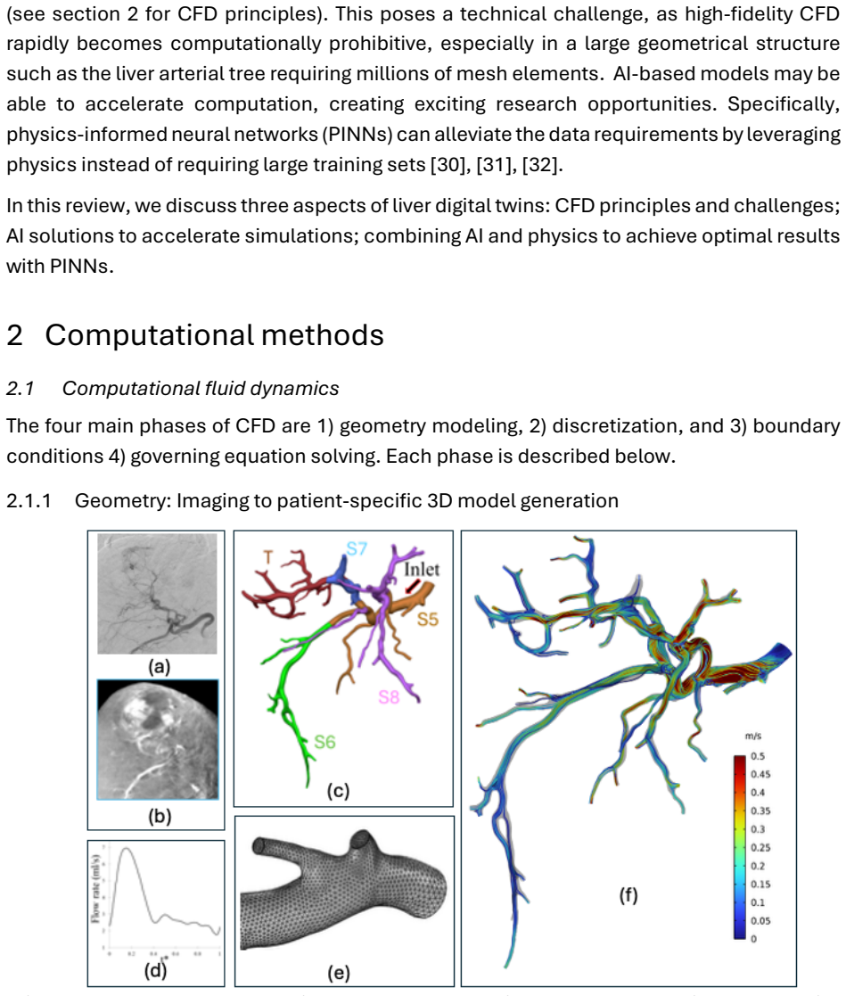
The central claim is that CFD and physics-informed AI together can form a dynamic, patient-specific digital twin of the liver that predicts Y-90 microsphere distribution and absorbed dose accurately enough to optimize radioembolization treatment planning. The paper's contribution is architectural: it specifies the pipeline from CBCT/CECT-derived hepatic artery geometry, meshing, patient-specific boundary conditions from Doppler ultrasound or Windkessel models, and Navier-Stokes flow solution with Lagrangian particle tracing, to replacing repeated expensive CFD solves with PINNs, PI-GANs, PI-DMs, or transformer-based surrogates. Around this pipeline it places an optimization loop in which inj
What carries the argument
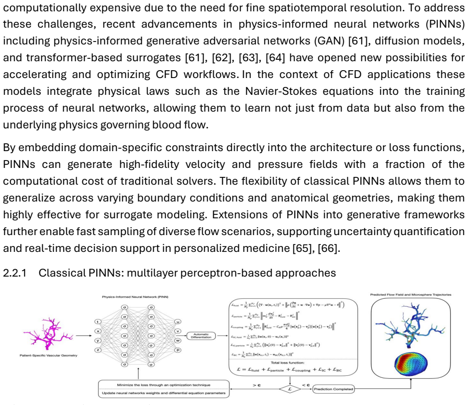
The load-bearing mechanism is the patient-specific hepatic arterial digital twin: a CFD model that solves the Navier-Stokes equations and tracks 20–60 µm microspheres in a Lagrangian frame, with one-way, two-way, or four-way coupling, generating ground-truth flow and dose distributions; and physics-informed neural networks that encode the same governing equations as loss terms or architectural constraints, serving as mesh-free surrogates that reproduce CFD-level outputs fast enough for iterative treatment planning. The named architectures—classical PINNs, PI-GANs, PI-DMs, and PINNsFormer—all carry the physical equations into the network so that the surrogate inherits physical fidelity while
Load-bearing premise
Patient-specific CFD, built from cone-beam CT geometry and Doppler-measured inlet flow, can accurately predict where 30-micron microspheres actually lodge in small distal liver vessels—despite the paper's own note that microspheres stop following blood flow there and that in vivo validation is limited.
What would settle it
Run the proposed CFD/PINN twin on a cohort of radioembolization patients and compare its predicted microsphere and dose distributions voxel-by-voxel or segment-by-segment against post-treatment Y-90 PET/CT. If the dose errors in tumors exceed the clinically relevant tolerance (for example, more than 100 Gy in lesions in a substantial fraction of patients, or segment-level mismatches that would change the injection plan), the twin's predictive core is falsified.
If this is right
- Treatment planning becomes an in silico search: sample injection sites and Y-90 activities, simulate the resulting dose distribution with the twin, and compare to physician-set targets for tumor and healthy liver.
- The pre-treatment vascular mapping procedure could be replaced or augmented by operating the digital twin before the first intervention.
- Between treatment sessions, the twin could be updated with post-treatment imaging to decide whether additional injections are needed and whether toxicity limits were respected.
- Physics-informed generative surrogates make repeated simulations fast enough for real-time or few-days decision support, which is the main barrier to clinical CFD use.
- The twin output could expand beyond liver dose to lung dose via arterio-venous shunt modeling and, with sufficient biomarkers, to tumor and liver response prediction.
Where Pith is reading between the lines
- Editorial inference: the same physics-informed surrogate stack could transfer to chemoembolization or to brain tumor radioembolization, where the vascular geometry is smaller and ground-truth validation may be more tractable.
- Editorial inference: because generative surrogates output distributions rather than single deterministic maps, they could supply confidence bounds on each treatment plan, which deterministic CFD cannot offer and which may be a practical route to regulatory acceptance.
- Editorial inference: the paper's own admission that microspheres stop following blood flow in small distal vessels suggests the twin's accuracy will hinge less on AI architecture and more on particle-level modeling—size, density, collisions, and stasis—so near-term validation should target bifurcation-level deposition data rather than whole-liver dose alone.
Editorial analysis
A structured set of objections, weighed in public.
Referee Report
Summary. This paper is a narrative review proposing a framework for a patient-specific digital twin of the liver for Y-90 radioembolization treatment planning. It combines computational fluid dynamics (CFD) for microsphere transport with physics-informed neural networks (PINNs) and generative AI surrogates (PI-GANs, diffusion models, transformers) to accelerate simulation. The review covers the imaging-to-mesh pipeline, boundary conditions, governing equations, particle transport coupling, mesh independence/validation, and clinical translation, concluding with a vision of an in silico treatment planning loop. The abstract and key points assert that CFD and physics-informed AI can maintain physical fidelity and accuracy while enabling real-time decision support.
Significance. If the proposed digital twin framework were validated, it would be a substantial step toward personalized, dosimetry-driven radioembolization planning. The review's contribution is organizational: it usefully consolidates CFD and physics-informed machine learning literature relevant to radioembolization, identifies the clinical workflow, and candidly acknowledges several validation gaps. It does not provide new experimental evidence, so its value depends on accurate representation of the cited literature and internal consistency of the claims. The paper also includes a number of self-citations, but the central framework does not rely exclusively on those works.
major comments (3)
- [Key Points and Abstract vs. Section 2.2.1] The key points claim that generative AI and PINNs 'maintain simulation accuracy,' and the abstract states these surrogates 'maintain physical fidelity.' This is directly contradicted by Section 2.2.1, which states that 'systematic benchmarks reporting their accuracy and computational performance remain limited' and that 'the accuracy of these methods has not been fully quantified beyond proof-of-concept.' Since the justification for using AI surrogates in treatment planning rests on accuracy, this is a load-bearing inconsistency. Please revise the abstract and key points to reflect the current limitations, or substantiate the accuracy claim with specific quantitative errors from the cited studies and clarify that these were obtained on simplified geometries.
- [Sections 1.3 and 3.3] The digital twin's central prediction—microsphere distribution in the distal hepatic arterial tree—is acknowledged in Section 1.3 to be insufficiently captured by blood-flow modeling alone, because microspheres do not follow blood flow in small vessels. Section 3.3 further admits that ground-truth imaging resolution (3–5 mm for 90Y PET and MAA SPECT) is far coarser than the vessels where deposition must be predicted. This is a load-bearing unknown for the entire framework. The review should explicitly frame this as an unresolved research question and propose a staged validation pathway (e.g., in vitro particle deposition in patient-derived geometries, comparison with post-treatment 90Y PET, or 4D flow MRI) before claiming the twin can 'optimize radioembolization planning.'
- [Section 2.1.4, Eq. (3)] Equation (3) models particle motion with only drag and gravity, while the surrounding text discusses two-way coupling and stasis. In the 200–500 µm vessels relevant to microsphere deposition, lift, added mass, and near-wall effects can be comparable to or greater than gravity for 20–60 µm particles. Presenting Eq. (3) as the governing particle equation overstates the fidelity of current CFD-based twins, especially since the paper later acknowledges that one-way coupling is an approximation. Please either include a more complete force balance or explicitly state the regime in which Eq. (3) is valid, and flag this as a limitation of existing CFD models.
minor comments (5)
- [Figure numbering] Figure numbering is inconsistent: Figure 2 appears in Section 2.1.1 (hepatic artery model) and again in Section 2.2.2 (PI-GAN overview, with a different caption). Figure 3 appears in Section 2.2.1 (PINN overview) and again in Section 3.2 (liver digital twin). Section 2.2.4 references 'Figure 4' but the displayed image is captioned 'Figure 2: Overview of the adapted PINNsFormer architecture.' Please renumber all figures and update cross-references.
- [References] References [48] and [49] are identical (Elghobashi 1994); remove the duplicate and renumber. Several references are incomplete or malformed: [23] lists author names as initials, [79] gives 'I. K et al.' with no full name, and [40] has inconsistent capitalization in the journal title. Please proofread all reference entries.
- [Equation (5)] The wall shear stress formula appears to be misprinted: the velocity gradient should be ∂u/∂y (or ∂u/∂n), not ∂u/∂t, if it is intended to represent the near-wall velocity gradient. Please correct the notation.
- [Terminology] The description of four-way coupling is imprecise. It says the model 'includes microsphere-microsphere interaction,' but four-way coupling typically also includes particle-to-fluid momentum feedback (i.e., two-way coupling) in addition to particle-particle collisions. Please define the term precisely to avoid confusion.
- [Abstract cost claim] The abstract/claim of 'reduced computational cost' for AI surrogates is not quantified anywhere in the review. Please provide representative speed-up factors or explicitly note that computational gains have not yet been systematically quantified for this application.
Circularity Check
No circularity; self-citations are illustrative and limitations are explicitly acknowledged.
full rationale
This is a narrative review and framework proposal rather than a derivation. The abstract's claim that 'CFD and physics-informed AI methods form the foundation of dynamic, patient-specific digital twin to optimize radioembolization planning' is a synthesis of existing literature, not a result computed from assumptions introduced in the paper. No parameter is fitted and no new predicted quantity is compared to data, so the fitted-input-called-prediction and self-definitional patterns do not apply. The paper does rely on several first-party citations (e.g., refs 14, 15, 19, 45, 58, 60, 92), but these are used as examples of prior CFD/hemodynamics work, not as uniqueness theorems or as the sole support for a derived conclusion; independent groups (Antón et al., Bomberna et al., Aramburu et al.) are also cited for the key CFD-modeling and validation claims. The manuscript repeatedly and explicitly flags its own limitations: Section 1.3 states that microspheres can be assumed to follow blood flow only in large arterial branches and that higher-fidelity models are likely required; Section 2.2.1 notes that systematic benchmarks 'remain limited' and large-scale patient-specific validations are 'still scarce'; Section 3.3 concedes that 'the accuracy is hard to quantify due to the lack of high-precision ground truth.' These are candid statements about predictive uncertainty and external validation, not circular reasoning. The review does not rename a known result as a new derivation, nor does it import a self-cited uniqueness theorem to force its choice of model. The only reason to give a nonzero score is the presence of multiple self-citations, but none is load-bearing in the sense of making a claimed prediction equivalent to its input by construction.
Axiom & Free-Parameter Ledger
axioms (3)
- domain assumption Blood flow in the hepatic arterial tree is governed by the incompressible Navier-Stokes equations.
- domain assumption Microsphere transport can be simulated as Lagrangian particles with one-way or two-way coupling to the fluid, without accounting for all particle-particle interactions.
- domain assumption Patient-specific anatomy and boundary conditions extracted from imaging are accurate enough for predictive simulation.
Cite this review
Pith. "Pith review of Towards Digital Twins for Optimal Radioembolization." pith.science (2026). https://pith.science/paper/XKHZQC5A
@misc{pith2026250902607,
  author       = {Pith},
  title        = {Pith review of: Towards Digital Twins for Optimal Radioembolization},
  year         = {2026},
  howpublished = {\url{https://pith.science/paper/XKHZQC5A}},
  note         = {Machine review of arXiv:2509.02607}
}
read the original abstract
Radioembolization is a localized liver cancer treatment that delivers radioactive microspheres (30 micron) to tumors via a catheter inserted in the hepatic arterial tree. The goal is to maximize therapeutic efficacy while minimizing damage to healthy liver tissue. However, optimization is challenging due to complex hepatic artery anatomy, variable blood flow, and uncertainty in microsphere transport. The creation of dynamic, patient-specific digital twins may provide a transformative solution to these challenges. This work outlines a framework for a liver radioembolization digital twin using high-fidelity computational fluid dynamics (CFD) and/or recent physics-informed machine learning approaches. The CFD approach involves microsphere transport calculations in the hepatic arterial tree with individual patient data, which enables personalized treatment planning. Although accurate, traditional CFD is computationally expensive and limits clinical applicability. To accelerate simulations, physics-informed neural networks (PINNs) and their generative extensions play an increasingly important role. PINNs integrate governing equations, such as the Navier-Stokes equations, directly into the neural network training process, enabling mesh-free, data-efficient approximation of blood flow and microsphere transport. Physics-informed generative adversarial networks (PI-GANs), diffusion models (PI-DMs), and transformer-based architectures further enable uncertainty-aware, temporally resolved predictions with reduced computational cost. These AI surrogates not only maintain physical fidelity but also support rapid sampling of diverse flow scenarios, facilitating real-time decision support. Together, CFD and physics-informed AI methods form the foundation of dynamic, patient-specific digital twin to optimize radioembolization planning and ultimately improve clinical outcomes.
Figures
Reference graph
Works this paper leans on
-
[1]
Cancer statistics, 2022 - Siegel - 2022 - CA: A Cancer Journal for Clinicians - Wiley Online Library
“Cancer statistics, 2022 - Siegel - 2022 - CA: A Cancer Journal for Clinicians - Wiley Online Library.” Accessed: Jun. 05, 2025. [Online]. Available: https://acsjournals.onlinelibrary.wiley.com/doi/full/10.3322/caac.21708
-
[2]
R. Murthy et al., “Yttrium-90 Microsphere Therapy for Hepatic Malignancy: Devices, Indications, Technical Considerations, and Potential Complications,” RadioGraphics, vol. 25, no. suppl_1, pp. S41–S55, Oct. 2005, doi: 10.1148/rg.25si055515
-
[3]
Trial ID ACTRN1261000069005.http://australiancancertrials.gov.au”
“Sirtex Technology Pty Ltd: Pilot study of selective internal radiation therapy (SIRT) with yttrium-90 resin microspheres (SIR-Spheres microspheres) in patients with renal cell carcinoma (STX0110, ‘RESIRT’). Trial ID ACTRN1261000069005.http://australiancancertrials.gov.au”
-
[4]
Package Insert TheraSphere® Yttrium-90 Glass Microspheres. Rev 14,
BTG, “Package Insert TheraSphere® Yttrium-90 Glass Microspheres. Rev 14,” 2019
2019
-
[5]
Chemoembolization for Primary and Metastatic Liver Cancer,
E. Liapi and J.-F. H. Geschwind, “Chemoembolization for Primary and Metastatic Liver Cancer,” The Cancer Journal, vol. 16, no. 2, p. 156, Apr. 2010, doi: 10.1097/PPO.0b013e3181d7e905
-
[6]
M. F. Georgiou, R. A. Kuker, M. T. Studenski, P. P. Ahlman, M. Witte, and L. Portelance, “Lung shunt fraction calculation using 99mTc-MAA SPECT/CT imaging for 90Y microsphere selective internal radiation therapy of liver tumors,” EJNMMI Res, vol. 11, no. 1, p. 96, Sep. 2021, doi: 10.1186/s13550-021-00837-z
-
[7]
Hepatic Radioembolization: A Multistep Theragnostic Procedure,
K. Ramdhani, M. G. E. H. Lam, A. J. A. T. Braat, M. L. J. Smits, and G. El-Haddad, “Hepatic Radioembolization: A Multistep Theragnostic Procedure,” PET Clinics, vol. 19, no. 3, pp. 431–446, Jul. 2024, doi: 10.1016/j.cpet.2024.03.010
-
[8]
A. Riaz et al., “Complications Following Radioembolization with Yttrium-90 Microspheres: A Comprehensive Literature Review,” Journal of Vascular and Interventional Radiology, vol. 20, no. 9, pp. 1121–1130, Sep. 2009, doi: 10.1016/j.jvir.2009.05.030
-
[9]
M. Seidensticker et al., “Impact of Pharmaceutical Prophylaxis on Radiation- Induced Liver Disease Following Radioembolization,” Cancers, vol. 13, no. 9, Art. no. 9, Jan. 2021, doi: 10.3390/cancers13091992
-
[10]
C. Chiesa et al., “Radioembolization of hepatocarcinoma with 90Y glass microspheres: treatment optimization using the dose-toxicity relationship,” Eur J Nucl Med Mol Imaging, vol. 47, no. 13, pp. 3018–3032, Dec. 2020, doi: 10.1007/s00259-020-04845-4
-
[11]
A. Jadoul et al., “Comparative dosimetry between 99mTc-MAA SPECT/CT and 90Y PET/CT in primary and metastatic liver tumors,” Eur J Nucl Med Mol Imaging, vol. 47, no. 4, pp. 828–837, Apr. 2020, doi: 10.1007/s00259-019-04465-7
-
[12]
Radiation Segmentectomy: Potential Curative Therapy for Early Hepatocellular Carcinoma,
R. J. Lewandowski et al., “Radiation Segmentectomy: Potential Curative Therapy for Early Hepatocellular Carcinoma,” Radiology, vol. 287, no. 3, pp. 1050–1058, Jun. 2018, doi: 10.1148/radiol.2018171768
-
[13]
A. Gabr et al., “Correlation of Y90-absorbed radiation dose to pathological necrosis in hepatocellular carcinoma: confirmatory multicenter analysis in 45 explants,” Eur J Nucl Med Mol Imaging, vol. 48, no. 2, pp. 580–583, Feb. 2021, doi: 10.1007/s00259-020-04976- 8
-
[14]
A. Taebi, N. Janibek, R. Goldman, R. Pillai, C. T. Vu, and E. Roncali, “The Impact of Injection Distance to Bifurcations on Yttrium-90 Distribution in Liver Cancer Radioembolization,” Journal of Vascular and Interventional Radiology, vol. 33, no. 6, pp. 668-677.e1, Jun. 2022, doi: 10.1016/j.jvir.2022.03.006
-
[15]
E. Roncali, A. Taebi, C. Foster, and C. T. Vu, “Personalized Dosimetry for Liver Cancer Y-90 Radioembolization Using Computational Fluid Dynamics and Monte Carlo Simulation,” Ann Biomed Eng, vol. 48, no. 5, pp. 1499–1510, May 2020, doi: 10.1007/s10439-020-02469-1
-
[16]
T. Bomberna, G. Maleux, and C. Debbaut, “Simplification strategies for a patient- specific CFD model of particle transport during liver radioembolization,” Computers in Biology and Medicine, vol. 178, p. 108732, Aug. 2024, doi: 10.1016/j.compbiomed.2024.108732
-
[17]
R. Antón et al., “A proof-of-concept study of the in-vivo validation of a computational fluid dynamics model of personalized radioembolization,” Sci Rep, vol. 11, no. 1, p. 3895, Feb. 2021, doi: 10.1038/s41598-021-83414-7
-
[18]
J. Aramburu, R. Antón, A. Rivas, J. C. Ramos, B. Sangro, and J. I. Bilbao, “Liver Radioembolization: An Analysis of Parameters that Influence the Catheter-Based Particle- Delivery via CFD,” Curr Med Chem, vol. 27, no. 10, pp. 1600–1615, 2020, doi: 10.2174/0929867325666180622145647
-
[19]
A. Taebi, R. M. Pillai, B. S. Roudsari, C. T. Vu, and E. Roncali, “Computational Modeling of the Liver Arterial Blood Flow for Microsphere Therapy: Effect of Boundary Conditions,” Bioengineering (Basel), vol. 7, no. 3, p. 64, Jun. 2020, doi: 10.3390/bioengineering7030064
-
[20]
The Value of Partnership in Technology-Enabled Facility Optimization,
Siemens Healthineers, “The Value of Partnership in Technology-Enabled Facility Optimization,” 2022
2022
-
[21]
Digital Twins and Living Models at NASA. B. Danette,
B. D. Allen, “Digital Twins and Living Models at NASA. B. Danette,” ASME, 2021
2021
-
[22]
Editorial,
Editorial, “Editorial,” Nat Comput Sci, vol. 4, no. 3, pp. 145–146
-
[23]
Z. Nrr, B. R, H. R, and S. G, “Mathematic Modeling of Tumor Growth During [177Lu]Lu-PSMA Therapy: Insights into Treatment Optimization,” Journal of nuclear medicine : official publication, Society of Nuclear Medicine, vol. 66, no. 1, Jan. 2025, doi: 10.2967/jnumed.124.268457
-
[24]
Digital twins for predictive oncology will be a paradigm shift for precision cancer care,
T. Hernandez-Boussard et al., “Digital twins for predictive oncology will be a paradigm shift for precision cancer care,” Nature medicine, vol. 27, no. 12, pp. 2065–2066, 2021
2065
-
[25]
Integrating mechanism-based modeling with biomedical imaging to build practical digital twins for clinical oncology,
C. Wu et al., “Integrating mechanism-based modeling with biomedical imaging to build practical digital twins for clinical oncology,” Biophysics reviews, vol. 3, no. 2, 2022
2022
-
[26]
Patient-specific characterization of breast cancer hemodynamics using image-guided computational fluid dynamics,
C. Wu et al., “Patient-specific characterization of breast cancer hemodynamics using image-guided computational fluid dynamics,” IEEE transactions on medical imaging, vol. 39, no. 9, pp. 2760–2771, 2020
2020
-
[27]
Personalized radiotherapy design for glioblastoma: integrating mathematical tumor models, multimodal scans, and Bayesian inference,
J. Lipková et al., “Personalized radiotherapy design for glioblastoma: integrating mathematical tumor models, multimodal scans, and Bayesian inference,” IEEE transactions on medical imaging, vol. 38, no. 8, pp. 1875–1884, 2019
2019
-
[28]
Building digital twins of the human immune system: toward a roadmap,
R. Laubenbacher et al., “Building digital twins of the human immune system: toward a roadmap,” NPJ digital medicine, vol. 5, no. 1, p. 64, 2022
2022
-
[29]
J. Brosch-Lenz et al., “Role of Artificial Intelligence in Theranostics: Toward Routine Personalized Radiopharmaceutical Therapies,” PET Clinics, vol. 16, no. 4, pp. 627–641, Oct. 2021, doi: 10.1016/j.cpet.2021.06.002
-
[30]
G. Kissas, Y. Yang, E. Hwuang, W. R. Witschey, J. A. Detre, and P. Perdikaris, “Machine learning in cardiovascular flows modeling: Predicting arterial blood pressure from non-invasive 4D flow MRI data using physics-informed neural networks,” Computer Methods in Applied Mechanics and Engineering, vol. 358, p. 112623, Jan. 2020, doi: 10.1016/j.cma.2019.112623
-
[31]
P. Moser, W. Fenz, S. Thumfart, I. Ganitzer, and M. Giretzlehner, “Modeling of 3D Blood Flows with Physics-Informed Neural Networks: Comparison of Network Architectures,” Fluids, vol. 8, no. 2, Art. no. 2, Feb. 2023, doi: 10.3390/fluids8020046
-
[32]
Hidden fluid mechanics: Learning velocity and pressure fields from flow visualizations,
M. Raissi, A. Yazdani, and G. E. Karniadakis, “Hidden fluid mechanics: Learning velocity and pressure fields from flow visualizations,” Science, vol. 367, no. 6481, pp. 1026–1030, 2020
2020
-
[33]
Anatomy of liver arteries for interventional radiology,
S. Favelier et al., “Anatomy of liver arteries for interventional radiology,” Diagnostic and interventional imaging, vol. 96, no. 6, pp. 537–546, 2015
work page 2015
-
[34]
Computational Fluid Dynamics Modeling of Liver Radioembolization: A Review,
J. Aramburu, R. Antón, M. Rodríguez-Fraile, B. Sangro, and J. I. Bilbao, “Computational Fluid Dynamics Modeling of Liver Radioembolization: A Review,” Cardiovasc Intervent Radiol, vol. 45, no. 1, pp. 12–20, Jan. 2022, doi: 10.1007/s00270-021- 02956-5
-
[35]
Computational fluid dynamics modelling of hemodynamics in aortic aneurysm and dissection: a review,
M. Hu, B. Chen, and Y. Luo, “Computational fluid dynamics modelling of hemodynamics in aortic aneurysm and dissection: a review,” Front. Bioeng. Biotechnol., vol. 13, Mar. 2025, doi: 10.3389/fbioe.2025.1556091
-
[36]
Explainable and Robust Deep Learning for Liver Segmentation Through U-Net Network,
M. C. Brunese, A. Rocca, A. Santone, M. Cesarelli, L. Brunese, and F. Mercaldo, “Explainable and Robust Deep Learning for Liver Segmentation Through U-Net Network,” Diagnostics (Basel), vol. 15, no. 7, p. 878, Mar. 2025, doi: 10.3390/diagnostics15070878
-
[37]
V-Net: Fully Convolutional Neural Networks for Volumetric Medical Image Segmentation,
F. Milletari, N. Navab, and S.-A. Ahmadi, “V-Net: Fully Convolutional Neural Networks for Volumetric Medical Image Segmentation,” in 2016 Fourth International Conference on 3D Vision (3DV), Oct. 2016, pp. 565–571. doi: 10.1109/3DV.2016.79
-
[38]
nnU-Net: a self-configuring method for deep learning-based biomedical image segmentation,
F. Isensee, P. F. Jaeger, S. A. A. Kohl, J. Petersen, and K. H. Maier-Hein, “nnU-Net: a self-configuring method for deep learning-based biomedical image segmentation,” Nat Methods, vol. 18, no. 2, pp. 203–211, Feb. 2021, doi: 10.1038/s41592-020-01008-z
-
[39]
Attention U-Net: Learning Where to Look for the Pancreas,
O. Oktay et al., “Attention U-Net: Learning Where to Look for the Pancreas,” May 20, 2018, arXiv: arXiv:1804.03999. doi: 10.48550/arXiv.1804.03999
-
[40]
Y. A. R. Shah, H. A. Qureshi, S. M. Qureshi, S. U. R. Shah, A. Shiwlani, and A. Ahmad, “A Review of Evaluating Deep Learning Techniques in Hepatic Cancer Imaging: Automated Segmentation and Tumor Quantification,” International Journal For Multidisciplinary Research, vol. 6, no. 5, 2024
work page 2024
-
[41]
Semi-automatic surface and volume mesh generation for subject-specific biomedical geometries,
I. Sazonov and P. Nithiarasu, “Semi-automatic surface and volume mesh generation for subject-specific biomedical geometries,” International Journal for Numerical Methods in Biomedical Engineering, vol. 28, no. 1, pp. 133–157, 2012, doi: 10.1002/cnm.1470
-
[42]
J. Aramburu et al., “Physiological outflow boundary conditions methodology for small arteries with multiple outlets: a patient-specific hepatic artery haemodynamics case study,” Proceedings of the Institution of Mechanical Engineers, Part H: Journal of Engineering in Medicine, vol. 229, no. 4, pp. 291–306, 2015
work page 2015
-
[43]
G. H. Hübner, N. Steudel, G. Kleber, C. Behrmann, E. Lotterer, and W. E. Fleig, “Hepatic arterial blood flow velocities: assessment by transcutaneous and intravascular Doppler sonography,” Journal of Hepatology, vol. 32, no. 6, pp. 893–899, Jun. 2000, doi: 10.1016/S0168-8278(00)80093-8
-
[44]
Optimizing Doppler and Color Flow US: Application to Hepatic Sonography,
J. B. Kruskal, P. A. Newman, L. G. Sammons, and R. A. Kane, “Optimizing Doppler and Color Flow US: Application to Hepatic Sonography,” RadioGraphics, vol. 24, no. 3, pp. 657–675, May 2004, doi: 10.1148/rg.243035139
-
[45]
A. Taebi, C. T. Vu, and E. Roncali, “Estimation of Yttrium-90 Distribution in Liver Radioembolization using Computational Fluid Dynamics and Deep Neural Networks,” in 2020 42nd Annual International Conference of the IEEE Engineering in Medicine & Biology Society (EMBC), Jul. 2020, pp. 4974–4977. doi: 10.1109/EMBC44109.2020.9176328
arXiv 2020
-
[46]
J. I. Bilbao et al., “Biocompatibility, Inflammatory Response, and Recannalization Characteristics of Nonradioactive Resin Microspheres: Histological Findings,” Cardiovasc Intervent Radiol, vol. 32, no. 4, pp. 727–736, Jul. 2009, doi: 10.1007/s00270-009-9592-9
-
[47]
Bubbles, drops, and particles,
W. M. E, “Bubbles, drops, and particles,” Prog. Energy Combust. Sci., vol. 12, p. 163, 1978
work page 1978
-
[49]
On predicting particle-laden turbulent flows,
S. Elghobashi, “On predicting particle-laden turbulent flows,” Appl. Sci. Res., vol. 52, no. 4, pp. 309–329, Jun. 1994, doi: 10.1007/BF00936835
-
[50]
J. Y. Moon, D. C. Suh, Y. S. Lee, Y. W. Kim, and J. S. Lee, “Considerations of Blood Properties, Outlet Boundary Conditions and Energy Loss Approaches in Computational Fluid Dynamics Modeling,” Neurointervention, vol. 9, no. 1, pp. 1–8, Feb. 2014, doi: 10.5469/neuroint.2014.9.1.1
-
[51]
Computational Modeling of Radioembolization: How to Calculate Infinity,
B. Toskich and R. J. Lewandowski, “Computational Modeling of Radioembolization: How to Calculate Infinity,” Cardiovasc Intervent Radiol, vol. 44, no. 12, pp. 2020–2021, Dec. 2021, doi: 10.1007/s00270-021-02989-w
-
[52]
Modeling dynamics of the cardiovascular system using fluid-structure interaction methods,
F. Syed, S. Khan, and M. Toma, “Modeling dynamics of the cardiovascular system using fluid-structure interaction methods,” Biology, vol. 12, no. 7, p. 1026, 2023
work page 2023
-
[53]
A. K. Alagan et al., “Histopathology-based near-realistic arterial wall reconstruction of a patient-specific cerebral aneurysm for fluid-structure interaction studies,” Computers in Biology and Medicine, vol. 185, p. 109579, Feb. 2025, doi: 10.1016/j.compbiomed.2024.109579
-
[54]
CFD-DEM model of plugging in flow with cohesive particles,
N. Saparbayeva and B. V. Balakin, “CFD-DEM model of plugging in flow with cohesive particles,” Sci Rep, vol. 13, p. 17188, Oct. 2023, doi: 10.1038/s41598-023-44202- 7
-
[55]
H. Sun, S. Xu, X. Pan, L. Shi, X. Geng, and Y. Cai, “Investigating the jamming of particles in a three-dimensional fluid-driven flow via coupled CFD–DEM simulations,” International Journal of Multiphase Flow, vol. 114, pp. 140–153, May 2019, doi: 10.1016/j.ijmultiphaseflow.2019.01.017
-
[56]
J. Aramburu, R. Antón, A. Rivas, J. C. Ramos, B. Sangro, and J. I. Bilbao, “Computational particle–haemodynamics analysis of liver radioembolization pretreatment as an actual treatment surrogate,” International Journal for Numerical Methods in Biomedical Engineering, vol. 33, no. 2, p. e02791, 2017, doi: 10.1002/cnm.2791
-
[57]
T. Bomberna, Koudehi ,Ghazal Adeli, Claerebout ,Charlotte, Verslype ,Chris, Maleux ,Geert, and C. and Debbaut, “Transarterial drug delivery for liver cancer: numerical simulations and experimental validation of particle distribution in patient-specific livers,” Expert Opinion on Drug Delivery, vol. 18, no. 3, pp. 409–422, Mar. 2021, doi: 10.1080/17425247....
-
[58]
N. K. Panneerselvam, B. J. Sudhir, S. K. Kannath, and B. S. V. Patnaik, “Hemodynamic analysis of coil filled patient-specific middle cerebral artery aneurysm using porous medium approach,” Physics of Fluids, vol. 35, no. 11, p. 111906, Nov. 2023, doi: 10.1063/5.0173688
-
[59]
Hemodynamic Shear Stress and Its Role in Atherosclerosis,
A. M. Malek, S. L. Alper, and S. Izumo, “Hemodynamic Shear Stress and Its Role in Atherosclerosis,” JAMA, vol. 282, no. 21, pp. 2035–2042, Dec. 1999, doi: 10.1001/jama.282.21.2035
-
[60]
N. K. Panneerselvam, B. Akade, A. A. Kumar, B. J. Sudhir, S. K. Kannath, and B. S. V. Patnaik, “Hemodynamic investigation of hemorrhagic stroke treatment options for a patient-specific aneurysm using a porous medium model,” Physics of Fluids, vol. 36, no. 7, p. 071915, Jul. 2024, doi: 10.1063/5.0214784
-
[61]
Physics-Informed Generative Adversarial Networks for Stochastic Differential Equations,
L. Yang, D. Zhang, and G. E. Karniadakis, “Physics-Informed Generative Adversarial Networks for Stochastic Differential Equations,” SIAM J. Sci. Comput., vol. 42, no. 1, pp. A292–A317, Jan. 2020, doi: 10.1137/18M1225409
-
[62]
A physics-informed diffusion model for high- fidelity flow field reconstruction,
D. Shu, Z. Li, and A. Barati Farimani, “A physics-informed diffusion model for high- fidelity flow field reconstruction,” Journal of Computational Physics, vol. 478, p. 111972, Apr. 2023, doi: 10.1016/j.jcp.2023.111972
-
[63]
Physics informed token transformer for solving partial differential equations,
C. Lorsung, Z. Li, and A. Barati Farimani, “Physics informed token transformer for solving partial differential equations,” Mach. Learn.: Sci. Technol., vol. 5, no. 1, p. 015032, Mar. 2024, doi: 10.1088/2632-2153/ad27e3
-
[64]
PINNsFormer: A Transformer-Based Framework For Physics-Informed Neural Networks,
Z. Zhao, X. Ding, and B. A. Prakash, “PINNsFormer: A Transformer-Based Framework For Physics-Informed Neural Networks,” 2023, arXiv. doi: 10.48550/ARXIV.2307.11833
-
[65]
Physics-Informed Neural Networks for Brain Hemodynamic Predictions Using Medical Imaging,
M. Sarabian, H. Babaee, and K. Laksari, “Physics-Informed Neural Networks for Brain Hemodynamic Predictions Using Medical Imaging,” IEEE Trans. Med. Imaging, vol. 41, no. 9, pp. 2285–2303, Sep. 2022, doi: 10.1109/TMI.2022.3161653
arXiv 2022
-
[66]
From PINNs to PIKANs: recent advances in physics-informed machine learning,
J. D. Toscano et al., “From PINNs to PIKANs: recent advances in physics-informed machine learning,” Mach. Learn. Comput. Sci. Eng, vol. 1, no. 1, p. 15, Jun. 2025, doi: 10.1007/s44379-025-00015-1
-
[67]
Δ -PINNs: Physics-informed neural networks on complex geometries,
F. Sahli Costabal, S. Pezzuto, and P. Perdikaris, “Δ -PINNs: Physics-informed neural networks on complex geometries,” Engineering Applications of Artificial Intelligence, vol. 127, p. 107324, Jan. 2024, doi: 10.1016/j.engappai.2023.107324
-
[68]
H. Zhang, R. Chan, and X.-C. Tai, “A Meshless Solver for Blood Flow Simulations in Elastic Vessels Using Physics-Informed Neural Network,” Jun. 06, 2024, arXiv: arXiv:2312.05601. doi: 10.48550/arXiv.2312.05601
work page internal anchor Pith review Pith/arXiv arXiv doi:10.48550/arxiv.2312.05601 2024
-
[69]
Physics informed neural networks for fluid flow analysis with repetitive parameter initialization,
J. Lee et al., “Physics informed neural networks for fluid flow analysis with repetitive parameter initialization,” Sci Rep, vol. 15, no. 1, p. 16740, May 2025, doi: 10.1038/s41598- 025-99354-5
doi:10.1038/s41598- 2025
-
[70]
Characterizing possible failure modes in physics-informed neural networks,
A. S. Krishnapriyan, A. Gholami, S. Zhe, R. M. Kirby, and M. W. Mahoney, “Characterizing possible failure modes in physics-informed neural networks,” 2021, doi: 10.48550/ARXIV.2109.01050
-
[71]
Z. Mo, Y. Fu, D. Xu, and X. Di, “TrafficFlowGAN: Physics-Informed Flow Based Generative Adversarial Network for Uncertainty Quantification,” in Machine Learning and Knowledge Discovery in Databases, M.-R. Amini, S. Canu, A. Fischer, T. Guns, P. Kralj Novak, and G. Tsoumakas, Eds., Cham: Springer Nature Switzerland, 2023, pp. 323–339. doi: 10.1007/978-3-03...
-
[72]
B. Siddani, S. Balachandar, W. C. Moore, Y. Yang, and R. Fang, “Machine learning for physics-informed generation of dispersed multiphase flow using generative adversarial networks,” Theor. Comput. Fluid Dyn., vol. 35, no. 6, pp. 807–830, Dec. 2021, doi: 10.1007/s00162-021-00593-9
-
[73]
Generative Adversarial Networks,
I. J. Goodfellow et al., “Generative Adversarial Networks,” Jun. 10, 2014, arXiv: arXiv:1406.2661. doi: 10.48550/arXiv.1406.2661
-
[74]
Denoising Diffusion Probabilistic Models,
J. Ho, A. Jain, and P. Abbeel, “Denoising Diffusion Probabilistic Models,” in Advances in Neural Information Processing Systems, Curran Associates, Inc., 2020, pp. 6840–6851. Accessed: Dec. 03, 2024. [Online]. Available: https://proceedings.neurips.cc/paper_files/paper/2020/hash/4c5bcfec8584af0d967f1ab1 0179ca4b-Abstract.html
work page 2020
-
[75]
G. Müller-Franzes et al., “A multimodal comparison of latent denoising diffusion probabilistic models and generative adversarial networks for medical image synthesis,” Sci Rep, vol. 13, no. 1, p. 12098, Jul. 2023, doi: 10.1038/s41598-023-39278-0
-
[76]
Diffusion Models Beat GANs on Image Synthesis,
P. Dhariwal and A. Nichol, “Diffusion Models Beat GANs on Image Synthesis,” arXiv.org. Accessed: Jun. 19, 2025. [Online]. Available: https://arxiv.org/abs/2105.05233v4
Pith/arXiv arXiv 2025
-
[77]
A Comparative Analysis Between GAN and Diffusion Models in Image Generation,
Y. Peng, “A Comparative Analysis Between GAN and Diffusion Models in Image Generation,” Transactions on Computer Science and Intelligent Systems Research, vol. 5, pp. 189–195, Aug. 2024, doi: 10.62051/0f1va465
-
[78]
Physics-Informed Diffusion Models,
J.-H. Bastek, W. Sun, and D. M. Kochmann, “Physics-Informed Diffusion Models,” Mar. 13, 2025, arXiv: arXiv:2403.14404. doi: 10.48550/arXiv.2403.14404
-
[79]
I. K et al., “New imaging tools in cardiovascular medicine: computational fluid dynamics and 4D flow MRI. - Abstract - Europe PMC”, Accessed: Aug. 21, 2025. [Online]. Available: https://europepmc.org/article/med/28929446
-
[80]
Respecting causality is all you need for training physics-informed neural networks,
S. Wang, S. Sankaran, and P. Perdikaris, “Respecting causality is all you need for training physics-informed neural networks,” arXiv.org. Accessed: Jun. 19, 2025. [Online]. Available: https://arxiv.org/abs/2203.07404v1
Pith/arXiv arXiv 2025
-
[81]
Stabilizing Generative Adversarial Networks: A Survey,
M. Wiatrak, S. V. Albrecht, and A. Nystrom, “Stabilizing Generative Adversarial Networks: A Survey,” Mar. 24, 2020, arXiv: arXiv:1910.00927. doi: 10.48550/arXiv.1910.00927
This paper was first reviewed by deepseek-v4-flash on August 5, 2026.
discussion (0)
Sign in with ORCID, Apple, or X to comment. Anyone can read and Pith papers without signing in.