REVIEW 4 major objections 6 minor 28 references
Temporally-Aware Diffusion Model for Brain Progression Modelling with Bidirectional Temporal Regularisation
T0 review · 4 major / 6 minor · reviewed 2026-08-05 · deepseek-v4-flash
Pith's one-line read TADM-3D predicts future brain MRIs as a baseline plus a diffusion-generated age-gap residual, and reports lower errors than DaniNet, CounterSynth, and BrLP on OASIS-3 and most NACC regions.
desk verdict Legitimate incremental extension of the authors' own 2D work, with a clean ablation and external validation; the central BAE loss is underspecified and the headline metric gaps are within noise, but the paper deserves serious review. read the letter →
The pith
A machine-rendered reading of the paper's core claim, the machinery that carries it, and where it could break.
The reading
What carries the argument
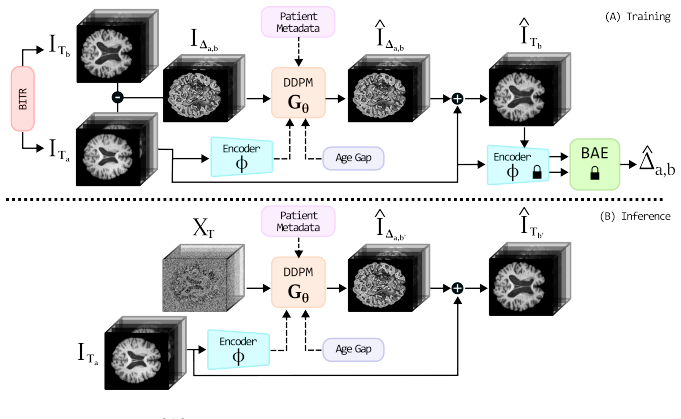
The central object is the predicted residual image IΔa,b = ITb − ITa, generated by a DDPM and added to the baseline. The load-bearing identity is that temporal progression can be learned as an intensity residual, so the model never has to regenerate whole-brain anatomy. Temporal control comes from conditioning on the age gap Δa,b rather than target age, and from the BAE loss L_BAE = (Δ̂a,b − Δa,b)², where Δ̂a,b is estimated by the frozen brain-age estimator. BITR makes time reversible during training by swapping baseline and follow-up at each step with probability 0.5. The residual formulation also keeps predictions anchored to the patient's own baseline anatomy.
What would settle it
For a fixed set of test subjects, run the BAE on real follow-up scans and on TADM-3D's predicted follow-ups from the same baselines, and compare the BAE's estimated age gaps against the true interval; if the BAE error on synthetic outputs is systematically different from its error on real scans, the L_BAE guidance is miscalibrated and the reported temporal gains need re-interpretation. Also, re-train with L_BAE computed through one-step DDIM denoising instead of whatever generation path Algorithm 1 uses, and check whether the MSE, SSIM, and volumetric gains persist.
Extended reading notes
Core claim
TADM-3D predicts the follow-up MRI as baseline plus a generated residual. The residual is produced by a DDPM conditioned on the baseline's latent encoding, the requested age gap, baseline age, and cognitive status. A frozen brain-age estimator maps baseline and predicted scans through a shared encoder into an estimated age gap, and the squared deviation from the true gap is added to the standard denoising loss. During training the roles of baseline and follow-up are swapped with probability 0.5, so the same model must predict forward and backward in time. On the OASIS-3 internal test set the method reaches MSE 0.004 and SSIM 0.902, beating BrLP (0.005 and 0.887) and reducing hippocampus volu
Load-bearing premise
The load-bearing premise is that the pre-trained brain-age estimator gives a trustworthy age-difference score when applied to synthetic diffusion outputs, and that the training loop can compute that score through a differentiable denoised residual—neither of which the paper verifies.
Editorial extensions
If this is right
- Forecasting a future MRI at arbitrary time intervals from a single baseline scan becomes an input parameter, because the model conditions on the age gap rather than a fixed follow-up schedule.
- Regional volumetric forecasts in AD-relevant structures improve, with hippocampus error roughly 40% lower than the strongest baseline on the internal test set.
- The model transfers to an out-of-distribution cohort: on NACC it leads on MSE and 4 of 5 regional volume metrics.
- Long-term prediction is the weak point: accuracy visibly drops for intervals beyond about 6.5 years, as the paper's own limitation example shows.
- Cognitive-status conditioning carries real signal: feeding the wrong cognitive label degrades predictions, most of all in the hippocampus.
Reading between the lines
- The age-gap conditioning trick is a candidate remedy for age-imbalanced longitudinal datasets; a direct test would train on a deliberately age-skewed split and measure whether performance degrades less than with age-conditioned baselines.
- BITR is a generic temporal regularizer that could be grafted onto other conditional generative pipelines; a cheap test is adding it to a latent diffusion baseline and checking temporal-consistency metrics without sacrificing image quality.
- Because the BAE is frozen while the encoder it consumes is trained end-to-end, the BAE's input distribution drifts during training; monitoring BAE age-gap error on generated images would reveal whether L_BAE remains a trustworthy guide.
- The residual formulation suggests a natural extension beyond T1-weighted brain MRI, e.g., predicting change in PET or other modalities as a residual shift, since static anatomy does not need to be regenerated.
Editorial analysis
A structured set of objections, weighed in public.
Referee Report
Summary. The paper proposes TADM-3D, a 3D denoising diffusion model that predicts brain MRI progression by learning voxel-wise residuals between baseline and follow-up scans. It conditions the diffusion process on the age gap between scans, patient metadata, and a baseline latent encoding, and introduces two temporal regularizers: a Brain-Age Estimator (BAE) loss (Eq. 7) that penalizes deviations between the predicted and true age gap, and a Back-In-Time Regularisation (BITR) that alternates forward and backward prediction during training. The method is evaluated on the OASIS-3 internal test set and on an external NACC test set, reporting MSE/SSIM and five regional volume errors against DaniNet, CounterSynth, and BrLP. Ablations isolate the contributions of age-gap conditioning, BAE, BITR, patient metadata, and 2.5D versus native 3D generation.
Significance. If the technical pipeline is made precise, the contribution is potentially valuable: learning 3D residual progression conditioned on age difference rather than absolute age is a sensible design, the external NACC evaluation is good practice, and the ablation study in Table 3 is informative. The main strengths are the 3D architecture, the residual formulation, and the explicit attempt to regularize temporal consistency with an age-estimation signal. However, the quantitative support is currently weakened by the absence of significance testing, by ambiguous implementation of the two central regularizers (BAE and BITR), and by some overstatement of external generalisation. These issues are fixable in revision, but they are load-bearing for the paper's main claims.
major comments (4)
- [§3.4, Eq. (7), §4.2, Algorithm 1] The BAE regularizer is the central novelty, but Eq. (7) is not well-defined as written. L_BAE requires a differentiable estimate of the generated residual ÎΔ; Algorithm 1 says only “Generate predicted residual bIΔ from the DDPM”, leaving open whether this is one-step denoising (x0 estimate), full reverse sampling, or DDIM. These choices give very different gradient signals. Additionally, Eq. (7) uses bΔ = Ψ(Φ(ÎTb)) − Ψ(Φ(ITa)), while §4.2 states Φ is trained end-to-end and §3.4 states the BAE parameters are frozen. If Φ is shared, the frozen BAE receives a drifting feature distribution and may be miscalibrated on synthetic scans. Since Table 3 shows removing BAE raises MSE from 0.004 to 0.009, this must be resolved: specify the differentiable path, clarify whether Φ is shared, and justify or calibrate the BAE on generated images.
- [§3.5, Algorithm 1] The BITR description and pseudocode are inconsistent. The text says the roles of ITa and ITb are randomly swapped, but Algorithm 1 keeps za = Φ(ITa) and computes ÎTb = ITa + ÎΔ, merely flipping “sign in Δ, A, and D”. A sign-flipped baseline age A and cognitive-status D do not implement a role swap; backward training should condition on Φ(ITb) and target residual ITa − ITb. As written, the pseudocode does not realize the proposed regularizer. Because Table 3 attributes a large drop to removing BITR (MSE 0.004 vs 0.008), this must be corrected or explicitly implemented as described.
- [Tables 1–3] No statistical significance testing is provided, and several headline differences are within one standard deviation: internal MSE 0.004±0.001 vs BrLP 0.005±0.002; SSIM 0.902±0.014 vs 0.887±0.017. Paired per-subject tests (e.g., Wilcoxon signed-rank or bootstrap confidence intervals on paired differences) are required to support the claim that TADM-3D overcomes previous methods. The same applies to the ablation rows and to the cognitive-conditioning experiment in Table 4.
- [§4.4, Table 2] The external generalisation claim is overstated. On the NACC set, BrLP achieves higher SSIM (0.909±0.023 vs 0.902±0.017) and lower lateral-ventricle volume error (0.213±0.350 vs 0.235±0.200); TADM-3D is best on MSE and on four of five regions. The text should qualify the external performance explicitly and should not present the external results as uniform superiority without significance testing.
minor comments (6)
- [§4.4] “reducing the MSE and increasing the SSIM by +0.001 and +0.15” should read “reducing MSE by 0.001 and increasing SSIM by 0.015” (Table 1: 0.004 vs 0.005; 0.902 vs 0.887).
- [Table 3] The TADM-3D w/o BITR row reports “874±0.021” for SSIM; this should be “0.874±0.021”.
- [§4.4] The text says comparisons include SADM [8], but SADM results are absent from Tables 1–2. If SADM requires a longitudinal sequence and is therefore not directly comparable, state this explicitly.
- [Algorithm 1] The notation B(0.5) is not defined (presumably Bernoulli). Also, “flip sign in D” is meaningless if D is categorical cognitive status; the pseudocode should be cleaned up.
- [Eq. (8)] No weight or scaling is specified between L_DM and L_BAE. Please report whether the two losses are weighted or normalized, since their scales differ substantially.
- [§4.6, Table 4] The cognitive-conditioning experiment would benefit from specifying the number of subjects, the paired setup, and significance testing; the current aggregate mean±std does not by itself support the conclusion that the model is not biased toward healthy ageing.
Circularity Check
No significant circularity: the central derivation is self-contained, and the BAE regularizer uses an externally pre-trained model with a data-derived target.
full rationale
TADM-3D's core prediction (Eq. 6) is a standard DDPM noise-prediction loss on the residual IΔ = ITb − ITa, supervised by ground-truth longitudinal pairs. The BAE regularizer (Eq. 7) compares a frozen, externally published brain-age estimator (Jónsson et al. [23]) on the generated follow-up against the chronological age gap Δa,b taken from the data; this is an auxiliary training loss, not a fitted parameter renamed as a prediction. The Back-In-Time Regularisation only swaps the roles of baseline and follow-up with probability p=0.5 during training, which is a data-augmentation strategy rather than a circular constraint. The comparative evaluation uses independently published baselines with public implementations; co-authorship on some baselines (DaniNet [2], BrLP [11]) and on the Sec. 4.6 evaluation protocol [28] is not load-bearing to the derivation, which is benchmarked against external ground-truth scans (OASIS-3 and NACC). The skeptic's concerns about BAE calibration on synthetic scans and the unspecified differentiable path for L_BAE in Algorithm 1 are substantive correctness risks, and the paper's own Limitations section concedes long-interval accuracy loss, but none of these make an equation equivalent to its inputs. No circular step can be quoted from the paper.
Assumptions & free parameters
free parameters (3)
- BAE loss weight in L_Tot =
1.0 (implicit)
- BITR swap probability p =
0.5
- Pre-trained BAE parameters =
Not reported
assumptions (5)
- standard math The DDPM forward and reverse processes (Eqs. 1-5) correctly model the residual image distribution.
- domain assumption After linear MNI152 registration and skull stripping, the voxel difference I_Δ = I_Tb - I_Ta is a well-posed target whose addition to I_Ta reconstructs a valid follow-up MRI.
- domain assumption The frozen BAE remains well-calibrated on synthetic DDPM outputs and on features from the jointly-trained encoder Φ.
- ad hoc to paper Backward training (BITR) with swapped roles is implementable by flipping the sign of Δ, A, and D.
- domain assumption Cognitive status labels are available and reliable at inference time.
Cite this review
Pith. "Pith review of Temporally-Aware Diffusion Model for Brain Progression Modelling with Bidirectional Temporal Regularisation." pith.science (2026). https://pith.science/paper/BPRJDATI
@misc{pith2026250903141,
  author       = {Pith},
  title        = {Pith review of: Temporally-Aware Diffusion Model for Brain Progression Modelling with Bidirectional Temporal Regularisation},
  year         = {2026},
  howpublished = {\url{https://pith.science/paper/BPRJDATI}},
  note         = {Machine review of arXiv:2509.03141}
}
read the original abstract
Generating realistic MRIs to accurately predict future changes in the structure of brain is an invaluable tool for clinicians in assessing clinical outcomes and analysing the disease progression at the patient level. However, current existing methods present some limitations: (i) some approaches fail to explicitly capture the relationship between structural changes and time intervals, especially when trained on age-imbalanced datasets; (ii) others rely only on scan interpolation, which lack clinical utility, as they generate intermediate images between timepoints rather than future pathological progression; and (iii) most approaches rely on 2D slice-based architectures, thereby disregarding full 3D anatomical context, which is essential for accurate longitudinal predictions. We propose a 3D Temporally-Aware Diffusion Model (TADM-3D), which accurately predicts brain progression on MRI volumes. To better model the relationship between time interval and brain changes, TADM-3D uses a pre-trained Brain-Age Estimator (BAE) that guides the diffusion model in the generation of MRIs that accurately reflect the expected age difference between baseline and generated follow-up scans. Additionally, to further improve the temporal awareness of TADM-3D, we propose the Back-In-Time Regularisation (BITR), by training TADM-3D to predict bidirectionally from the baseline to follow-up (forward), as well as from the follow-up to baseline (backward). Although predicting past scans has limited clinical applications, this regularisation helps the model generate temporally more accurate scans. We train and evaluate TADM-3D on the OASIS-3 dataset, and we validate the generalisation performance on an external test set from the NACC dataset. The code will be available upon acceptance.
Figures


Reference graph
Works this paper leans on
-
[1]
D. Ravi, D. C. Alexander, N. P. Oxtoby, A. D. N. Initiative, Degenera- tive adversarial neuroimage nets: generating images that mimic disease progression, in: International Conference on Medical Image Computing and Computer-Assisted Intervention, Springer, 2019, pp. 164–172
work page 2019
-
[2]
D. Ravi, S. B. Blumberg, S. Ingala, F. Barkhof, D. C. Alexander, N. P. Oxtoby, A. D. N. Initiative, et al., Degenerative adversarial neuroimage nets for brain scan simulations: Application in ageing and dementia, Medical Image Analysis 75 (2022) 102257
work page 2022
-
[3]
G. Pombo, R. Gray, M. J. Cardoso, S. Ourselin, G. Rees, J. Ash- burner, P. Nachev, Equitable modelling of brain imaging by counterfactual augmentation with morphologically constrained 3d deep generative models, Medical Image Analysis 84 (2023) 102723. doi:https://doi.org/10.1016/j.media.2022.102723. URL https://www.sciencedirect.com/science/article/pii/S1...
-
[4]
A. L. Young, N. P. Oxtoby, S. Garbarino, N. C. Fox, F. Barkhof, J. M. Schott, D. C. Alexander, Data-driven modelling of neurodegenerative disease progression: thinking outside the black box, Nature Reviews Neuroscience 25 (2) (2024) 111–130. doi:10.1038/s41583-023-00779-6. URL http://dx.doi.org/10.1038/s41583-023-00779-6
- [5]
-
[6]
C. R. Jack Jr, D. A. Bennett, K. Blennow, M. C. Carrillo, B. Dunn, S. B. Haeberlein, D. M. Holtzman, W. Jagust, F. Jessen, J. Karlawish, et al., Nia-aa research framework: toward a biological definition of alzheimer’s disease, Alzheimer’s & Dementia 14 (4) (2018) 535–562
work page 2018
-
[7]
D. Liu, M. Kelly, P. Gong, A spatial–temporal approach to monitor- ing forest disease spread using multi-temporal high spatial resolution imagery, Remote sensing of environment 101 (2) (2006) 167–180
work page 2006
-
[8]
J. S. Yoon, C. Zhang, H.-I. Suk, J. Guo, X. Li, Sadm: Sequence-aware diffusion model for longitudinal medical image generation, in: Informa- tion Processing in Medical Imaging, 2022. URL https://api.semanticscholar.org/CorpusID:254823541
work page 2022
Show all 28 references
-
[9]
B. Kim, J. C. Ye, Diffusion deformable model for 4d temporal medical image generation, in: Medical Image Computing and Computer Assisted Intervention – MICCAI 2022: 25th International Conference, Singapore, September 18–22, 2022, Proceedings, Part I, Springer-Verlag, Berlin, H...
2022
-
[10]
B. Kim, I. Han, J. C. Ye, Diffusemorph: Unsupervised deformable im- age registration using diffusion model, in: Computer Vision – ECCV 2022: 17th European Conference, Tel Aviv, Israel, October 23–27, 2022, Proceedings, Part XXXI, Springer-Verlag, Berlin, Heidelberg, 2022, p. 347–364
2022
-
[11]
Puglisi, D
L. Puglisi, D. C. Alexander, D. Rav ` ı, Enhancing spatiotemporal dis- ease progression models via latent diffusion and prior knowledge, in: M. G. Linguraru, Q. Dou, A. Feragen, S. Giannarou, B. Glocker, K. Lekadir, J. A. Schnabel (Eds.), Medical Image Computing and Com- puter...
2024
-
[12]
T. Xia, A. Chartsias, S. A. Tsaftaris, Consistent brain ageing synthesis, in: D. Shen, T. Liu, T. M. Peters, L. H. Staib, C. Essert, S. Zhou, P.-T. Yap, A. Khan (Eds.), Medical Image Computing and Computer Assisted Intervention – MICCAI 2019, Springer International Publishing,...
2019
-
[13]
T. Xia, A. Chartsias, C. Wang, S. A. Tsaftaris, Learning to synthesise the ageing brain without longitudinal data, Medical Image Analysis 73 (2021) 102169. doi:https://doi.org/10.1016/j.media.2021.102169. URL https://www.sciencedirect.com/science/article/pii/S1361841521002152
2021
-
[14]
P. J. LaMontagne, T. L. Benzinger, J. C. Morris, S. Keefe, R. Hornbeck, C. Xiong, E. Grant, J. Hassenstab, K. Moulder, A. G. Vlassenko, et al., Oasis-3: longitudinal neuroimaging, clinical, and cognitive dataset for normal aging and alzheimer disease, MedRxiv (2019) 2019–12
2019
-
[15]
Litrico, F
M. Litrico, F. Guarnera, M. V. Giuffrida, D. Rav ` ı, S. Battiato, TADM: Temporally-Aware Diffusion Model for Neurodegenerative Progression on Brain MRI , in: proceedings of Medical Image Computing and Com- puter Assisted Intervention – MICCAI 2024, Vol. LNCS 15002, Springer N...
2024
-
[16]
J. H. Cole, S. J. Ritchie, M. E. Bastin, V. Hern´ andez, S. Mu˜ noz Maniega, N. Royle, J. Corley, A. Pattie, S. E. Harris, Q. Zhang, et al., Brain age predicts mortality, Molecular psychiatry 23 (5) (2018) 1385–1392
2018
-
[17]
Rondinella, F
A. Rondinella, F. Guarnera, O. Giudice, A. Ortis, G. Russo, E. Crispino, F. Pappalardo, S. Battiato, Enhancing multiple sclerosis lesion segmen- tation in multimodal mri scans with diffusion models, in: 2023 IEEE International Conference on Bioinformatics and Biomedicine (BIBM...
2023
-
[18]
T. Xia, A. Chartsias, C. Wang, S. A. Tsaftaris, Learning to synthesise the ageing brain without longitudinal data, Medical image analysis 73 (2019) 102169. URL https://api.semanticscholar.org/CorpusID:208637395
2019
-
[19]
R. He, G. Ang, D. Tward, Individualized multi-horizon mri trajectory prediction for alzheimer’s disease (2024). arXiv:2408.02018. URL https://arxiv.org/abs/2408.02018
2024 arXiv
-
[20]
Wilms, J
M. Wilms, J. J. Bannister, P. Mouches, M. E. MacDonald, D. Ra- jashekar, S. Langner, N. D. Forkert, Invertible modeling of bidirectional relationships in neuroimaging with normalizing flows: Application to brain aging, IEEE Trans. Med. Imaging 41 (9) (2022) 2331–2347. 21
2022
-
[21]
J. Ho, A. Jain, P. Abbeel, Denoising diffusion probabilistic models, Ad- vances in neural information processing systems 33 (2020) 6840–6851
2020
-
[22]
Vaswani, N
A. Vaswani, N. Shazeer, N. Parmar, J. Uszkoreit, L. Jones, A. N. Gomez, L. u. Kaiser, I. Polosukhin, Attention is all you need, in: I. Guyon, U. V. Luxburg, S. Bengio, H. Wallach, R. Fergus, S. Vishwanathan, R. Garnett (Eds.), Advances in Neural Information Processing Systems,...
2017
-
[23]
B. A. J´ onsson, G. Bjornsdottir, T. Thorgeirsson, L. M. Ellingsen, G. B. Walters, D. Gudbjartsson, H. Stefansson, K. Stefansson, M. Ulfarsson, Brain age prediction using deep learning uncovers associated sequence variants, Nature communications 10 (1) (2019) 5409
2019
-
[24]
D. L. Beekly, E. M. Ramos, W. W. Lee, W. D. Deitrich, M. E. Jacka, J. Wu, J. L. Hubbard, T. D. Koepsell, J. C. Morris, W. A. Kukull, NIA Alzheimer’s Disease Centers, The national alzheimer’s coordinating center (NACC) database: the uniform data set, Alzheimer Dis Assoc Disord ...
2007
-
[25]
Jenkinson, C
M. Jenkinson, C. F. Beckmann, T. E. Behrens, M. W. Woolrich, S. M. Smith, FSL, Neuroimage 62 (2) (2012) 782–790
2012
-
[26]
H. Li, Y. Yang, M. Chang, S. Chen, H. Feng, Z. Xu, Q. Li, Y. Chen, Srdiff: Single image super-resolution with diffusion probabilistic models, Neurocomputing 479 (2022) 47–59
2022
-
[27]
Billot, D
B. Billot, D. N. Greve, O. Puonti, A. Thielscher, K. Van Leemput, B. Fischl, A. V. Dalca, J. E. Iglesias, Synthseg: Segmentation of brain MRI scans of any contrast and resolution without retraining, Medical Image Analysis 86 (2023) 102789. doi:10.1016/j.media.2023.102789
2023
-
[28]
Puglisi, D
L. Puglisi, D. C. Alexander, D. Ravi, Brain latent progression: Individual-based spatiotemporal disease progression on 3d brain mris via latent diffusion, arXiv preprint arXiv:2502.08560 (2025). 22
2025 arXiv
Reviewed August 5, 2026 · model on record in the stance chip above.
Discussion (0). Continue with ORCID to comment.