Elasticity and plasticity of epithelial gap closure
Pith reviewed 2026-05-18 18:46 UTC · model grok-4.3
The pith
A minimal continuum model reveals that epithelial gap closure switches between cell intercalation into the gap and deintercalation from the boundary depending on the relative size of the intercalation energy barrier and released energy.
A machine-rendered reading of the paper's core claim, the machinery that carries it, and where it could break.
Core claim
In the minimal continuum model of the closure of a circular gap bounded by a contractile actomyosin cable, the interplay between elasticity and plasticity is governed by the energy barrier Eb to cell intercalation and the energy ΔE released by it: if Eb ≫ ΔE, cells intercalate into the gap to close it, whereas for a fluidised tissue with Eb ≪ ΔE, cells deintercalate from the boundary into the bulk and inhomogeneities of the actomyosin cable acquire an emergent mechanical role.
What carries the argument
The single energy barrier Eb and released energy ΔE for cell intercalation in the minimal continuum description, which sets whether the tissue closes by adding cells at the edge or by ejecting them from the boundary.
If this is right
- When Eb greatly exceeds ΔE, cells intercalate into the gap and close it through addition at the edge.
- When Eb is much smaller than ΔE in a fluidised tissue, cells deintercalate from the boundary into the bulk.
- In the fluidised regime, inhomogeneities in the actomyosin cable play an emergent mechanical role in closure.
- The model explains the mechanical contribution of tissue fluidisation to serosa closure in Tribolium and to epiboly and wound healing more generally.
Where Pith is reading between the lines
- Modulating tissue fluidity might offer a way to steer gap closure outcomes in wound-healing contexts.
- The same energy-barrier logic could apply to gap closure in other epithelial systems beyond the circular geometry studied here.
- Testing the model on tissues with controlled actomyosin cable tension variations would check whether inhomogeneities indeed dominate only in the low-barrier regime.
Load-bearing premise
A minimal continuum model that uses only one energy barrier Eb and one released energy ΔE for intercalation is enough to capture the main mechanical competition between elasticity and plasticity in real epithelial tissues.
What would settle it
Live imaging of cell trajectories at the gap boundary in an epithelial tissue whose fluidity (and thus Eb relative to ΔE) can be tuned, checking whether cells move into the gap or outward into the bulk during closure.
Figures


read the original abstract
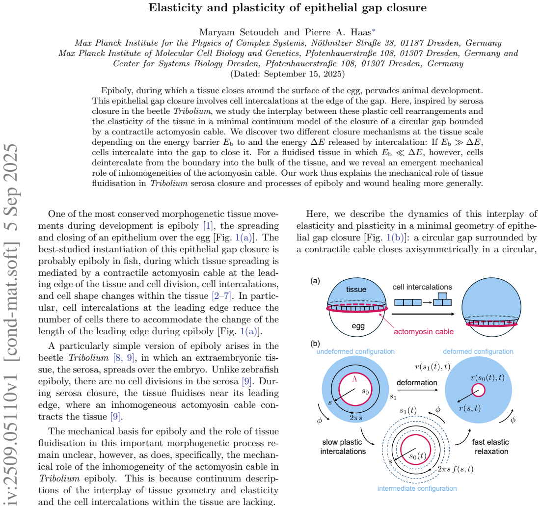
Epiboly, during which a tissue closes around the surface of the egg, pervades animal development. This epithelial gap closure involves cell intercalations at the edge of the gap. Here, inspired by serosa closure in the beetle Tribolium, we study the interplay between these plastic cell rearrangements and the elasticity of the tissue in a minimal continuum model of the closure of a circular gap bounded by a contractile actomyosin cable. We discover two different closure mechanisms at the tissue scale depending on the energy barrier $E_\text{b}$ to and the energy $\Delta E$ released by intercalation: If $E_\text{b}\gg\Delta E$, cells intercalate into the gap to close it. For a fluidised tissue in which $E_\text{b}\ll\Delta E$, however, cells deintercalate from the boundary into the bulk of the tissue, and we reveal an emergent mechanical role of inhomogeneities of the actomyosin cable. Our work thus explains the mechanical role of tissue fluidisation in Tribolium serosa closure and processes of epiboly and wound healing more generally.
Editorial analysis
A structured set of objections, weighed in public.
Referee Report
Summary. The manuscript develops a minimal continuum model of circular epithelial gap closure bounded by a contractile actomyosin cable, motivated by serosa closure in Tribolium. Plasticity is encoded via a single energy barrier Eb to cell intercalation and an energy release ΔE upon intercalation. The central result is a bifurcation in closure mechanism: when Eb ≫ ΔE cells intercalate into the gap, whereas when Eb ≪ ΔE (fluidised regime) cells deintercalate from the boundary into the tissue bulk, with inhomogeneities in the actomyosin cable acquiring an emergent mechanical role.
Significance. If the two-regime distinction and the emergent role of cable inhomogeneities survive scrutiny, the work supplies a compact mechanical explanation for how tissue fluidisation modulates gap closure in epiboly and wound healing, linking a simple energy-barrier model to observable cell behaviours at the tissue scale.
major comments (1)
- [Model setup] Model setup / continuum limit: the claim that cells deintercalate from the gap boundary when Eb ≪ ΔE is generated inside a coarse-grained description that ties the plastic strain rate to an effective potential whose minimum shifts with the Eb/ΔE ratio. Because the continuum smoothing erases discrete cell–cell and cell–cable contacts that set the local force balance at the free edge, it is unclear whether the predicted direction of plastic flow is robust or an artefact of the coarse-graining. This issue is load-bearing for the fluidised-regime mechanism.
minor comments (1)
- [Abstract] The abstract states the qualitative outcomes clearly but does not indicate the explicit form of the energy functional or the constitutive relation for the plastic strain rate; adding one sentence on these points would improve accessibility without lengthening the abstract.
Simulated Author's Rebuttal
We thank the referee for their detailed reading of our manuscript and for identifying a key point concerning the robustness of the continuum description in the fluidised regime. We address this comment below and have revised the manuscript accordingly to strengthen the justification of our approach.
read point-by-point responses
-
Referee: [Model setup] Model setup / continuum limit: the claim that cells deintercalate from the gap boundary when Eb ≪ ΔE is generated inside a coarse-grained description that ties the plastic strain rate to an effective potential whose minimum shifts with the Eb/ΔE ratio. Because the continuum smoothing erases discrete cell–cell and cell–cable contacts that set the local force balance at the free edge, it is unclear whether the predicted direction of plastic flow is robust or an artefact of the coarse-graining. This issue is load-bearing for the fluidised-regime mechanism.
Authors: We agree that the continuum limit requires careful justification, as averaging necessarily smooths discrete cell–cell and cell–cable interactions. In our model the plastic strain rate is obtained from the derivative of an effective potential whose minima are set by the ratio Eb/ΔE; this potential is constructed by coarse-graining the microscopic energy landscape of intercalation events. When Eb ≪ ΔE the minimum shifts to favour negative plastic strain (deintercalation), reflecting a thermodynamic preference for reducing the number of boundary cells. The contractile cable enters through the boundary stress condition, which remains well-defined after averaging. While local force fluctuations at individual contacts are lost, the net direction of plastic flow is preserved because it is dictated by the global energy balance rather than by any single contact geometry. To make this explicit we have added a paragraph in the Model section together with a supplementary derivation that starts from a discrete energy functional and shows that the sign of the coarse-grained flow is unchanged under spatial averaging. We have also included a brief discussion of the regime of validity of the continuum approximation. revision: partial
Circularity Check
No significant circularity; outcomes follow from independent model parameters
full rationale
The paper presents a minimal continuum model in which Eb and ΔE are introduced as independent parameters whose ratio determines the direction of plastic flow (intercalation into the gap when Eb ≫ ΔE versus deintercalation when Eb ≪ ΔE). These regimes and the emergent role of actomyosin inhomogeneities are obtained by solving the model's equations rather than by fitting to data or by any self-referential definition. No load-bearing self-citations, uniqueness theorems, or ansatzes imported from prior work are invoked to force the central claims; the derivation chain remains self-contained against the stated modeling assumptions.
Axiom & Free-Parameter Ledger
free parameters (2)
- Eb
- ΔE
axioms (1)
- domain assumption The tissue can be described by a minimal continuum model that couples elasticity to discrete plastic rearrangements via an energy barrier.
Lean theorems connected to this paper
-
IndisputableMonolith/Cost/FunctionalEquation.leanwashburn_uniqueness_aczel unclear?
unclearRelation between the paper passage and the cited Recognition theorem.
We discover two different closure mechanisms ... depending on the energy barrier Eb to and the energy Delta E released by intercalation: If Eb ≫ Delta E, cells intercalate into the gap ... For a fluidised tissue in which Eb ≪ Delta E, however, cells deintercalate ...
-
IndisputableMonolith/Foundation/AlphaCoordinateFixation.leancostAlphaLog_high_calibrated_iff unclear?
unclearRelation between the paper passage and the cited Recognition theorem.
the elastic energy density ... e(s,t)=C/2[Es^2 + Es Ephi + Ephi^2] f(s,t)
What do these tags mean?
- matches
- The paper's claim is directly supported by a theorem in the formal canon.
- supports
- The theorem supports part of the paper's argument, but the paper may add assumptions or extra steps.
- extends
- The paper goes beyond the formal theorem; the theorem is a base layer rather than the whole result.
- uses
- The paper appears to rely on the theorem as machinery.
- contradicts
- The paper's claim conflicts with a theorem or certificate in the canon.
- unclear
- Pith found a possible connection, but the passage is too broad, indirect, or ambiguous to say the theorem truly supports the claim.
Reference graph
Works this paper leans on
-
[1]
Solnica-Krezel, Conserved patterns of cell movements during vertebrate gastrulation, Curr
L. Solnica-Krezel, Conserved patterns of cell movements during vertebrate gastrulation, Curr. Biol. 15, R213 (2005)
work page 2005
-
[2]
R. E. Keller and J. P. Trinkaus, Rearrangement of en- veloping layer cells without disruption of the epithelial permeability barrier as a factor in Fundulus epiboly, Dev. Biol. 120, 12 (1987)
work page 1987
-
[3]
M. K¨ oppen, B. G. Fern´ andez, L. Carvalho, A. Jacinto, and C.-P. Heisenberg, Coordinated cell-shape changes control epithelial movement in zebrafish and Drosophila, Development 133, 2671 (2006)
work page 2006
-
[4]
S. E. Lepage and A. E. E. Bruce, Zebrafish epiboly: mechanics and mechanisms, Int. J. Dev. Biol. 54, 1213 (2010)
work page 2010
-
[5]
M. Behrndt, G. Salbreux, P. Campinho, R. Hauschild, F. Oswald, J. Roensch, S. W. Grill, and C.-P. Heisenberg, Forces driving epithelial spreading in zebrafish gastrula- tion, Science 338, 257 (2012)
work page 2012
-
[6]
P. Campinho, M. Behrndt, J. Ranft, T. Risler, N. Minc, and C.-P. Heisenberg, Tension-oriented cell divisions limit anisotropic tissue tension in epithelial spreading during zebrafish epiboly, Nat. Cell Biol. 15, 1405 (2013)
work page 2013
-
[7]
A. E. E. Bruce and C.-P. Heisenberg, Mechanisms of ze- brafish epiboly: A current view, in Gastrulation: From Embryonic Pattern to Form , Current Topics in De- velopmental Biology, Vol. 136, edited by L. Solnica- Krezel (Academic Press, Elsevier, London, England,
-
[8]
Chap. 11, pp. 319–341
-
[9]
M. A. Benton, M. Akam, and A. Pavlopoulos, Cell and tissue dynamics during Tribolium embryogenesis revealed by versatile fluorescence labeling approaches, Develop- ment 140, 3210 (2013)
work page 2013
-
[10]
A. Jain, V. Ulman, A. Mukherjee, M. Prakash, M. B. Cuenca, L. G. Pimpale, S. M¨ unster, R. Haase, K. A. Pan- filio, F. Jug, S. W. Grill, P. Tomanˇ c´ ak, and A. Pavlopou- los, Regionalized tissue fluidization is required for epithe- lial gap closure during insect gastrulation, Nat. Commun. 11, 5604 (2020)
work page 2020
- [11]
-
[12]
P. Marmottant, A. Mgharbel, J. K¨ afer, B. Audren, J.- P. Rieu, J.-C. Vial, B. van der Sanden, A. F. M. Mar´ ee, F. Graner, and H. Delano¨ e-Ayari, The role of fluctuations and stress on the effective viscosity of cell aggregates, Proc. Natl. Acad. Sci. USA 106, 17271 (2009)
work page 2009
-
[13]
D. Bi, J. H. Lopez, J. M. Schwarz, and M. L. Manning, Energy barriers and cell migration in densely packed tis- sues, Soft Matter 10, 1885 (2014)
work page 2014
- [14]
-
[15]
M. Popovi´ c, V. Druelle, N. A. Dye, F. J¨ ulicher, and M. Wyart, Inferring the flow properties of epithelial tissues from their geometry, New J. Phys. 23, 033004 (2021)
work page 2021
- [16]
-
[17]
D. Bi, J. H. Lopez, J. M. Schwarz, and M. L. Manning, A density-independent rigidity transition in biological tis- sues, Nat. Phys. 11, 1074 (2015)
work page 2015
- [18]
-
[19]
M. Krajnc, Solid–fluid transition and cell sorting in ep- ithelia with junctional tension fluctuations, Soft Matter 16, 3209 (2020)
work page 2020
- [20]
- [21]
-
[22]
T. Yamamoto, D. M. Sussman, T. Shibata, and M. L. Manning, Non-monotonic fluidization generated by fluc- 6 tuating edge tensions in confluent tissues, Soft Matter 18, 2168 (2022)
work page 2022
-
[23]
H. P. Jain, A. Voigt, and L. Angheluta, From cell inter- calation to flow, the importance of T1 transitions, Phys. Rev. Res. 6, 033176 (2024)
work page 2024
-
[24]
M. F. Staddon and C. D. Modes, Curved-edge vertex models and increased tissue fluidity, Phys. Rev. Res. 7, 013218 (2025)
work page 2025
-
[25]
E. Hannezo and C.-P. Heisenberg, Rigidity transitions in development and disease, Trends Cell Biol. 32, 433 (2022)
work page 2022
-
[26]
P.-F. Lenne and V. Trivedi, Sculpting tissues by phase transitions, Nat. Commun. 13, 664 (2022)
work page 2022
-
[27]
Lubliner, Plasticity Theory , revised ed
J. Lubliner, Plasticity Theory , revised ed. (Dover, Mine- ola, NY, 2008) Chap. 8, pp. 465–499
work page 2008
- [28]
-
[29]
Goriely, The Mathematics and Mechanics of Biological Growth (Springer, Berlin, Germany, 2017) Chap
A. Goriely, The Mathematics and Mechanics of Biological Growth (Springer, Berlin, Germany, 2017) Chap. 11–12, pp. 261–373
work page 2017
-
[30]
See Supplemental Material at [url to be inserted], which includes Refs. [26–28, 30 –38], for (i) the derivation of the elastic energy density, (ii) details of the calculation of the intercalation rates, and (iii) details of the numerical solution of the governing equations, including additional simulation results
-
[31]
D. J. Steigmann, Koiter’s shell theory from the perspec- tive of three-dimensional nonlinear elasticity, J. Elasticity 111, 91 (2013)
work page 2013
-
[32]
P. A. Haas and R. E. Goldstein, Morphoelasticity of large bending deformations of cell sheets during development, Phys. Rev. E 103, 022411 (2021)
work page 2021
-
[33]
R. G. Ramachandran, R. Alert, and P. A. Haas, Buckling by disordered growth, Phys. Rev. E 110, 054405 (2024)
work page 2024
-
[34]
H. A. Erbay, On the asymptotic membrane theory of thin hyperelastic plates, Int. J. Eng. Sci. 35, 151 (1997)
work page 1997
-
[35]
J. Dervaux and M. Ben Amar, Morphogenesis of growing soft tissues, Phys. Rev. Lett. 101, 068101 (2008)
work page 2008
-
[36]
R. W. Ogden, Non-linear elastic deformations (Dover, Mineola, NY, 1997) Chap. 2.2, 3.4, pp. 83–121, 152–155
work page 1997
-
[37]
J. Dervaux, P. Ciarletta, and M. Ben Amar, Morpho- genesis of thin hyperelastic plates: A constitutive theory of biological growth in the F¨ oppl–von K´ arm´ an limit, J. Mech. Phys. Solids 57, 458 (2009)
work page 2009
-
[38]
M. Abramowitz and I. A. Stegun, Handbook of Mathe- matical Functions, Applied Mathematics Series, Vol. 55 (National Bureau of Standards, Washington, DC, 1964) Chap. 25.5, pp. 896–897
work page 1964
-
[39]
W. H. Press, S. A. Teukolsky, W. T. Vetterling, and B. P. Flannery, Numerical Recipes: The Art of Scientific Com- puting, 3rd ed. (Cambridge University Press, Cambridge, England, 2007) Chap. 20.1, pp. 1031–1043
work page 2007
- [40]
-
[41]
J. H. Espenson, Chemical Kinetics and Reaction Mecha- nisms (McGraw-Hill, New York, NY, 1981) Chap. 8, pp. 150–165
work page 1981
-
[42]
Stirzaker, Elementary Probability, 2nd ed
D. Stirzaker, Elementary Probability, 2nd ed. (Cambridge University Press, Cambridge, UK, 2003) Chap. 4.6, pp. 131–134
work page 2003
-
[43]
A. Brugu´ es, E. Anon, V. Conte, J. H. Veldhuis, M. Gupta, J. Colombelli, J. J. Mu˜ noz, G. W. Brod- land, B. Ladoux, and X. Trepat, Forces driving epithelial wound healing, Nat. Phys. 10, 683 (2014)
work page 2014
-
[44]
S. R. K. Vedula, G. Peyret, I. Cheddadi, T. Chen, A. Brugu´ es, H. Hirata, H. Lopez-Menendez, Y. Toyama, L. Neves de Almeida, X. Trepat, C. T. Lim, and B. Ladoux, Mechanics of epithelial closure over non- adherent environments, Nat. Commun. 6, 6111 (2015)
work page 2015
-
[45]
S. Begnaud, T. Chen, D. Delacour, R.-M. M` ege, and B. Ladoux, Mechanics of epithelial tissues during gap clo- sure, Curr. Opin. Cell Biol. 42, 52 (2016)
work page 2016
-
[46]
N. K. Babu, M. Sreepadmanabh, S. Dutta, and T. Bhat- tacharjee, Interplay of geometry and mechanics in epithe- lial wound healing, Phys. Rev. E 110, 054411 (2024)
work page 2024
-
[47]
S. E. Lim, P. Vicente-Munuera, and Y. Mao, Forced back into shape: Mechanics of epithelial wound repair, Curr. Opin. Cell Biol. 87, 102324 (2024)
work page 2024
-
[48]
V. Movrin and M. Krajnc, Initiation of epithelial wound closure by an active instability at the purse string, Bio- phys. J. 124, 107 (2025)
work page 2025
-
[49]
R. J. Tetley, M. F. Staddon, D. Heller, A. Hoppe, S. Banerjee, and Y. Mao, Tissue fluidity promotes ep- ithelial wound healing, Nat. Phys. 15, 1195 (2019)
work page 2019
-
[50]
R. M. Sarate, J. Hochstetter, M. Valet, A. Hallou, Y. Song, N. Bansaccal, M. Ligare, M. Aragona, D. En- gelman, A. Bauduin, O. Camp` as, B. D. Simons, and C. Blanpain, Dynamic regulation of tissue fluidity con- trols skin repair during wound healing, Cell 187, 5298 (2024)
work page 2024
-
[51]
Code is available at zenodo:17062742. Elasticity and plasticity of epithelial gap closure • Supplemental Material • Maryam Setoudeh and Pierre A. Haas Max Planck Institute for the Physics of Complex Systems, N ¨othnitzer Straße 38, 01187 Dresden, Germany Max Planck Institute of Molecular Cell Biology and Genetics, Pfotenhauerstraße 108, 01307 Dresden, Ger...
-
[52]
Results for this boundary conditions are shown in Fig. S2 and only differ at a quantitative level: the boundary conditions thus have but a slight effect on the dynamics of epiboly. [S1] D. J. Steigmann, Koiter’s shell theory from the perspective of three-dimensional nonlinear elasticity, J. Elasticity 111, 91 (2013). [S2] P . A. Haas and R. E. Goldstein, Mo...
work page 2013
- [53]
discussion (0)
Sign in with ORCID, Apple, or X to comment. Anyone can read and Pith papers without signing in.