REVIEW 6 major objections 5 minor 38 references
A continuous-variable quantum model matches classical medical-image classifiers
Reviewed by Pith at T0; open to challenge. T0 means a machine referee read the full paper against a public rubric. the ladder, T0–T4 →
T0 review · deepseek-v4-flash
2026-08-04 00:14 UTC pith:YVH5766N
load-bearing objection A plausible feasibility result undercut by misreported tables: the comparable-performance claim cannot be trusted until Table 3 is reconciled with the text. the 6 major comments →
Towards Continuous-variable Quantum Neural Networks for Biomedical Imaging
The pith
A machine-rendered reading of the paper's core claim, the machinery that carries it, and where it could break.
Core claim
On its own terms, the paper's discovery is that a 4-qumode Gaussian circuit—displacement gates encoding four PCA components, alternating rotation and squeezing gates, beamsplitter entangling layers, and position-quadrature measurements feeding a linear head—produces test-set F1 scores of about 85% on pneumonia chest X-rays, 75% on breast ultrasound, and 45% on an 11-class organ CT task. These numbers sit within a few points of a same-parameter classical network and a same-parameter discrete-variable quantum circuit, and the CV model is the most robust of the three when Gaussian noise is added to the images. The authors interpret this as evidence that continuous-variable encoding offers compa
What carries the argument
The central object is a continuous-variable variational circuit whose modes are quantum harmonic oscillators (qumodes). Data enters through displacement gates; trainable rotation and squeezing gates reshape the phase-space state; beamsplitter gates entangle adjacent modes; and the model output is the vector of position-quadrature expectation values. All gates are Gaussian, so the circuit functions as a learned linear-optical feature map, and the trainable parameters number 42 including the classical output layer.
Load-bearing premise
The experiments stand or fall on the assumption that four PCA components keep enough diagnostic information: the paper itself reports retaining only about 60% of variance for the two binary datasets and 48% for the organ dataset.
What would settle it
Train the same classical linear head on the original 784-pixel images, or on PCA with 8–16 components, and compare accuracy on the organ dataset; if accuracy climbs well above 47%, the reported parity among CV, DV, and classical models is an artifact of the compression bottleneck rather than a property of the quantum circuits.
If this is right
- CV and DV quantum models perform within statistical noise of a classical network on all three imaging tasks at the same 42-parameter scale.
- The CV model holds up better than the DV model when Gaussian noise is added, staying closer to the classical model's stability across the full noise range.
- On the 11-class organ task, the CV model beats the DV model by roughly six to seven points in F1, suggesting continuous encoding helps as class count grows.
- On the small imbalanced breast dataset, the CV model correctly identifies substantially more true negatives than the DV or classical models, pointing to a minority-class focus.
- Decision heatmaps differ across paradigms, with the CV model attending to more localized regions, which the authors link to better interpretability for clinical use.
Where Pith is reading between the lines
- Because the circuit uses only Gaussian gates, it is efficiently simulable classically; the value shown here is representational, not computational speedup. A non-Gaussian gate, such as a cubic-phase gate, would be needed to test whether an actual quantum advantage appears.
- The PCA bottleneck at 48–60% retained variance likely bounds every result; a natural test is to rerun the same models with 8 or 16 components, or on raw images, to see whether the classical baseline pulls ahead once more signal is available.
- If the CV model's minority-class advantage holds under less aggressive compression, it could matter for screening tasks where false negatives are costly, since averaged accuracy hides that asymmetry.
- The Gaussian layer is essentially a learned linear-optical transformation; the right control is a classical linear model on the same PCA features, and a useful extension would be a nonlinear classical model to clarify what the quantum feature map adds.
Editorial analysis
A structured set of objections, weighed in public.
Referee Report
Summary. This paper reports a feasibility study of a continuous-variable (CV) quantum neural network for biomedical image classification. The model is a 4-qumode photonic circuit composed of Gaussian gates (displacement, rotation, squeezing, beamsplitter) with position-quadrature expectation measurements feeding a 10-parameter classical linear head (42 parameters total). Inputs are 4-dimensional PCA projections of 28x28 grayscale images from three MedMNIST subsets: PneumoniaMNIST (binary), BreastMNIST (small, imbalanced binary), and OrganAMNIST (11-class multiclass). The CV model is compared with a nominally matched 4-qubit DV quantum circuit and a classical 'same-scale' network under identical hyperparameters, using accuracy/precision/recall/F1, AUROC/AUPRC, Gaussian noise robustness, Grad-CAM heatmaps, and Friedman/Wilcoxon hypothesis tests. The central claim (Section 6) is that the CV QNN attains performance comparable to the classical and DV counterparts while showing higher noise robustness, minority-class focus, and multiclass classification advantages; the paper explicitly disclaims quantum advantage.
Significance. Strengths: the comparison is controlled at a fixed parameter scale with identical hyperparameters, fixed seeds, and the same PCA-compressed inputs across three datasets of different task structure; the paper honestly states that no quantum advantage is demonstrated; and the topic — CV quantum models on medical imaging — is genuinely underexplored, so a careful feasibility benchmark would be useful if the numbers are reliable. The significance is currently bounded by four structural limitations: the Gaussian circuit is affine in its inputs and classically simulable (so the model is effectively linear); the 4-component PCA retains only 48-60% of the variance; test metrics come from a single chosen fold; and most importantly the reported numbers are internally inconsistent — Table 3 disagrees with the text and Table 2 contains impossible p-values. The stress-test concern about Table 3 is confirmed on reading the manuscript. Until these points are resolved, the quantitative content of the central claim is not established.
major comments (6)
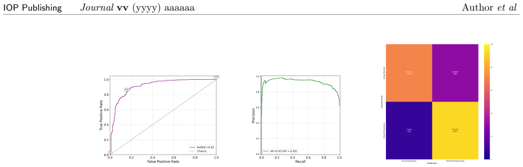
- [Section 4, Experiments 1-3; Table 3] Table 3 cannot be reconciled with the prose of Experiments 1-3, and both conflict with Table 2 in places. (i) Breast CV: text gives ACC=R=F1=75.64%, P=73.17%; Table 3 lists P=0.7564, R=0.7317 (P/R swapped). (ii) Pneumonia CV: text gives ACC=R=F1=84.29%, P=84.37%; Table 3 lists P=0.8429, R=0.8437 (again swapped). (iii) Organ CV: text gives ACC=R=F1=45.63%; Table 3 lists R=0.4257. (iv) Breast DV: text reports P=80.67%; 0.8067 appears nowhere in Table 3. (v) Experiment 1 reports CV Pneumonia validation accuracy 89.67%, while Table 2 lists 0.864±0.059. Since Section 6's quantitative claims ('comparable performance,' 'minority class focus,' '+7% F1') are built on these tables, the metrics must be recomputed from the confusion matrices and reconciled with the text before the central claim is evaluable. The 'PENDING' code repository prevents independent verification.
- [Section 4, Experiments 1-3; Table 3] All test-set metrics are reported for the single best fold of the threefold cross-validation, with no variance or confidence intervals. The Section 6 claims rest on small between-model differences (e.g., 84.29% vs 85.40% on Pneumonia; 75.64% vs 76.28% on Breast) that are comparable to the fold-to-fold standard deviations in Table 2 (e.g., CV Pneumonia ACC 0.864±0.059). Selecting the best fold post hoc inflates expected performance and makes the claimed parity/advantages unquantified. Report test metrics per fold or as mean±std across folds, as is already done for the validation metrics in Table 2.
- [Section 4, Experiment 6; Table 2] The statistical analysis has three problems. First, the p column of Table 2 repeats the chi-square statistic in the Pneumonia and Organ rows (e.g., χ²=0.441, p=0.441; the true p for χ²(2)=0.441 is ≈0.80). Second, with only three folds the exact two-sided Wilcoxon signed-rank p can take only the values 0.25, 0.5, 0.75, 1.0; the table reports 0.111, 0.180, 0.273, 0.778, which are impossible for n=3. Third, the Bonferroni-corrected threshold 0.0167 is below the minimum achievable Wilcoxon p at n=3, so non-rejection is guaranteed by construction. 'No statistically significant difference' is therefore vacuous and cannot serve as evidence for the 'comparable performance' conclusion in Section 6.
- [Section 3 vs Section 4, Experiment 1] The classical baseline on which the central parity claim rests is never defined. Section 3 specifies the CV and DV circuits (42 parameters each) but contains no classical architecture; Section 4 states only that 'a classical counterpart with an identical architecture and number of parameters is trained.' A 42-parameter 'identical' classical network is not described (a 4x2 linear head has only 10 parameters), so the reader cannot determine what function class the classical model represents, how it compares structurally with the affine Gaussian circuit, or whether the comparison is fair. The classical architecture (layers, nonlinearities, parameter count) must be specified.
- [Section 3.1, Table 1; Section 6; Abstract] The 4-component PCA projection retains only ~60% (Breast, Pneumonia) and ~48% (Organ) of the variance, and Section 3.1 itself notes that OrganAMNIST 'cannot be represented with only 4 components per sample.' Because all three models consume the same 4-dimensional projection, the reported accuracies largely measure the PCA bottleneck, and the near-parity across models is partly forced by the shared input. This does not invalidate the comparative claim 'under this configuration,' but it does not support the abstract's 'viability for future computer-aided diagnosis systems' or Section 6's 'promise in clinical practice' at test accuracies of 45-85%, far below common MedMNIST baselines. Re-scope the conclusions or add experiments at higher k.
- [Section 3.2; Section 4, Experiment 6; Abstract] The CV circuit uses only Gaussian gates (D, R, S, BS) and position-quadrature measurements, so as a function of the 4 PCA inputs it implements an affine map (followed by the affine classical head) and is efficiently classically simulable. The paper acknowledges at the end of Experiment 6 that 'the results fail to demonstrate quantum advantage,' yet the same paragraph asserts that 'the CV quantum model offers higher representational capacity for structured and imbalanced biomedical data,' and the abstract lists 'model expressiveness' as an evaluated property. No expressivity measure is reported, and a Gaussian circuit has the same function class as a linear model; the 'feature extraction'/'feature amplification' language of Section 3.2 is correspondingly overstated. Recommend stating the affine/simulable nature explicitly and tempering these claims, or adding non-Gaussian gates.
minor comments (5)
- [Section 3.2, Figure 2] The qumode diagram appears garbled in the text: the gate sequences shown for different modes are inconsistent with each other and with the stated two-layer repeated structure. Please redraw and align with the parameter count.
- [Sections 3.2 and 3.3, parameter tables] The displacement gate (CV) and first RY gate (DV) are described as data encoding, yet the per-layer gate count (2×4×4) counts them as trainable parameters; the beamsplitter is two-mode but is counted per-mode. Clarify the split between data-encoding and trainable parameters in both quantum models.
- [Section 4, Experiment 5; Section 6] Applying Grad-CAM to 4-dimensional PCA inputs produces heatmaps that are at most linear combinations of the four retained eigenimages; the Section 6 claims of a 'more comprehensible heatmap' and 'better output interpretability' should be tempered or the method validated on the PCA bottleneck.
- [Declarations, Code availability] The code repository is listed as 'PENDING' although the introduction promises release of 'all code, trained weights, and logs to ensure transparency and replication.' This must be resolved for the reproducibility claim to hold.
- [Throughout] Typos and infelicities: 'apodt' (Eq. 19), 'Simlarly' (Section 2), 'analogoues' (Section 3.2), 'as as the following' (Eq. 12), 'OrganMNIST' for OrganAMNIST (Section 4, Experiment 6), and a missing closing bracket in Eq. (17): d(FPR(σ).
Circularity Check
No circularity: the paper is an empirical feasibility study on external MedMNIST benchmarks; central claims rest on measured results and statistical tests, not on construction.
full rationale
The paper makes no formal derivation claim that could reduce to its inputs. Section 3 constructs the CV and DV circuits from standard Gaussian and discrete-variable gates, and Section 4 reports measured train/validation/test metrics. The central claim of comparable performance is supported by empirical results and by Friedman/Wilcoxon hypothesis tests, not by a derivation from the model definitions. The PCA encoder in Section 3.1 is fitted on the training data only, and the reported test-set metrics are predictions on held-out data, so no fitted parameter is renamed as a prediction. No load-bearing step relies on a self-citation or an imported uniqueness theorem; the cited prior work on CV QML provides background and architecture inspiration, but the proposed circuits, training, and evaluation are self-contained. The observed inconsistencies between Table 3 and the experimental text, and the 'PENDING' code repository, are serious reproducibility and correctness concerns, but they are not circularity: they do not show that any claimed result is equivalent to its inputs by construction. Therefore no circular steps are identified.
Axiom & Free-Parameter Ledger
free parameters (4)
- Number of PCA components (k=4) =
4
- Trainable circuit and classical-head parameters =
42 per model
- Circuit depth / number of variational layers =
2
- Noise range for robustness test =
sigma in [0.1, 1.0] in steps of 0.05
axioms (4)
- domain assumption PennyLane's Gaussian backend (with Strawberry Fields plugin) faithfully simulates the CV quantum circuit dynamics.
- domain assumption 4 PCA components preserve enough class-discriminative information for classification.
- domain assumption Threefold cross-validation F1 scores provide enough statistical power for Friedman/Wilcoxon comparisons.
- ad hoc to paper Grad-CAM can be meaningfully applied to PCA-reconstructed 4-dimensional inputs through quantum circuits.
read the original abstract
Continuous-variable (CV) quantum computing offers a promising framework for scalable quantum machine learning, leveraging optical systems with infinite-dimensional Hilbert spaces. While discrete-variable (DV) quantum neural networks have shown remarkable progress in various computer vision tasks, CV quantum models remain comparatively underexplored. In this work, we present a feasibility study of continuous-variable quantum neural networks (CV-QCNNs) applied to biomedical image classification. Utilizing photonic circuit simulation frameworks, we construct CV quantum circuits composed of Gaussian gates, such as displacement, squeezing, rotation, and beamsplitters to emulate convolutional behavior. Our experiments are conducted on the MedMNIST dataset collection, a set of annotated medical image benchmarks for multiple diagnostic tasks. We evaluate CV-QCNN's performance in terms of classification accuracy, model expressiveness, and resilience to Gaussian noise, comparing against classical CNNs and equivalent DV quantum circuits. This study aims to identify trade-offs between DV and CV paradigms for quantum-enhanced medical imaging. Our results highlight the potential of continuous-variable models and their viability for future computer-aided diagnosis systems.
Figures




Reference graph
Works this paper leans on
-
[1]
Esteva A, Kuprel B, Novoa R A, Ko J, Swetter S M, Blau H M and Thrun S 2017Nature 542(7639) 115–118 ISSN 1476-4687 URLhttps://pubmed.ncbi.nlm.nih.gov/28117445/
-
[3]
Wang L, Lin Z Q and Wong A 2020Scientific Reports 2020 10:110(1) 1–12 ISSN 2045-2322 URLhttps://www.nature.com/articles/s41598-020-76550-z
2020
-
[4]
Lecun Y, Bengio Y and Hinton G 2015Nature 2015 521:7553521(7553) 436–444 ISSN 1476-4687 URLhttps://www.nature.com/articles/nature14539
2015
-
[5]
Dunjko V and Briegel H J 2017 URLhttps://arxiv.org/abs/1709.02779v1 18 IOP PublishingJournalvv(yyyy) aaaaaa Authoret al
Pith/arXiv arXiv 2017
-
[6]
Nielsen M A and Chuang I L 2010Quantum Computation and Quantum Information
-
[7]
Shor P W 1996SIAM Journal on Computing26(5) 1484–1509 URLhttp: //arxiv.org/abs/quant-ph/9508027http://dx.doi.org/10.1137/S0097539795293172
-
[8]
Grover L K 1996Proceedings of the Annual ACM Symposium on Theory of ComputingPart F129452212–219 ISSN 07378017 URLhttps://arxiv.org/abs/quant-ph/9605043v3
-
[9]
15, Page 9415(3) 94 ISSN 1999-5903 URLhttps: //www.mdpi.com/1999-5903/15/3/94/htmhttps://www.mdpi.com/1999-5903/15/3/94
Rasool R U, Ahmad H F, Rafique W, Qayyum A, Qadir J and Anwar Z 2023Future Internet 2023, Vol. 15, Page 9415(3) 94 ISSN 1999-5903 URLhttps: //www.mdpi.com/1999-5903/15/3/94/htmhttps://www.mdpi.com/1999-5903/15/3/94
2023
-
[10]
Ullah U and Garcia-Zapirain B 2024IEEE Access1211423–11450 ISSN 21693536
-
[11]
Schuld M, Sinayskiy I and Petruccione F 2014Contemporary Physics56(2) 172–185 URL http://arxiv.org/abs/1409.3097http://dx.doi.org/10.1080/00107514.2014.964942
Pith/arXiv arXiv 2014
-
[12]
Schuld M and Petruccione F 2018 URL https://link.springer.com/10.1007/978-3-319-96424-9
-
[13]
Killoran N, Bromley T R, Arrazola J M, Schuld M, Quesada N and Lloyd S 2018Physical Review Research1(3) URLhttp://arxiv.org/abs/1806.06871http: //dx.doi.org/10.1103/PhysRevResearch.1.033063
-
[14]
Al-Dhabyani W, Gomaa M, Khaled H and Fahmy A 2020Data in Brief28104863 ISSN 2352-3409
-
[15]
Bilic P, Christ P, Li H B, Vorontsov E, Ben-Cohen A, Kaissis G, Szeskin A, Jacobs C, Mamani G E H, Chartrand G, Loh¨ ofer F, Holch J W, Sommer W, Hofmann F, Hostettler A, Lev-Cohain N, Drozdzal M, Amitai M M, Vivantik R, Sosna J, Ezhov I, Sekuboyina A, Navarro F, Kofler F, Paetzold J C, Shit S, Hu X, Lipkov´ a J, Rempfler M, Piraud M, Kirschke J, Wiestler...
Pith/arXiv arXiv 1901
-
[16]
Kermany D S, Goldbaum M, Cai W, Valentim C C, Liang H, Baxter S L, McKeown A, Yang G, Wu X, Yan F, Dong J, Prasadha M K, Pei J, Ting M, Zhu J, Li C, Hewett S, Dong J, Ziyar I, Shi A, Zhang R, Zheng L, Hou R, Shi W, Fu X, Duan Y, Huu V A, Wen C, Zhang E D, Zhang C L, Li O, Wang X, Singer M A, Sun X, Xu J, Tafreshi A, Lewis M A, Xia H and Zhang K 2018Cell17...
-
[17]
Yang J, Shi R, Wei D, Liu Z, Zhao L, Ke B, Pfister H and Ni B 2023Scientific Data1041
-
[18]
Schuld M and Killoran N 2019Physical Review Letters122(4) 040504 ISSN 10797114 URL https://journals.aps.org/prl/abstract/10.1103/PhysRevLett.122.040504
-
[19]
Sengupta K and Srivastava P R 2021BMC Medical Informatics and Decision Making21(1) 1–14 ISSN 14726947 URLhttps: //bmcmedinformdecismak.biomedcentral.com/articles/10.1186/s12911-021-01588-6
-
[20]
Abdulsalam G, Meshoul S and Shaiba H 2022Intelligent Automation & Soft Computing36(1) 761–779 ISSN 1079-8587 URL https://www.techscience.com/iasc/v36n1/50016.htmlhttps: //www.techscience.com/iasc/v36n1/50016
-
[21]
Sagingalieva A, Kordzanganeh M, Kenbayev N, Kosichkina D, Tomashuk T and Melnikov A 2023Cancers 2023, Vol. 15, Page 270515(10) 2705 ISSN 2072-6694 URL https://www.mdpi.com/2072-6694/15/10/2705/htmhttps: //www.mdpi.com/2072-6694/15/10/2705 19 IOP PublishingJournalvv(yyyy) aaaaaa Authoret al
2023
-
[22]
Reka S S, Karthikeyan H L, Shakil A J, Venugopal P and Muniraj M 2024IEEE Access12 104568–104584 ISSN 21693536
-
[23]
Ara T, Mishra V P, Bali M and Yenkikar A 2025MethodsX15103605 ISSN 2215-0161
-
[24]
5, Page 515(3) 51 ISSN 2673-7426 URL https://www.mdpi.com/2673-7426/5/3/51/htmhttps://www.mdpi.com/2673-7426/5/3/51
Marzoug N, Halab K, Meslouhi O E, Elassad Z E A and Akhloufi M A 2025 BioMedInformatics 2025, Vol. 5, Page 515(3) 51 ISSN 2673-7426 URL https://www.mdpi.com/2673-7426/5/3/51/htmhttps://www.mdpi.com/2673-7426/5/3/51
2025
-
[25]
Lau H K, Pooser R, Siopsis G and Weedbrook C 2017Physical Review Letters118(8) 080501 ISSN 10797114 URL https://journals.aps.org/prl/abstract/10.1103/PhysRevLett.118.080501
-
[26]
Moody G, Sorger V J, Blumenthal D J, al, Ding C, Xu X Y, Niu Y F, Arrazola J M, Bromley T R, Izaac J, Myers C R, Br´ adler K and Killoran N 2019Quantum Science and Technology 4(2) 024004 ISSN 2058-9565 URL https://iopscience.iop.org/article/10.1088/2058-9565/aaf59ehttps: //iopscience.iop.org/article/10.1088/2058-9565/aaf59e/meta
-
[27]
Li L H, Zhang D B and Wang Z D 2022Physics Letters A436128088 ISSN 0375-9601
-
[28]
Ghasemian E, Razminia A and Rostami H 2023Quantum Information Processing22(10) 1–22 ISSN 15731332 URLhttps://link.springer.com/article/10.1007/s11128-023-04137-4
-
[29]
Kairon P and Bhattacharyya S 2021Advances in Intelligent Systems and Computing1279 113–123 ISSN 2194-5365 URL https://link.springer.com/chapter/10.1007/978-981-15-9290-4_12
-
[30]
Choe S 2022 URLhttps://arxiv.org/abs/2204.01194v1
Pith/arXiv arXiv 2022
-
[31]
Anand P, Chandra M G and Khandelwal A 20242024 16th International Conference on COMmunication Systems and NETworkS, COMSNETS 2024994–999
-
[32]
Hotelling H 1933Journal of Educational Psychology24417–441
-
[33]
Jolliffe I T and Cadima J 2016Philosophical Transactions of the Royal Society A: Mathematical, Physical and Engineering Sciences37420150202
-
[34]
Grandini M, Bagli E and Visani G 2020 URLhttps://arxiv.org/abs/2008.05756v1
Pith/arXiv arXiv 2020
-
[35]
Davis J and Goadrich M 2006 URLhttps://minds.wisconsin.edu/handle/1793/60482
2006
-
[36]
Villard´ on G, Zhao X, Le P B and Nguyen Z T 2022 URL https://doi.org/10.3390/math10091410
-
[37]
Selvaraju R R, Cogswell M, Das A, Vedantam R, Parikh D and Batra D 2017 Grad-cam: Visual explanations from deep networks via gradient-based localization2017 IEEE International Conference on Computer Vision (ICCV)pp 618–626
2017
-
[38]
Pineau J, Pu P, Snoek J, Wilson A Get al.2021Journal of Machine Learning Research22 1–20
-
[39]
Kingma D P and Ba J 2017International Conference on Learning Representations (ICLR) (Preprint1412.6980) 20 IOP PublishingJournalvv(yyyy) aaaaaa Authoret al (a) PneumoniaMNIST heatmaps (b) OrganAMNIST heatmaps (c) BreastMNIST heatmaps CV QNN heatmaps DV QNN heatmaps Classical NN heatmaps Figure 17.Comparison of Grad-CAM heatmaps across models and datasets....
discussion (0)
Sign in with ORCID, Apple, or X to comment. Anyone can read and Pith papers without signing in.