Formation of Light-Emitting Defects in Ag-based Memristors
Pith reviewed 2026-05-17 04:31 UTC · model grok-4.3
The pith
Defects formed during initial activation in silver-based memristors produce the observed light emission.
A machine-rendered reading of the paper's core claim, the machinery that carries it, and where it could break.
Core claim
In Ag-based in-plane memristors the electroluminescence originates from defects generated inside the switching matrix during device activation, and the early-stage formation together with the subsequent evolution of these defects can be followed by combining electrical stimulation with correlated electroluminescence and photoluminescence measurements.
What carries the argument
Correlated electrical stimulation with electroluminescence and photoluminescence measurements that track defect formation and evolution from the first activation cycle.
If this is right
- Tuning the activation voltage or pulse duration can adjust the density of light-emitting defects and thereby the emission brightness.
- Stabilizing the defects at an early stage enables repeatable multilevel optical memory states.
- Controlling defect formation supports integration of light emission into neuromorphic circuits that combine memory and optical signaling.
Where Pith is reading between the lines
- The same early-defect mechanism may operate in other metal-ion memristors, pointing to a general route for adding optical output to resistive switching devices.
- Repeating the correlated measurements over many switching cycles could show whether the defects anneal, migrate, or accumulate and thereby limit device lifetime.
- Material choices that slow or accelerate defect creation could be tested to set the emission wavelength range for specific neuromorphic applications.
Load-bearing premise
The observed electroluminescence is produced by defects created inside the switching matrix specifically during the initial device activation.
What would settle it
No detectable change in photoluminescence spectra that matches the onset of electroluminescence when electrical switching first occurs.
Figures
read the original abstract
Optical memristors are innovative devices that enable the integration of electro-optical functionalities - such as light modulation, multilevel optical memory, and nonvolatile reprogramming - into neuromorphic networks. Recently, their capabilities have expanded with the development of light-emitting memristors, which operate through various emission mechanisms. One notable process involves the electroluminescence of defects generated within the switching matrix during device activation. In this study, we explore the early-stage formation and evolution of the species responsible for light emission in Ag-based in-plane memristors. Our approach combines electrical stimulation with correlated optical electroluminescence and photoluminescence measurements. The findings provide valuable insights into controlling emission processes in memristors, paving the way for their integration as essential components in neuromorphic circuits.
Editorial analysis
A structured set of objections, weighed in public.
Referee Report
Summary. The manuscript reports an experimental investigation of Ag-based in-plane memristors in which correlated electrical stimulation, electroluminescence (EL), and photoluminescence (PL) measurements are used to track the early-stage formation and evolution of light-emitting defects generated inside the switching matrix during device activation. The authors conclude that these observations furnish insights into controlling emission processes and support integration of such devices into neuromorphic circuits.
Significance. If the attribution of the observed EL to activation-induced matrix defects can be placed on a secure experimental footing, the work would add a useful data point to the growing literature on light-emitting memristors. The combination of in-plane geometry with simultaneous electrical-optical probing is a reasonable experimental approach for the mesoscopic-physics community, and the topic aligns with current interest in multifunctional neuromorphic hardware.
major comments (2)
- [Abstract / Results] Abstract and Results sections: the central claim that the electroluminescence originates from defects generated within the switching matrix during activation is asserted without the supporting data required to establish causality. Specifically, the manuscript must show (i) that measurable EL appears only after the activation step, (ii) that the EL spectrum matches established defect signatures rather than Ag electrode or interface states, and (iii) that appropriate controls exclude pre-existing or extrinsic emission sources. None of these elements are supplied in the current text.
- [Methods / Figures] Methods and Figure captions: no raw spectra, error bars, or quantitative thresholds for activation are provided. Without these, it is impossible to assess whether the reported correlation between electrical switching and optical emission is statistically robust or reproducible across devices.
minor comments (2)
- [Abstract] The abstract is concise but would benefit from one or two quantitative statements (e.g., onset voltage, spectral peak position) to give readers an immediate sense of the key experimental result.
- [Introduction] Notation for the switching matrix and electrode materials should be defined consistently the first time each term appears.
Simulated Author's Rebuttal
We thank the referee for their careful reading of the manuscript and for the constructive comments, which have helped us identify areas where the presentation of evidence can be strengthened. We have revised the manuscript to address the concerns regarding causality and data presentation while preserving the original scientific content.
read point-by-point responses
-
Referee: [Abstract / Results] Abstract and Results sections: the central claim that the electroluminescence originates from defects generated within the switching matrix during activation is asserted without the supporting data required to establish causality. Specifically, the manuscript must show (i) that measurable EL appears only after the activation step, (ii) that the EL spectrum matches established defect signatures rather than Ag electrode or interface states, and (iii) that appropriate controls exclude pre-existing or extrinsic emission sources. None of these elements are supplied in the current text.
Authors: We thank the referee for this detailed critique. The time-resolved electrical-optical data in the Results section already demonstrate the emergence of EL concurrent with the activation event, but we agree that explicit before-and-after comparisons and spectral attribution can be made more prominent. In the revised manuscript we have added a new panel to Figure 2 that directly compares EL intensity immediately before and after the activation pulse, confirming that measurable emission appears only post-activation. We have also inserted a spectral overlay (new Figure S3) showing that the observed EL peak positions align with literature-reported defect emission in the Ag-based matrix while differing from both bare Ag electrode emission and interface-state spectra obtained from control devices. Finally, we have included data from unactivated reference devices and substrate-only controls in the revised Results section, which exhibit no detectable EL under identical excitation conditions. These additions establish the required causal link without altering the original conclusions. revision: yes
-
Referee: [Methods / Figures] Methods and Figure captions: no raw spectra, error bars, or quantitative thresholds for activation are provided. Without these, it is impossible to assess whether the reported correlation between electrical switching and optical emission is statistically robust or reproducible across devices.
Authors: We agree that quantitative details and raw data improve transparency. In the revised version we have added the raw EL and PL spectra to the Supplementary Information (new Figures S1 and S2), included error bars on all averaged intensity and voltage plots in the main figures, and explicitly stated the activation threshold criteria (voltage ramp rate, compliance current, and resistance drop threshold) in the Methods section. We have also added a statement reporting the number of devices tested (N = 12) and the fraction that exhibited reproducible EL after activation (10/12), thereby addressing reproducibility across devices. revision: yes
Circularity Check
No circularity: experimental observations with no derivation chain
full rationale
This is an experimental paper reporting direct measurements of electroluminescence and photoluminescence in Ag-based memristors under electrical stimulation. No equations, fitted parameters, predictions, or first-principles derivations are present that could reduce to inputs by construction. Claims rest on observed data correlations rather than self-definitional loops, fitted-input predictions, or load-bearing self-citations. The analysis is self-contained against external benchmarks with no mathematical circularity.
Axiom & Free-Parameter Ledger
Lean theorems connected to this paper
-
IndisputableMonolith/Cost/FunctionalEquation.leanwashburn_uniqueness_aczel unclear?
unclearRelation between the paper passage and the cited Recognition theorem.
One notable process involves the electroluminescence of defects generated within the switching matrix during device activation... correlated optical electroluminescence and photoluminescence measurements.
-
IndisputableMonolith/Foundation/ArithmeticFromLogic.leanembed_injective unclear?
unclearRelation between the paper passage and the cited Recognition theorem.
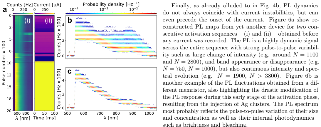
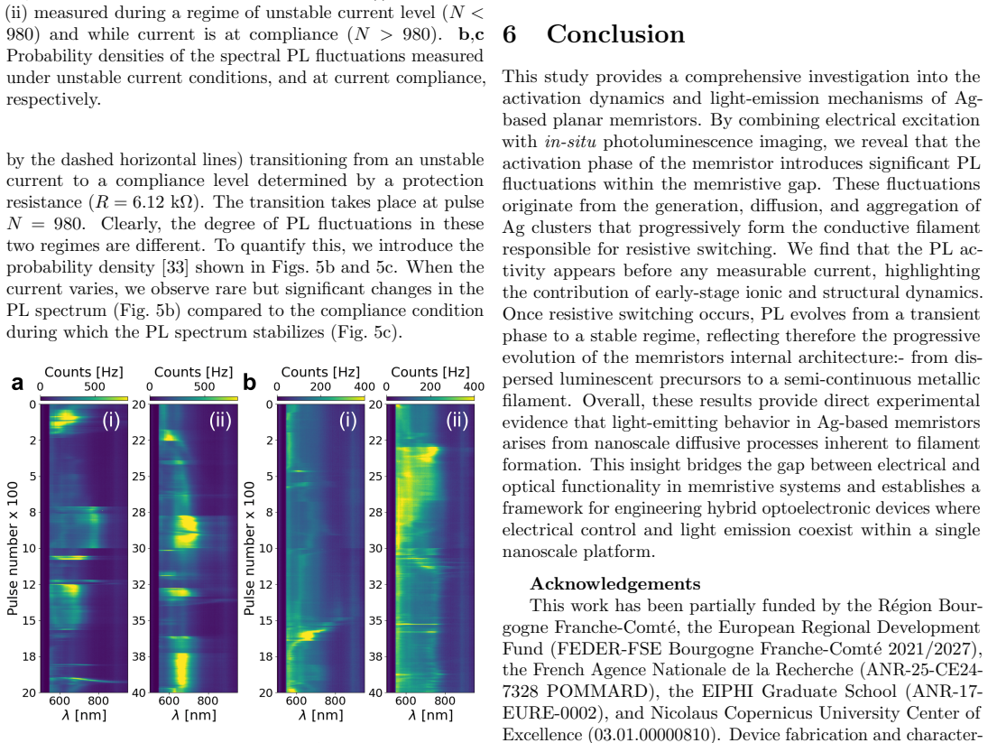
PL activity appears before any measurable current... Ag clusters that progressively form the conductive filament
What do these tags mean?
- matches
- The paper's claim is directly supported by a theorem in the formal canon.
- supports
- The theorem supports part of the paper's argument, but the paper may add assumptions or extra steps.
- extends
- The paper goes beyond the formal theorem; the theorem is a base layer rather than the whole result.
- uses
- The paper appears to rely on the theorem as machinery.
- contradicts
- The paper's claim conflicts with a theorem or certificate in the canon.
- unclear
- Pith found a possible connection, but the passage is too broad, indirect, or ambiguous to say the theorem truly supports the claim.
Reference graph
Works this paper leans on
- [1]
-
[2]
M.-K. Song, J.-H. Kang, X. Zhang, W. Ji, A. Ascoli, I. Messaris, A. S. Demirkol, B. Dong, S. Aggarwal, W. Wan, S.-M. Hong, S. G. Cardwell, I. Boybat, J.-s. Seo, J.-S. Lee, M. Lanza, H. Yeon, M. Onen, J. Li, B. Yildiz, J. A. del Alamo, S. Kim, S. Choi, G. Milano, C. Ricciardi, L. Alff, Y. Chai, Z. Wang, H. Bhaskaran, M. C. Hersam, D. Strukov, H.-S. P. Wong...
- [3]
-
[4]
M. Le Gallo, R. Khaddam-Aljameh, M. Stanisavljevic, A. Vasilopoulos, B. Kersting, M. Dazzi, G. Karunaratne, M. Brndli, A. Singh, S. M. Mller, J. Bchel, X. Tim- oneda, V. Joshi, M. J. Rasch, U. Egger, A. Garofalo, A. Petropoulos, T. Antonakopoulos, K. Brew, S. Choi, I. Ok, T. Philip, V. Chan, C. Silvestre, I. Ahsan, N. Saulnier, V. Narayanan, P. A. Frances...
-
[5]
L. Ye, Z. Gao, J. Fu, W. Ren, C. Yang, J. Wen, X. Wan, Q. Ren, S. Gu, X. Liu, X. Lian, L. Wang,Frontiers in Physics2022,10
-
[6]
C. Weilenmann, A. N. Ziogas, T. Zellweger, K. Portner, M. Mladenovi, M. Kaniselvan, T. Moraitis, M. Luisier, A. Emboras,Nature Communications2024,15, 1
-
[7]
A. H. Atabaki, S. Moazeni, F. Pavanello, H. Gevorgyan, J. Notaros, L. Alloatti, M. T. Wade, C. Sun, S. A. Kruger, H. Meng, K. Al Qubaisi, I. Wang, B. Zhang, A. Khilo, C. V. Baiocco, M. A. Popovi, V. M. Stojanovi, R. J. Ram, Nature2018,556, 7701 349
-
[8]
D. Kramnik, I. Wang, A. Ramesh, J. M. Fargas Cabanil- las, . Gluhovi, S. Buchbinder, P. Zarkos, C. Adamopoulos, P. Kumar, V. M. Stojanovi, M. A. Popovi,Nature Elec- tronics2025,8, 7 620
- [9]
-
[10]
N. Youngblood, C. A. Ros Ocampo, W. H. P. Pernice, H. Bhaskaran,Nature Photonics2023,17, 7 561
- [11]
-
[12]
C.-W. Chang, W.-C. Tan, M.-L. Lu, T.-C. Pan, Y.-J. Yang, Y.-F. Chen,Scientific Reports2014,4, 1 5121
-
[13]
N. Farmakidis, N. Youngblood, X. Li, J. Tan, J. L. Swett, Z. Cheng, C. D. Wright, W. H. P. Pernice, H. Bhaskaran, Science Advances2019,5, 11
-
[14]
M. E. Pereira, R. Martins, E. Fortunato, P. Barquinha, A. Kiazadeh,Neuromorphic Computing and Engineering 2023,3, 2 022002
work page 2023
-
[15]
H. Tan, S. van Dijken,Nature Communications2023, 14, 1
-
[16]
A. A. Zakhidov, B. Jung, J. D. Slinker, H. D. Abrua, G. G. Malliaras,Organic Electronics2010,11, 1 150
- [17]
-
[18]
Y. Zhu, C. Wu, Z. Xu, Y. Liu, H. Hu, T. Guo, T. W. Kim, Y. Chai, F. Li,Nano Letters2021,21, 14 6087
- [19]
-
[20]
C. Zhu, W. Huang, W. Li, X. Yu, X. Li,Research2022, 2022
work page 2022
-
[21]
C. He, J. Li, X. Wu, P. Chen, J. Zhao, K. Yin, M. Cheng, W. Yang, G. Xie, D. Wang, D. Liu, R. Yang, D. Shi, Z. Li, L. Sun, G. Zhang,Advanced Materials2013,25, 39 5593
- [22]
- [23]
-
[24]
Y. Yang, P. Gao, S. Gaba, T. Chang, X. Pan, W. Lu, Nature communications2012,3732
- [25]
-
[26]
K. Malchow, T. Zellweger, B. Cheng, A. Leray, J. Leuthold, A. Bouhelier,ACS Nano2024,18, 35 24004
-
[27]
J. Mangalam, S. Agarwal, A. Resmi, M. Sundararajan, K. Jinesh,Organic Electronics2016,2933
-
[28]
C. Wolf, S. Nau, S. Sax, Y. Busby, J.-J. Pireaux, E. J. W. List-Kratochvil,Japanese Journal of Applied Physics 2015,54, 12 120301
work page 2015
-
[29]
Y. Yang, W. D. Lu,IEEE Transactions on Nanotechnol- ogy2016,15, 3 465
-
[30]
R. Salh,Defect Related Luminescence in Silicon Dioxide Network: A Review, InTech, ISBN 9789533075877,2011
work page 2011
-
[31]
A. Phuruangrat, J. Rodr´ ıguez, M. A. V´ asquez-Agust´ ın, A. Morales-S´ anchez, M. Aceves-Mijares,Journal of Nano- materials2014,2014409482
-
[32]
T.-H. Lee, J. I. Gonzalez, J. Zheng, R. M. Dickson,Ac- counts of Chemical Research2004,38, 7 534
-
[33]
J. Margueritat, A. Bouhelier, L. Markey, G. Colas des Francs, A. Dereux, S. Lau-Truong, J. Grand, G. Lvi, N. Flidj, J. Aubard, E. Finot,The Journal of Physical Chemistry C2012,116, 51 26919. 7
discussion (0)
Sign in with ORCID, Apple, or X to comment. Anyone can read and Pith papers without signing in.