REVIEW 4 major objections 4 minor 2 cited by
A single depth image of the body surface can reveal the 3D locations and shapes of 41 internal organs, the paper claims.
Reviewed by Pith at T0; open to challenge. T0 means a machine referee read the full paper against a public rubric. the ladder, T0–T4 →
T0 review · deepseek-v4-flash
2026-08-03 08:00 UTC pith:IIKTPTBR
load-bearing objection A genuinely new depth-to-volume system with credible in-distribution results, but the clinical transfer claim rests on unvalidated synthetic co-generation and a metric inconsistency. the 4 major comments →
Depth to Anatomy: Organ Localization from Depth Images for Automated Patient Table Positioning in Radiology Workflow
The pith
A machine-rendered reading of the paper's core claim, the machinery that carries it, and where it could break.
Core claim
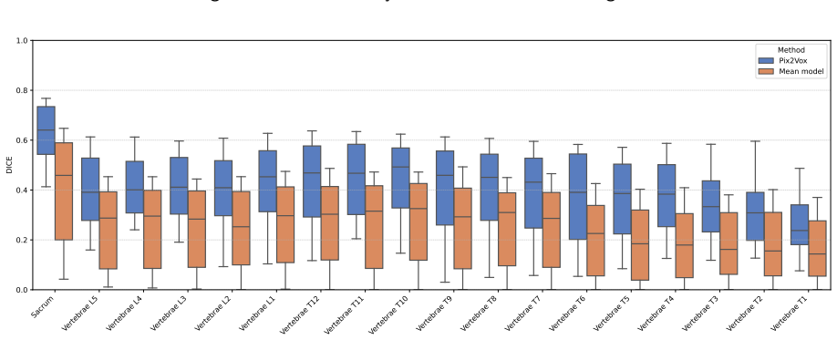
The central claim is that 3D volumetric segmentations of 41 internal anatomical structures can be predicted end-to-end from a single anterior depth image, without any body-model fitting or intermediate surface reconstruction. Using a U-shaped architecture that converts 2D encoder features into a 3D decoder via learnable 2D-to-3D layers, the model achieves mean Dice 0.48±0.20 and mean symmetric surface distance 8.34±8.11 mm on a held-out set of 998 synthetic examples. Bounding boxes derived from the predictions have a mean absolute detection offset of about 11 mm. The authors further claim that qualitative results on real-world depth images of volunteers in an MRI scanner indicate generalizat
What carries the argument
The load-bearing component is the hybrid 2D-to-3D conversion block: at the encoder bottleneck and each skip connection, 2D feature maps gain a singleton depth axis and pass through a 3D convolution with a kernel of size k_l×1×1, expanding the feature depth so the 3D decoder can reconstruct volumetric masks. Equally important is the synthetic generation pipeline that renders orthographic anterior depth images from MRI body masks and applies stochastic fabric-like augmentations (line folds and low-frequency clusters with up to 30 mm displacement) to mimic clothing and blankets, forcing the network to rely on global body proportions rather than surface texture.
Load-bearing premise
The entire pipeline rests on the assumption that synthetic depth images rendered from MRI body masks, with cloth-like noise added, faithfully represent real depth-camera images of clothed patients on a scanner table; if real perspective distortion, pose variation, or sensor noise lies outside the simulated range, the reported accuracy will not carry over to clinical practice.
What would settle it
Acquire paired real depth images and volumetric ground truth (CT or MRI) for the same patients in a clinical scanner setup, run the model, and compute the mean absolute bounding-box offset and Dice. If the offsets exceed roughly 20 mm or Dice falls below 0.3 on major organs, the claim that synthetic training transfers to practice is refuted.
If this is right
- If the claim holds, a depth sensor mounted above the scanner bore can predict organ positions in scanner coordinates, enabling automatic table translation to the imaging isocenter.
- The ~11 mm bounding-box offset is within the range that could eliminate or reduce the need for scout-based planning in many routine exams.
- The method requires no patient-specific mesh or template, so it could work in a single snapshot at the table.
- Because training data are synthetic, the framework can be extended to larger cohorts and new organs without new manual annotations — only MRI volumes are needed.
- The performance hierarchy (large organs strong, small organs weaker but well-centered) suggests that the approach is useful for scan-range planning even when fine boundary fidelity is lacking.
Where Pith is reading between the lines
- A natural next validation is to obtain paired real depth images and CT/MRI ground truth for the same patients; the paper's qualitative real-world evidence would then be checked quantitatively, and the reported 11 mm offset would likely change depending on pose and clothing variability.
- The synthetic-to-real gap could be narrowed by fine-tuning on a modest set of real sensor images, or by applying the augmentation parameters to simulate perspective distortion and self-occlusion, which the current orthographic rendering omits.
- The authors' own observation that the anterior-posterior axis is hardest suggests that adding a second depth view (e.g., lateral) would improve the bounding-box estimate most where the system is weakest.
- The architecture's success at predicting bones (femur, sacrum around 0.6 Dice) hints that the same surface-to-volume map could support radiation therapy planning or surgical navigation, not just table positioning.
Editorial analysis
A structured set of objections, weighed in public.
Referee Report
Summary. The paper proposes a learning-based framework to predict 3D volumetric segmentations of 41 anatomical structures from a single anterior 2D depth image, intended for automated patient table positioning in radiology. Training data are synthetically generated from 10,020 NAKO whole-body MRI scans: depth images are rendered by ray-casting the MRI body surface, and ground-truth labels are produced with TotalSegmentatorMRI. A hybrid 2D-to-3D CNN (Pix2Vox) is trained and evaluated on a held-out synthetic test set, reporting mean Dice 0.48±0.20, ASSD 8.34±8.11 mm, and bounding-box detection offsets around 11 mm. The authors also present qualitative results on real-world Kinect v2 depth images and claim generalization to clinical settings.
Significance. If the results hold, the approach could substantially streamline radiology workflows by replacing scout-based planning with a single depth image. The paper's strengths include a large-scale synthetic training pipeline, a held-out test set, comparison against a mean-model baseline (0.34 Dice), and publicly available code and pretrained models. The in-distribution results show meaningful improvement over baseline and plausible localization accuracy. However, the load-bearing claim of clinical generalization rests on qualitative evidence only, and the ground-truth labels are auto-generated without manual validation. These issues limit the strength of the conclusions as currently stated.
major comments (4)
- [§3.4.3, §4.2] The real-world generalization claim is not supported quantitatively. The Kinect v2 evaluation (Figure 7) is purely qualitative, with no ground-truth volumes, landmarks, or metrics, and §4.2 explicitly concedes a domain gap (occlusions, sensor-specific distortions, clothing/blanket effects). Because the synthetic test set is co-generated from the same MRI volumes used for labels, it cannot test transfer under real perspective geometry, sensor noise, or non-rigid surface deformation. To support the abstract's claim of 'generalize to practical clinical settings,' the authors must either provide quantitative real-world evaluation (e.g., against CT/MRI ground truth or manual annotations) or substantially weaken the conclusion to 'qualitative feasibility'.
- [§2.3] Ground-truth labels come from TotalSegmentatorMRI auto-segmentation with no validation against manual annotations. The post-processing (hole filling, connected component analysis) and the structure selection based on 'qualitative visual assessment' are heuristic. If auto-segmentation errors are systematic—particularly for small organs like the thyroid or trachea—the reported Dice and ASSD values may be biased. The authors should validate labels on a subset (e.g., against manual segmentation or a second automated tool) or at least quantify the uncertainty of the auto-segmentation for the selected structures.
- [Abstract vs §3.4.2] The reported metrics are inconsistent between the abstract and the results section. The abstract states mean Dice 0.44±0.2 and ASSD 7.69±5.68 mm, while §3.4.2 reports 0.48±0.20 and 8.34±8.11 mm. If the difference arises from averaging per-organ vs per-volume or from a different test-set split, this should be explicitly stated. As written, the discrepancy undermines reproducibility and needs clarification.
- [§2.2, Eq. (4)] The stochastic augmentation in Eq. (4) perturbs only depth values within the patient silhouette; it does not modify the label geometry or simulate non-rigid misalignment between the body surface and internal anatomy caused by clothing, blankets, or pose shifts. Thus the training data cannot teach the model to be invariant to real-world surface-anatomy misalignment, which is precisely the mechanism of the domain gap acknowledged in §4.2. The authors should either extend the augmentation to include joint surface-label deformations or provide a dedicated experiment quantifying sensitivity to such misalignment.
minor comments (4)
- [Abstract] Typo: 'and and' appears in the results sentence.
- [§2.2, Eq. (2)] The notation d(x,z)=argmax_y(M_{x,y,z}) is ambiguous for a binary mask; using 'max{y: M=1}' or 'first encountered foreground index' would be clearer.
- [§3.2] The mean model's thresholding at 50% of the maximum voxel intensity is ad hoc; if this parameter were tuned, it should be reported. Also, the mean model's strong performance in the anterior-posterior axis (DOE ~13 mm) is interesting and deserves more analysis.
- [Figures 3–5] The figures lack axis labels for the side abbreviations (e.g., 'left', 'right'), making them hard to read; please add a caption note or expand the abbreviations.
Circularity Check
No significant circularity: the core depth-to-volume prediction is an empirical supervised mapping evaluated on held-out synthetic subjects, and the only self-citations are non-load-bearing context.
full rationale
The paper's central derivation is an empirical supervised learning pipeline, not a closed-form derivation. Input depth maps are ray-cast from MRI body masks (§2.2) and labels are obtained by TotalSegmentatorMRI on the same volumes (§2.3), so the paired training data are co-generated. However, the learned mapping is not defined in terms of the labels, nor are the predicted volumes a fitted re-expression of the depth input: the depth image is a lossy 2D anterior projection while the target is a full 3D segmentation, so the task is genuinely underdetermined and must be learned. The held-out test set of 998 subjects is a true prediction, and the Pix2Vox result (Dice 0.48) exceeds the mean-model baseline (0.34), showing the prediction is not trivially forced by the training labels. The only self-citation appears in the design of the 2.5D CNN baseline, which is not the central claim and is reimplemented in this paper; no uniqueness theorem or load-bearing result is imported from the authors' prior work. The qualitative real-world evaluation (§3.4.3) and the acknowledged domain gap (§4.2) are external-validity limitations, not circularity: the paper explicitly concedes that synthetic depth may not fully capture real sensor variability and that no quantitative real-world ground truth was assessed. Thus there is no step in the claimed derivation chain that reduces to its own inputs by construction.
Axiom & Free-Parameter Ledger
free parameters (8)
- MRI body mask threshold ε =
0.02
- Depth clipping threshold =
0.3
- Augmentation displacement maximum =
30 mm
- Augmentation scale factor S =
100
- Loss weights (Dice, BCE) =
0.5, 0.5
- Training hyperparameters (lr, warmup, batch, steps) =
0.001, 1000, 8, 15000
- Layer-wise 2D-to-3D conversion dimensions (kl, sl, fl) =
not reported
- Mean model binarization threshold =
50% of max voxel intensity
axioms (3)
- domain assumption TotalSegmentatorMRI produces anatomically correct segmentations suitable as ground truth for the 41 structures.
- domain assumption Orthographic ray-casting from an MRI body mask faithfully mimics the top-down depth sensor used clinically.
- domain assumption The training distribution (supine, standardized NAKO positioning, minimal pose variation) matches the target clinical distribution.
read the original abstract
In clinical radiology, accurate patient table positioning is essential to align specific internal organs of interest with the scanner imaging isocenter, ensuring image quality and diagnostic reliability. Automated patient positioning can streamline this process and improve radiology workflow efficiency by reducing the time required for manual table adjustments and scout-based scan planning. We propose a learning-based framework that predicts 3D organ locations and shapes for 41 anatomical structures, including both bones and soft tissues, directly from a single 2D depth image of the body surface. Leveraging $10,020$ whole-body MRI scans from the German National Cohort (NAKO) dataset, we synthetically generate depth images paired with anatomical segmentations to train a convolutional neural network for volumetric organ prediction. Our method achieves a mean dice similarity coefficient of $0.44\pm0.2$ and and a symmetric average surface distance of $7.69\pm5.68$ mm across all structures. Furthermore, the model derives organ bounding boxes with a mean absolute detection offset of $10.99\pm5.54$ mm. Qualitative results on real-world depth images indicate the ability of the model to generalize to practical clinical settings. These findings suggest that depth-only organ localization can support automated patient positioning reducing setup time, minimizing operator variability, and improving patient comfort.
Figures




Forward citations
Cited by 2 Pith papers
-
SAMe: A Semantic Anatomy Mapping Engine for Robotic Ultrasound
SAMe grounds clinical complaints to target organs, builds a patient-specific anatomical map from a single external image, and outputs probe initialization poses, reaching 97.3% liver and 81.7% kidney hit rates on a re...
-
SAMe: A Semantic Anatomy Mapping Engine for Robotic Ultrasound
SAMe grounds complaints to organs, builds a lightweight patient anatomy model from one body image, and outputs probe initialization poses, outperforming keypoint baselines in real-robot liver and kidney trials.
Reference graph
Works this paper leans on
-
[2005]
Tugba Akinci D’Antonoli, Lucas K Berger, Ashraya K In- drakanti, Nathan Vishwanathan, Jakob Weiß, Matthias Jung, Zeynep Berkarda, Alexander Rau, Marco Reisert, Thomas K¨ustner, et al. Totalsegmentator mri: Sequence- independent segmentation of 59 anatomical structures in mr images.arXiv preprint arXiv:2405.19492,
-
[2022]
Accuracy of automated patient positioning in ct using a 3d camera for body contour detection.European Radiology, 29(4):2079–2088,
Ronald Booij, Ricardo PJ Budde, Marcel L Dijkshoorn, and Marcel van Straten. Accuracy of automated patient positioning in ct using a 3d camera for body contour detection.European Radiology, 29(4):2079–2088,
2079
-
[2024]
Predicting anatomical structures from 2d depth images of patients
Kai Geißler, Daniel Mensing, Temke Kohlbrandt, Jochen G Hirsch, and Stefan Heldmann. Predicting anatomical structures from 2d depth images of patients. InMedical Imaging 2025: Image Processing, volume 13406, pages 407–411. SPIE,
2025
discussion (0)
Sign in with ORCID, Apple, or X to comment. Anyone can read and Pith papers without signing in.