REVIEW 2 major objections 5 minor 1 cited by
VesselTok turns large vessel graphs into compact tokens that reconstruct, generate, and repair tubular anatomy.
Reviewed by Pith at T0; open to challenge. T0 means a machine referee read the full paper against a public rubric. the ladder, T0–T4 →
T0 review · grok-4.5
2026-07-13 22:23 UTC pith:YSNA3B7E
load-bearing objection Solid, usable tokenizer for large tubular biomedical graphs; the fixed-pseudo-radius bet is load-bearing but already ablated and the multi-task evidence holds. the 2 major comments →
VesselTok: Tokenizing Vessel-like 3D Biomedical Graph Representations for Reconstruction and Generation
The pith
A machine-rendered reading of the paper's core claim, the machinery that carries it, and where it could break.
Core claim
A VAE-style encoder that attends over centerline points (not surface samples) can compress topologically complex 3D vessel graphs into fixed-length continuous tokens; decoding those tokens back to an occupancy field and skeletonizing it recovers graphs whose connectivity and geometry outperform surface-based baselines, and the same tokens support generation and link prediction.
What carries the argument
VesselTok: a transformer VAE that maps centerline points plus a fixed pseudo-radius occupancy field into a compact latent token sequence Z, then reconstructs the field by cross-attention to query points.
Load-bearing premise
A single fixed pseudo-radius around every centerline is enough to capture real vessel topology, so that skeletonization of the decoded occupancy field recovers a faithful discrete graph and true radius variation can be left as a later regression step.
What would settle it
Train and decode the same model on graphs whose true radii vary sharply or that contain loops thinner than the chosen pseudo-radius; if the recovered Betti numbers and centerline Dice collapse relative to surface-based baselines, the fixed-radius premise fails.
Editorial analysis
A structured set of objections, weighed in public.
Referee Report
Summary. VesselTok is a VAE-style tokenizer that maps large 3D spatial graphs of tubular biomedical structures (airways, pulmonary vessels, cerebral vessels) to compact continuous latent tokens. It represents each graph as a continuous occupancy field obtained by dilating centerline segments with a fixed pseudo-radius, encodes the centerline point cloud with a transformer VAE, and decodes an occupancy field from which a discrete graph is recovered by skeletonization. The paper reports improved reconstruction (clDice, Chamfer, Betti errors) relative to 3DShape2VecSet and Hunyuan3D 2.0 across multiple anatomies, generalization to unseen scales and topologies (TopCoW, renal vasculature, sagittal cuts), unconditional/conditional generation via EDM diffusion in token space, and transfer to a link-prediction inverse problem. Ablations cover token count K, channel dimension C, and pseudo-radius r; a post-hoc radius regressor is shown in the supplement.
Significance. If the empirical claims hold, VesselTok supplies a practical, domain-adapted latent interface for topologically complex vessel-like graphs that prior graph or surface tokenizers struggle to scale. The multi-anatomy training set, OOD tests (including order-of-magnitude larger renal graphs via chunking), generative metrics (FID/MMD/Betti/Coverage), link-prediction comparison, and explicit ablations on the free parameters constitute a solid experimental package. The centerline-plus-pseudo-radius design is a clear, falsifiable modeling bet that concentrates capacity on topology and large-scale geometry; the promised code release further raises the work’s utility for the community. These strengths make the contribution relevant for biomedical shape analysis and generative modeling of curvilinear networks.
major comments (2)
- Table 1 (and the corresponding average row): while VesselTok improves clDice and Chamfer Distance on every dataset, |Δβ1| is higher than at least one baseline on AIIB, PARSE, HiPas and Pulmonary-AV. The abstract and introduction claim that the tokens “robustly encode complex topologies.” The mixed loop-error results indicate a geometry–topology trade-off that is not discussed; a short analysis of when and why loops are lost or hallucinated would make the topology claim precise rather than overstated.
- Section 3.1 Eq. (1) and Section 4.6: the entire pipeline rests on a single fixed pseudo-radius r = 0.016. The ablation on ATM is useful, yet the main multi-anatomy tables, OOD experiments and generative models all use this one value. Because true radius variation is deferred to a post-hoc regressor (Supp. B), it remains unclear whether the latent itself is invariant to realistic radius changes (e.g., stenoses or aneurysms). A controlled experiment that re-renders the same centerlines at several radii and measures latent/reconstruction stability would directly test the load-bearing modeling assumption.
minor comments (5)
- Table and figure captions occasionally contain spacing artifacts (“T able 1”, “Fig. 1:VesselTok”). Clean these for production.
- Section 4.4 / Table 4: MMD-CD and MMD-EMD are reported ×10^{-2}; state the scaling factor explicitly in the table header for readability.
- Inference (Section 3.2): the occupancy threshold τ = 0.5 and the skeletonization algorithm are fixed; a one-sentence sensitivity note (or reference to the modular claim) would help readers who wish to substitute other extractors.
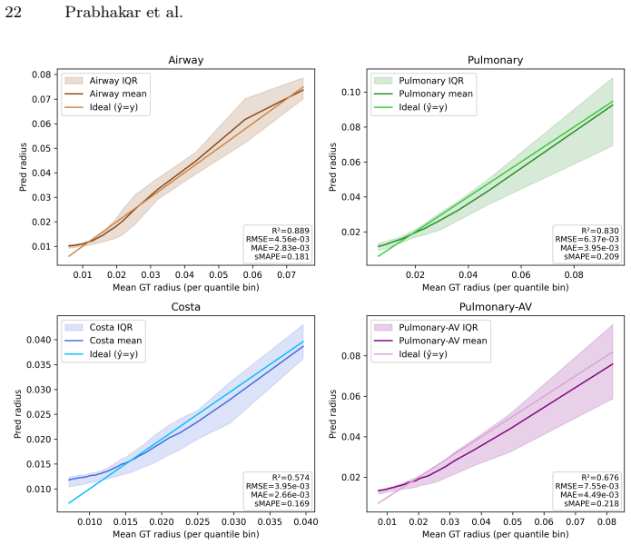
- Supplementary Sec. B reports R² and sMAPE for radius regression; adding the same metrics on the held-out OOD sets (TopCoW, renal) would strengthen the claim that radius can safely be treated as post-hoc.
- Figure 3 and Figure 5 would benefit from a small inset or color legend that distinguishes true-positive, false-positive and false-negative branches, making qualitative topology differences easier to judge.
Circularity Check
No significant circularity: empirical tokenizer trained by reconstruction+KL and evaluated on held-out multi-anatomy, OOD, generative, and inverse-task metrics against external baselines.
full rationale
VesselTok is a standard VAE-style shape tokenizer (centerline points + fixed pseudo-radius occupancy field, transformer encoder/decoder, BCE+KL loss) whose latents are then used for EDM diffusion and conditional flow-matching. All quantitative claims (clDice/CD/Betti, FID/MMD/COV, link-prediction) are measured on held-out or OOD data against independently reimplemented external baselines (3DShape2VecSet, Hunyuan3D 2.0, VesselGPT, autodecoder). The pseudo-radius r=0.016 is an explicit hyper-parameter chosen by ablation (Sec. 4.6, Fig. 6), not a fitted constant later re-labeled as a prediction; true radius is deferred to a separate post-hoc regressor whose R^{2}/sMAPE are reported only as supporting evidence (Supp. B). Self-citations to the authors’ prior vessel-generation papers appear only as related-work baselines that the present method is claimed to improve upon, never as uniqueness theorems or load-bearing premises. No equation reduces a claimed first-principles result to its own inputs by construction. The derivation chain is therefore self-contained and non-circular.
Axiom & Free-Parameter Ledger
free parameters (5)
- pseudo-radius r =
0.016
- token count K =
512
- latent channel dimension C =
4
- KL weight λ =
1e-3
- occupancy threshold τ =
0.5
axioms (3)
- ad hoc to paper A fixed pseudo-radius occupancy field around centerline segments is a topology-preserving representation of real vessel graphs whose true radii vary.
- domain assumption Skeletonization of a thresholded occupancy field followed by neighborhood connectivity recovers a discrete graph whose Betti numbers and geometry can be fairly compared across methods.
- standard math Standard transformer VAE + EDM diffusion machinery is a valid generative model once a continuous latent of fixed length is obtained.
invented entities (2)
-
graph occupancy field ϕ_r with fixed pseudo-radius
no independent evidence
-
VesselTok continuous token sequence Z ∈ R^{l×c}
no independent evidence
read the original abstract
Spatial graphs provide a lightweight and elegant representation of curvilinear anatomical structures such as blood vessels, lung airways, and neuronal networks. Accurately modeling these graphs is crucial in clinical and (bio-)medical research. However, the high spatial resolution of large networks drastically increases their complexity, resulting in significant computational challenges. In this work, we aim to tackle these challenges by proposing VesselTok, a framework that approaches spatially dense graphs from a parametric shape perspective to learn latent representations (tokens). VesselTok leverages centerline points with a pseudo radius to effectively encode tubular geometry. Specifically, we learn a novel latent representation conditioned on centerline points to encode neural implicit representations of vessel-like, tubular structures. We demonstrate VesselTok's performance across diverse anatomies, including lung airways, lung vessels, and brain vessels, highlighting its ability to robustly encode complex topologies. To prove the effectiveness of VesselTok's learnt latent representations, we show that they (i) generalize to unseen anatomies, (ii) support generative modeling of plausible anatomical graphs, and (iii) transfer effectively to downstream inverse problems, such as link prediction.
Figures




Forward citations
Cited by 1 Pith paper
-
SynVA: A Modular Toolkit for Vessel Generation and Aneurysm Editing
SynVA toolkit generates realistic vascular meshes and anatomically plausible aneurysms, releasing 50,000 labeled samples for medical vision tasks.
Reference graph
Works this paper leans on
-
[1]
Medical Image Analysis94, 103099 (2024)
Amiranashvili, T., Lüdke, D., Li, H.B., Zachow, S., Menze, B.H.: Learning contin- uous shape priors from sparse data with neural implicit functions. Medical Image Analysis94, 103099 (2024)
2024
-
[2]
arXiv preprint arXiv:2506.11163 (2025)
Batten, J., Schaap, M., Sinclair, M., Bai, Y., Glocker, B.: Vector representations of vessel trees. arXiv preprint arXiv:2506.11163 (2025)
Pith/arXiv arXiv 2025
-
[3]
Scientific reports8(1), 5341 (2018)
Belchi, F., Pirashvili, M., Conway, J., Bennett, M., Djukanovic, R., Brodzki, J.: Lung topology characteristics in patients with chronic obstructive pulmonary dis- ease. Scientific reports8(1), 5341 (2018)
2018
-
[4]
arXiv preprint arXiv:2407.02599 (2024)
Bensadoun, R., Monnier, T., Kleiman, Y., Kokkinos, F., Siddiqui, Y., Kariya, M., Harosh, O., Shapovalov, R., Graham, B., Garreau, E., et al.: Meta 3d gen. arXiv preprint arXiv:2407.02599 (2024)
Pith/arXiv arXiv 2024
-
[5]
Cell reports methods2(4) (2022)
Bumgarner, J.R., Nelson, R.J.: Open-source analysis and visualization of seg- mented vasculature datasets with VesselVio. Cell reports methods2(4) (2022)
2022
-
[6]
arXiv preprint arXiv:2412.15618 (2024)
Chang,J.H.R.,Wang,Y.,Martin,M.A.B.,Gu,J.,Zhao,X.,Susskind,J.,Tuzel,O.: 3d shape tokenization via latent flow matching. arXiv preprint arXiv:2412.15618 (2024)
Pith/arXiv arXiv 2024
-
[7]
Journal of biomedical informatics56, 395–405 (2015)
Chapman, B.E., Berty, H.P., Schulthies, S.L.: Automated generation of directed graphs from vascular segmentations. Journal of biomedical informatics56, 395–405 (2015)
2015
-
[8]
In: Proceedings of the Computer Vision and Pattern Recognition Conference
Chen, R., Zhang, J., Liang, Y., Luo, G., Li, W., Liu, J., Li, X., Long, X., Feng, J., Tan, P.: Dora: Sampling and benchmarking for 3d shape variational auto-encoders. In: Proceedings of the Computer Vision and Pattern Recognition Conference. pp. 16251–16261 (2025)
2025
-
[9]
In: International Conference on Medical Image Computing and Computer-Assisted Intervention
Chen, S., Zhang, G., Lai, J., Shen, B., Zhang, S., Dong, C., Chen, X., Li, Y.: Hier- archical part-based generative model for realistic 3d blood vessel. In: International Conference on Medical Image Computing and Computer-Assisted Intervention. pp. 257–267. Springer (2025)
2025
-
[10]
In: International Conference on Pattern Recognition
Cheng, H., Zheng, L., Yan, Z., Zhang, H., Meng, B., Xu, X.: Fusion of machine learninganddeepneuralnetworksforpulmonaryarteriesandveinssegmentationin lung cancer surgery planning. In: International Conference on Pattern Recognition. pp. 422–438. Springer (2024)
2024
-
[11]
Nature Com- munications16(1), 2262 (2025)
Chu, Y., Luo, G., Zhou, L., Cao, S., Ma, G., Meng, X., Zhou, J., Yang, C., Xie, D., Mu, D., et al.: Deep learning-driven pulmonary artery and vein segmentation reveals demography-associated vasculature anatomical differences. Nature Com- munications16(1), 2262 (2025)
2025
-
[12]
Medical image analysis15(5), 738–747 (2011)
Dabbah, M.A., Graham, J., Petropoulos, I.N., Tavakoli, M., Malik, R.A.: Auto- matic analysis of diabetic peripheral neuropathy using multi-scale quantitative morphology of nerve fibres in corneal confocal microscopy imaging. Medical image analysis15(5), 738–747 (2011)
2011
-
[13]
PLoS computational biology19(10), e1011496 (2023)
Epp, R., Glück, C., Binder, N.F., El Amki, M., Weber, B., Wegener, S., Jenny, P., Schmid, F.: The role of leptomeningeal collaterals in redistributing blood flow during stroke. PLoS computational biology19(10), e1011496 (2023)
2023
-
[14]
In: International Con- ference on Medical Image Computing and Computer-Assisted Intervention
Feldman, P., Fainstein, M., Siless, V., Delrieux, C., Iarussi, E.: Vesselvae: Recur- sive variational autoencoders for 3d blood vessel synthesis. In: International Con- ference on Medical Image Computing and Computer-Assisted Intervention. pp. 67–76. Springer (2023)
2023
-
[15]
In: International Conference on Medi- VesselTok 17 cal Image Computing and Computer-Assisted Intervention
Feldman, P., Sinnona, M., Delrieux, C., Siless, V., Iarussi, E.: Vesselgpt: Autore- gressive modeling of vascular geometry. In: International Conference on Medi- VesselTok 17 cal Image Computing and Computer-Assisted Intervention. pp. 662–672. Springer (2025)
2025
-
[16]
arXiv preprint arXiv:2305.02463 (2023)
Jun, H., Nichol, A.: Shap-e: Generating conditional 3d implicit functions. arXiv preprint arXiv:2305.02463 (2023)
Pith/arXiv arXiv 2023
-
[17]
Advances in neural information processing systems35, 26565–26577 (2022)
Karras,T.,Aittala,M.,Aila,T.,Laine,S.:Elucidatingthedesignspaceofdiffusion- based generative models. Advances in neural information processing systems35, 26565–26577 (2022)
2022
-
[18]
Cell180(4), 780–795 (2020)
Kirst, C., Skriabine, S., Vieites-Prado, A., Topilko, T., Bertin, P., Gerschenfeld, G., Verny, F., Topilko, P., Michalski, N., Tessier-Lavigne, M., et al.: Mapping the fine-scale organization and plasticity of the brain vasculature. Cell180(4), 780–795 (2020)
2020
-
[19]
In: Medical Imaging with Deep Learning (2025)
Kuipers, T.P., Konduri, P.R., Bekkers, E.J., Marquering, H.: Self-supervised syn- thetic cerebral vessel tree generation using semantic signed distance fields. In: Medical Imaging with Deep Learning (2025)
2025
-
[20]
In: Medical Imaging with Deep Learning (2024)
Kuipers, T.P., Konduri, P.R., Marquering, H., Bekkers, E.J.: Generating cerebral vessel trees of acute ischemic stroke patients using conditional set-diffusion. In: Medical Imaging with Deep Learning (2024)
2024
-
[21]
CVGIP: graphical models and image processing 56(6), 462–478 (1994)
Lee, T.C., Kashyap, R.L., Chu, C.N.: Building skeleton models via 3-d medial surface axis thinning algorithms. CVGIP: graphical models and image processing 56(6), 462–478 (1994)
1994
-
[22]
European Journal of Radiology 153, 110383 (2022)
Lin, E., Kamel, H., Gupta, A., RoyChoudhury, A., Girgis, P., Glodzik, L.: Incom- plete circle of Willis variants and stroke outcome. European Journal of Radiology 153, 110383 (2022)
2022
-
[23]
arXiv preprint arXiv:2210.02747 (2022)
Lipman, Y., Chen, R.T., Ben-Hamu, H., Nickel, M., Le, M.: Flow matching for generative modeling. arXiv preprint arXiv:2210.02747 (2022)
Pith/arXiv arXiv 2022
-
[24]
Advances in Neural Information Processing Systems36, 25854–25875 (2023)
Liu, Z., Shi, Y., Zhang, A., Zhang, E., Kawaguchi, K., Wang, X., Chua, T.S.: Re- thinking tokenizer and decoder in masked graph modeling for molecules. Advances in Neural Information Processing Systems36, 25854–25875 (2023)
2023
-
[25]
arXiv preprint arXiv:1711.05101 (2017)
Loshchilov, I., Hutter, F.: Decoupled weight decay regularization. arXiv preprint arXiv:1711.05101 (2017)
Pith/arXiv arXiv 2017
-
[26]
arXiv preprint arXiv:2304.03708 (2023)
Luo, G., Wang, K., Liu, J., Li, S., Liang, X., Li, X., Gan, S., Wang, W., Dong, S., Wang, W., et al.: Efficient automatic segmentation for multi-level pulmonary arteries: The parse challenge. arXiv preprint arXiv:2304.03708 (2023)
Pith/arXiv arXiv 2023
-
[27]
International Journal of Computer Vision133(4), 1855–1878 (2025)
Mello Rella, E., Chhatkuli, A., Konukoglu, E., Van Gool, L.: Neural vector fields for implicit surface representation and inference. International Journal of Computer Vision133(4), 1855–1878 (2025)
2025
-
[28]
IEEE Com- puter Graphics and Applications29(6), 6–13 (2009)
Meyer-Spradow, J., Ropinski, T., Mensmann, J., Hinrichs, K.: Voreen: A rapid- prototyping environment for ray-casting-based volume visualizations. IEEE Com- puter Graphics and Applications29(6), 6–13 (2009)
2009
-
[29]
IEEE transactions on med- ical imaging43(12), 4442–4456 (2024)
Mou, L., Lin, J., Zhao, Y., Liu, Y., Ma, S., Zhang, J., Lv, W., Zhou, T., Liu, J., Frangi, A.F., et al.: Costa: A multi-center tof-mra dataset and a style self- consistency network for cerebrovascular segmentation. IEEE transactions on med- ical imaging43(12), 4442–4456 (2024)
2024
-
[30]
Medical Image Analysis97, 103253 (2024)
Nan, Y., Xing, X., Wang, S., Tang, Z., Felder, F.N., Zhang, S., Ledda, R.E., Ding, X., Yu, R., Liu, W., et al.: Hunting imaging biomarkers in pulmonary fibrosis: benchmarks of the aiib23 challenge. Medical Image Analysis97, 103253 (2024)
2024
-
[31]
Frontiers in Bioengineering and Biotechnology11, 1271760 (2023) 18 Prabhakar et al
Ortiz-Puerta, D., Diaz, O., Retamal, J., Hurtado, D.E.: Morphometric analysis of airways in pre-copd and mild COPD lungs using continuous surface representations of the bronchial lumen. Frontiers in Bioengineering and Biotechnology11, 1271760 (2023) 18 Prabhakar et al
2023
-
[32]
In: Proceedings of the IEEE/CVF Conference on Computer Vision and Pattern Recognition (CVPR) (June 2019)
Park,J.J.,Florence,P.,Straub,J.,Newcombe,R.,Lovegrove,S.:Deepsdf:Learning continuous signed distance functions for shape representation. In: Proceedings of the IEEE/CVF Conference on Computer Vision and Pattern Recognition (CVPR) (June 2019)
2019
-
[33]
2023 ieee
Peebles, W.S., Xie, S.: Scalable diffusion models with transformers. 2023 ieee. In: CVF International Conference on Computer Vision (ICCV). vol. 4172 (2022)
2023
-
[34]
In: International Con- ference on Medical Image Computing and Computer-Assisted Intervention
Prabhakar, C., Shit, S., Amiranashvili, T., Li, H.B., Menze, B.: Semantically con- sistent discrete diffusion for 3d biological graph modeling. In: International Con- ference on Medical Image Computing and Computer-Assisted Intervention. pp. 594–604. Springer (2025)
2025
-
[35]
In: Interna- tional Conference on Medical Image Computing and Computer-Assisted Interven- tion
Prabhakar, C., Shit, S., Musio, F., Yang, K., Amiranashvili, T., Paetzold, J.C., Li, H.B., Menze, B.: 3d vessel graph generation using denoising diffusion. In: Interna- tional Conference on Medical Image Computing and Computer-Assisted Interven- tion. pp. 3–13. Springer (2024)
2024
-
[36]
Advances in neural information processing systems30(2017)
Qi, C.R., Yi, L., Su, H., Guibas, L.J.: Pointnet++: Deep hierarchical feature learn- ing on point sets in a metric space. Advances in neural information processing systems30(2017)
2017
-
[37]
Rampášek, L., Galkin, M., Dwivedi, V.P., Luu, A.T., Wolf, G., Beaini, D.: Recipe forageneral,powerful,scalablegraphtransformer.AdvancesinNeuralInformation Processing Systems35, 14501–14515 (2022)
2022
-
[38]
In: Proceedings of the IEEE/CVF conference on computer vision and pattern recognition
Shit, S., Paetzold, J.C., Sekuboyina, A., Ezhov, I., Unger, A., Zhylka, A., Pluim, J.P., Bauer, U., Menze, B.H.: cldice-a novel topology-preserving loss function for tubular structure segmentation. In: Proceedings of the IEEE/CVF conference on computer vision and pattern recognition. pp. 16560–16569 (2021)
2021
-
[39]
Advances in neural information processing systems33, 7462–7473 (2020)
Sitzmann, V., Martel, J., Bergman, A., Lindell, D., Wetzstein, G.: Implicit neural representations with periodic activation functions. Advances in neural information processing systems33, 7462–7473 (2020)
2020
-
[40]
arXiv preprint arXiv:2311.01138 (2023)
Støverud, K.H., Bouget, D., Pedersen, A., Leira, H.O., Langø, T., Hofstad, E.F.: Aeropath: An airway segmentation benchmark dataset with challenging pathology. arXiv preprint arXiv:2311.01138 (2023)
Pith/arXiv arXiv 2023
-
[41]
Nature methods17(4), 442–449 (2020)
Todorov, M.I., Paetzold, J.C., Schoppe, O., Tetteh, G., Shit, S., Efremov, V., Todorov-Völgyi, K., Düring, M., Dichgans, M., Piraud, M., et al.: Machine learning analysis of whole mouse brain vasculature. Nature methods17(4), 442–449 (2020)
2020
-
[42]
arXiv preprint arXiv:1710.10903 (2017)
Veličković, P., Cucurull, G., Casanova, A., Romero, A., Lio, P., Bengio, Y.: Graph attention networks. arXiv preprint arXiv:1710.10903 (2017)
Pith/arXiv arXiv 2017
-
[43]
In: Joint European Conference on Machine Learning and Knowledge Discovery in Databases
Vignac, C., Osman, N., Toni, L., Frossard, P.: Midi: Mixed graph and 3d denois- ing diffusion for molecule generation. In: Joint European Conference on Machine Learning and Knowledge Discovery in Databases. pp. 560–576. Springer (2023)
2023
-
[44]
Nature methods18(12), 1532–1541 (2021)
Walsh, C.L., Tafforeau, P., Wagner, W., Jafree, D., Bellier, A., Werlein, C., Küh- nel, M., Boller, E., Walker-Samuel, S., Robertus, J., et al.: Imaging intact human organs with local resolution of cellular structures using hierarchical phase-contrast tomography. Nature methods18(12), 1532–1541 (2021)
2021
-
[45]
Wang,L.,Hassani,K.,Zhang,S.,Fu,D.,Yuan,B.,Cong,W.,Hua,Z.,Wu,H.,Yao, N.,Long,B.:Learninggraphquantizedtokenizers.arXivpreprintarXiv:2410.13798 (2024)
Pith/arXiv arXiv 2024
-
[46]
BMC medical imaging16(1), 68 (2016)
Wang, X., Liu, E., Wu, Z., Zhai, F., Zhu, Y.C., Shui, W., Zhou, M.: Skeleton-based cerebrovascular quantitative analysis. BMC medical imaging16(1), 68 (2016)
2016
-
[47]
In: Proceedings of the Computer Vision and Pattern Recognition Conference
Wittmann, B., Wattenberg, Y., Amiranashvili, T., Shit, S., Menze, B.: vesselFM: A Foundation Model for Universal 3D Blood Vessel Segmentation. In: Proceedings of the Computer Vision and Pattern Recognition Conference. pp. 20874–20884 (2025) VesselTok 19
2025
-
[48]
arXiv preprint arXiv:1804.04381 (2018)
Wolterink, J.M., Leiner, T., Isgum, I.: Blood vessel geometry synthesis using gen- erative adversarial networks. arXiv preprint arXiv:1804.04381 (2018)
Pith/arXiv arXiv 2018
-
[49]
arXiv preprint arXiv:2403.01121 (2024)
Xia, L., Kao, B., Huang, C.: Opengraph: Towards open graph foundation models. arXiv preprint arXiv:2403.01121 (2024)
Pith/arXiv arXiv 2024
-
[50]
In: Proceedings of the Computer Vision and Pattern Recognition Conference
Xiang, J., Lv, Z., Xu, S., Deng, Y., Wang, R., Zhang, B., Chen, D., Tong, X., Yang, J.: Structured 3d latents for scalable and versatile 3d generation. In: Proceedings of the Computer Vision and Pattern Recognition Conference. pp. 21469–21480 (2025)
2025
-
[51]
ArXiv pp
Yang, K., Musio, F., Ma, Y., Juchler, N., Paetzold, J.C., Al-Maskari, R., Höher, L., Li, H.B., Hamamci, I.E., Sekuboyina, A., et al.: Benchmarking the cow with the topcow challenge: Topology-aware anatomical segmentation of the circle of willis for cta and mra. ArXiv pp. arXiv–2312 (2025)
2025
-
[52]
arXiv preprint arXiv:2308.02117 (2023)
Yang, L., Tian, Y., Xu, M., Liu, Z., Hong, S., Qu, W., Zhang, W., Cui, B., Zhang, M., Leskovec, J.: Vqgraph: Rethinking graph representation space for bridging gnns and mlps. arXiv preprint arXiv:2308.02117 (2023)
Pith/arXiv arXiv 2023
-
[53]
ACM Transactions On Graphics (TOG)42(4), 1–16 (2023)
Zhang, B., Tang, J., Niessner, M., Wonka, P.: 3dshape2vecset: A 3d shape repre- sentation for neural fields and generative diffusion models. ACM Transactions On Graphics (TOG)42(4), 1–16 (2023)
2023
-
[54]
arXiv preprint arXiv:2412.02202 (2024)
Zhang, J., Xiong, F., Xu, M.: 3d representation in 512-byte: Variational tokenizer is the key for autoregressive 3d generation. arXiv preprint arXiv:2412.02202 (2024)
Pith/arXiv arXiv 2024
-
[55]
arXiv preprint arXiv:2409.06322 (2024)
Zhang, J., Xiong, F., Xu, M.: G3pt: Unleash the power of autoregressive modeling in 3d generation via cross-scale querying transformer. arXiv preprint arXiv:2409.06322 (2024)
Pith/arXiv arXiv 2024
-
[56]
Medical image analysis90, 102957 (2023)
Zhang, M., Wu, Y., Zhang, H., Qin, Y., Zheng, H., Tang, W., Arnold, C., Pei, C., Yu, P., Nan, Y., et al.: Multi-site, multi-domain airway tree modeling. Medical image analysis90, 102957 (2023)
2023
-
[57]
In: International Conference on Medical Image Computing and Computer-Assisted Intervention
Zhang, M., Zhang, H., You, X., Yang, G.Z., Gu, Y.: Implicit representation em- braces challenging attributes of pulmonary airway tree structures. In: International Conference on Medical Image Computing and Computer-Assisted Intervention. pp. 546–556. Springer (2024)
2024
-
[58]
arXiv preprint arXiv:2501.12202 (2025)
Zhao, Z., Lai, Z., Lin, Q., Zhao, Y., Liu, H., Yang, S., Feng, Y., Yang, M., Zhang, S., Yang, X., et al.: Hunyuan3d 2.0: Scaling diffusion models for high resolution textured 3d assets generation. arXiv preprint arXiv:2501.12202 (2025)
Pith/arXiv arXiv 2025
-
[59]
Advances in neural information processing sys- tems36, 73969–73982 (2023) 20 Prabhakar et al
Zhao, Z., Liu, W., Chen, X., Zeng, X., Wang, R., Cheng, P., Fu, B., Chen, T., Yu, G., Gao, S.: Michelangelo: Conditional 3d shape generation based on shape-image- text aligned latent representation. Advances in neural information processing sys- tems36, 73969–73982 (2023) 20 Prabhakar et al. Fig. 8: Left:Representative samples from the training distributi...
2023
-
[60]
airway datasets (ATM [56], AIIB [30], AeroPath [40])
-
[61]
cerebral vasculature (COSTA [29])
-
[62]
8 reports the mean, median, minimum, and maximum numbers of nodes and edges for each dataset
pulmonary vessels (HiPas [11], PARSE [26], Pulmonary-AV [10]) Tab. 8 reports the mean, median, minimum, and maximum numbers of nodes and edges for each dataset. The ranges differ substantially across datasets, indi- cating variation in graph size. Further, across datasets, theβ0 statistics reveal marked heterogeneity in connectivity. Several airway and pu...
-
[63]
Airway: all samples from ATM, AIIB, and AeroPath datasets
-
[64]
Costa:samples(cerebralvasculature),separatedduetodistinctbrainanatomy
-
[65]
Full-Pulmonary: samples from HiPas and PARSE
-
[66]
Pulmonary-AV: contains only one vascular subsystem (arterial or venous) per case VesselTok 29 The anatomical category is encoded as a learnable embedding and used to con- dition the generative model. During training, we randomly drop the category embedding in 25% of the batches, which encourages the model to also support unconditional generation. Training...
discussion (0)
Sign in with ORCID, Apple, or X to comment. Anyone can read and Pith papers without signing in.