Energy-based Tissue Manifolds for Longitudinal Multiparametric MRI Analysis
Pith reviewed 2026-05-22 10:45 UTC · model grok-4.3
The pith
Energy landscape from one baseline MRI scan tracks tissue changes over time without retraining
A machine-rendered reading of the paper's core claim, the machinery that carries it, and where it could break.
Core claim
The central claim is that the baseline energy manifold, learned from a single scan via denoising score matching, encodes the set of observed contrast combinations as a fixed geometric reference. Longitudinal assessment is performed by evaluating how the distribution of MRI sequence vectors evolves under this energy function, with local minima defining tissue basins. In the presented paediatric case with later recurrence, follow-up scans showed progressive deviation in energy and directional displacement in sequence space toward the baseline tumour-associated regime before clear radiological reappearance. In contrast, a case with stable disease showed voxel distributions remaining confined to
What carries the argument
The energy function E_θ(u) over the multi-sequence intensity vector, trained by denoising score matching on the baseline scan alone, which supplies local minima as tissue basins, gradient magnitude as proximity to regime boundaries, and Laplacian curvature as local constraint structure.
Load-bearing premise
The baseline energy manifold learned from one scan remains a valid fixed reference across later scans, and shifts in voxel distributions under this function reflect biological tissue changes rather than imaging variability or artifacts.
What would settle it
A recurring tumor case in which follow-up voxel distributions show no progressive rise in energy or directional movement toward the baseline tumor regime before radiological reappearance would falsify the reported pattern.
Figures


read the original abstract
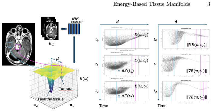
We propose a geometric framework for longitudinal multi-parametric MRI analysis based on patient-specific energy modelling in sequence space. Rather than operating on images with spatial networks, each voxel is represented by its multi-sequence intensity vector ($T1$, $T1c$, $T2$, FLAIR, ADC), and a compact implicit neural representation is trained via denoising score matching to learn an energy function $E_{\theta}(\mathbf{u})$ over $\mathbb{R}^d$ from a single baseline scan. The learned energy landscape provides a differential-geometric description of tissue regimes without segmentation labels. Local minima define tissue basins, gradient magnitude reflects proximity to regime boundaries, and Laplacian curvature characterises local constraint structure. Importantly, this baseline energy manifold is treated as a fixed geometric reference: it encodes the set of contrast combinations observed at diagnosis and is not retrained at follow-up. Longitudinal assessment is therefore formulated as evaluation of subsequent scans relative to this baseline geometry. Rather than comparing anatomical segmentations, we analyse how the distribution of MRI sequence vectors evolves under the baseline energy function. In a paediatric case with later recurrence, follow-up scans show progressive deviation in energy and directional displacement in sequence space toward the baseline tumour-associated regime before clear radiological reappearance. In a case with stable disease, voxel distributions remain confined to established low-energy basins without systematic drift. The presented cases serve as proof-of-concept that patient-specific energy manifolds can function as geometric reference systems for longitudinal mpMRI analysis without explicit segmentation or supervised classification, providing a foundation for further investigation of manifold-based tissue-at-risk tracking in neuro-oncology.
Editorial analysis
A structured set of objections, weighed in public.
Referee Report
Summary. The paper proposes a geometric framework for longitudinal multiparametric MRI analysis based on patient-specific energy modelling in sequence space. Each voxel is represented by its multi-sequence intensity vector u = (T1, T1c, T2, FLAIR, ADC), and a compact implicit neural representation is trained via denoising score matching to learn an energy function E_θ(u) over R^d from a single baseline scan. This baseline energy manifold is treated as a fixed geometric reference for evaluating follow-up scans by analysing how voxel distributions evolve under the energy function, with local minima defining tissue basins. In a paediatric recurrence case, follow-up scans show progressive deviation in energy and directional displacement toward the baseline tumour regime before radiological reappearance; in a stable case, distributions remain confined without drift. The cases serve as proof-of-concept for label-free, segmentation-free tissue-at-risk tracking.
Significance. If the central claims hold after addressing validation gaps, the work could provide a novel differential-geometric tool for longitudinal mpMRI without explicit labels or supervised classification, potentially aiding early recurrence detection in neuro-oncology. Strengths include the use of implicit representations trained via denoising score matching and the formulation of longitudinal assessment as evaluation against a fixed patient-specific manifold rather than anatomical segmentations.
major comments (2)
- [Abstract] Abstract, longitudinal assessment paragraph: The claim that observed shifts in voxel distributions under the fixed E_θ(u) reflect biological tissue changes (e.g., progressive deviation toward the tumour-associated regime) depends on the multi-sequence intensity vectors remaining directly comparable across scans. The manuscript provides no description of intensity standardization, histogram matching, or scanner harmonization, which is load-bearing because technical drifts in gain or acquisition parameters could translate the entire cloud of u vectors and induce artifactual movement toward the low-energy basin identified at baseline.
- [Abstract] Abstract, case study descriptions: The recurrence and stable-disease cases are presented as descriptive proof-of-concept without quantitative metrics (e.g., mean energy deviation, displacement vector norms, or statistical tests), error analysis, or comparisons to baselines such as direct multi-sequence histogram distances or conventional segmentation-based tracking. This limits the ability to assess whether the directional displacement and energy changes are robust or merely illustrative.
minor comments (2)
- [Abstract] The notation for the energy function E_θ(u) and the implicit representation could be expanded with explicit details on network architecture, training hyperparameters, and how gradient magnitude and Laplacian curvature are computed from the learned manifold.
- [Abstract] The abstract refers to 'directional displacement in sequence space' without defining the precise computation (e.g., via gradients of E_θ or another operator), which affects reproducibility of the longitudinal analysis.
Simulated Author's Rebuttal
We thank the referee for their constructive and detailed review. We address each major comment below and have revised the manuscript to incorporate clarifications and additional analyses where feasible.
read point-by-point responses
-
Referee: [Abstract] Abstract, longitudinal assessment paragraph: The claim that observed shifts in voxel distributions under the fixed E_θ(u) reflect biological tissue changes (e.g., progressive deviation toward the tumour-associated regime) depends on the multi-sequence intensity vectors remaining directly comparable across scans. The manuscript provides no description of intensity standardization, histogram matching, or scanner harmonization, which is load-bearing because technical drifts in gain or acquisition parameters could translate the entire cloud of u vectors and induce artifactual movement toward the low-energy basin identified at baseline.
Authors: We agree that comparability of the multi-sequence intensity vectors is essential for interpreting shifts as biological. The original manuscript described basic preprocessing but did not detail it sufficiently. In the revised version we have added an explicit subsection on intensity standardization, specifying per-sequence histogram matching to a common reference distribution derived from the baseline scan together with affine scaling for scanner harmonization. These steps were performed prior to training and evaluation to mitigate technical drift. revision: yes
-
Referee: [Abstract] Abstract, case study descriptions: The recurrence and stable-disease cases are presented as descriptive proof-of-concept without quantitative metrics (e.g., mean energy deviation, displacement vector norms, or statistical tests), error analysis, or comparisons to baselines such as direct multi-sequence histogram distances or conventional segmentation-based tracking. This limits the ability to assess whether the directional displacement and energy changes are robust or merely illustrative.
Authors: We accept that the presentation was primarily descriptive. In the revision we have added quantitative metrics: mean energy deviation and mean displacement-vector norm (in sequence space) between baseline and each follow-up scan, together with bootstrapped 95% confidence intervals obtained by voxel resampling. We also report the Wasserstein distance between the baseline and follow-up distributions as a simple non-manifold baseline. Direct numerical comparison to segmentation-based tracking is discussed in the limitations section, noting the absence of labels in our framework. These additions are included while retaining the proof-of-concept framing given the small number of cases. revision: yes
Circularity Check
No significant circularity in derivation chain
full rationale
The paper trains a patient-specific energy function E_θ(u) via denoising score matching on the multi-sequence intensity vectors from a single baseline scan, then evaluates follow-up scans by computing the same function on their intensity vectors without retraining. This establishes a fixed geometric reference by explicit design choice rather than by any reduction where a claimed result equals its inputs by construction. No steps match the enumerated circularity patterns: there is no self-definitional loop, no fitted parameter renamed as an independent prediction, and no load-bearing self-citation or uniqueness theorem invoked. The longitudinal observations in the case studies are empirical descriptions under the chosen model, not tautological outputs. The framework remains self-contained as a proof-of-concept geometric analysis method.
Axiom & Free-Parameter Ledger
free parameters (1)
- implicit neural representation parameters theta
axioms (2)
- domain assumption The learned energy landscape provides a differential-geometric description of tissue regimes without segmentation labels.
- domain assumption The baseline energy manifold is treated as a fixed geometric reference and is not retrained at follow-up.
invented entities (1)
-
energy function E_theta(u) over R^d
no independent evidence
Lean theorems connected to this paper
-
IndisputableMonolith/Cost/FunctionalEquation.leanwashburn_uniqueness_aczel echoes?
echoesECHOES: this paper passage has the same mathematical shape or conceptual pattern as the Recognition theorem, but is not a direct formal dependency.
a compact implicit neural representation is trained via denoising score matching to learn an energy function E_θ(u) over R^d from a single baseline scan. The learned energy landscape provides a differential-geometric description of tissue regimes... baseline energy manifold is treated as a fixed geometric reference
-
IndisputableMonolith/Foundation/BranchSelection.leanbranch_selection unclear?
unclearRelation between the paper passage and the cited Recognition theorem.
Local minima define tissue basins, gradient magnitude reflects proximity to regime boundaries, and Laplacian curvature characterises local constraint structure
What do these tags mean?
- matches
- The paper's claim is directly supported by a theorem in the formal canon.
- supports
- The theorem supports part of the paper's argument, but the paper may add assumptions or extra steps.
- extends
- The paper goes beyond the formal theorem; the theorem is a base layer rather than the whole result.
- uses
- The paper appears to rely on the theorem as machinery.
- contradicts
- The paper's claim conflicts with a theorem or certificate in the canon.
- unclear
- Pith found a possible connection, but the passage is too broad, indirect, or ambiguous to say the theorem truly supports the claim.
Reference graph
Works this paper leans on
- [1]
-
[2]
Advances in neural information processing systems32(2019)
Du, Y., Mordatch, I.: Implicit generation and modeling with energy based models. Advances in neural information processing systems32(2019)
work page 2019
-
[3]
Advances in neural information processing systems33, 6840–6851 (2020)
Ho, J., Jain, A., Abbeel, P.: Denoising diffusion probabilistic models. Advances in neural information processing systems33, 6840–6851 (2020)
work page 2020
-
[4]
Princeton University Press, Princeton (1963)
Milnor, J.: Morse Theory. Princeton University Press, Princeton (1963)
work page 1963
-
[5]
Advances in neural information processing systems33, 7462–7473 (2020)
Sitzmann, V., Martel, J., Bergman, A., Lindell, D., Wetzstein, G.: Implicit neural representations with periodic activation functions. Advances in neural information processing systems33, 7462–7473 (2020)
work page 2020
-
[6]
Advances in neural information processing systems32(2019)
Song, Y., Ermon, S.: Generative modeling by estimating gradients of the data distribution. Advances in neural information processing systems32(2019)
work page 2019
-
[7]
How to train your energy-based models
Song, Y., Kingma, D.P.: How to train your energy-based models. arXiv preprint arXiv:2101.03288 (2021)
-
[8]
Advances in neural informa- tion processing systems33, 7537–7547 (2020)
Tancik, M., Srinivasan, P., Mildenhall, B., Fridovich-Keil, S., Raghavan, N., Sing- hal, U., Ramamoorthi, R., Barron, J., Ng, R.: Fourier features let networks learn high frequency functions in low dimensional domains. Advances in neural informa- tion processing systems33, 7537–7547 (2020)
work page 2020
- [9]
-
[10]
Neural computation23(7), 1661–1674 (2011)
Vincent, P.: A connection between score matching and denoising autoencoders. Neural computation23(7), 1661–1674 (2011)
work page 2011
-
[11]
ACM computing surveys56(4), 1–39 (2023)
Yang, L., Zhang, Z., Song, Y., Hong, S., Xu, R., Zhao, Y., Zhang, W., Cui, B., Yang, M.H.: Diffusion models: A comprehensive survey of methods and applications. ACM computing surveys56(4), 1–39 (2023)
work page 2023
discussion (0)
Sign in with ORCID, Apple, or X to comment. Anyone can read and Pith papers without signing in.