Coherent Control of Nanoscale Nuclear Spin Ensembles in the Spin Noise Regime
Pith reviewed 2026-05-10 17:45 UTC · model grok-4.3
The pith
The initial RF phase and its orientation to the NV axis determine whether nuclear spin rotations produce full, partial, or zero contrast in the readout.
A machine-rendered reading of the paper's core claim, the machinery that carries it, and where it could break.
Core claim
In correlation spectroscopy of nanoscale nuclear spin ensembles driven by RF fields, the resulting nuclear spin evolution depends critically on the initial RF phase and its orientation relative to the NV crystalline axis; as a result, the same nuclear rotation can produce full, partial, or vanishing contrast in the subsequent NV-center readout.
What carries the argument
The phase- and orientation-dependent evolution of nuclear spins under RF irradiation in correlation spectroscopy, which directly maps the applied field's initial conditions onto the measured contrast.
If this is right
- Reliable calibration of RF phase and orientation becomes necessary for consistent contrast in multidimensional nanoscale NMR experiments.
- Failure to control these parameters can produce ambiguous signals that lead to misinterpretation of nuclear spin dynamics.
- Protocol design for correlation-based spin resonance must incorporate the relative orientation between the RF field and the NV axis to avoid vanishing signals.
- The same RF pulse sequence can be used to toggle between detectable and undetectable regimes by changing only the initial phase.
Where Pith is reading between the lines
- Similar phase-sensitivity effects may appear in other solid-state spin sensors when RF or microwave control is applied in the noise-dominated regime.
- Phase variation could be turned into an additional control knob to selectively enhance or suppress specific nuclear species in mixed ensembles.
- The finding suggests that existing calibration routines for NV-based NMR should be extended to include phase as a monitored variable rather than assumed constant.
Load-bearing premise
The measured changes in contrast are produced primarily by the RF phase and its alignment with the NV axis rather than by decoherence, pulse imperfections, or ensemble inhomogeneities.
What would settle it
Systematically varying the initial RF phase while keeping orientation fixed and checking whether the NV readout contrast follows the predicted full-to-zero variation pattern.
Figures
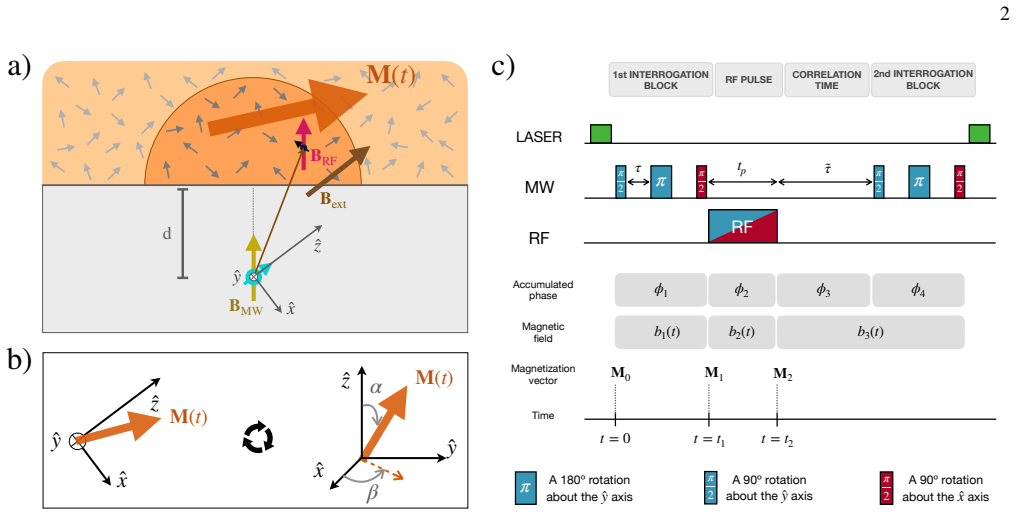

read the original abstract
Spin defects in solids, such as the nitrogen-vacancy (NV) center in diamond, have emerged as a key tool for detecting nuclear spins at the nanoscale. While active nuclear spin control via radio-frequency (RF) irradiation is often unnecessary for standard spin-noise detection, it becomes essential for advanced protocols like multidimensional nanoscale NMR. In this work, we investigate nuclear spin control using correlation spectroscopy techniques. We demonstrate, both theoretically and experimentally, that the resulting nuclear spin dynamics depend critically on the initial RF phase and its orientation relative to the NV crystalline axis. Depending on these parameters, identical nuclear rotations can yield full, partial, or even vanishing contrast in the NV readout. These findings highlight a previously underappreciated aspect of spin manipulation in the spin-noise regime: the link between the phase and direction of the applied RF field and its direct impact on correlation-based experiments. Consequently, imperfect calibration of these parameters can lead to ambiguous signal contrasts and misinterpretation of the underlying nuclear spin dynamics. Our results provide deeper insight into nanoscale spin control and pave the way toward reliable multidimensional spin resonance experiments.
Editorial analysis
A structured set of objections, weighed in public.
Referee Report
Summary. The manuscript investigates the coherent control of nanoscale nuclear spin ensembles using NV centers in diamond within the spin noise regime. Through correlation spectroscopy, the authors demonstrate both theoretically and experimentally that nuclear spin dynamics under RF irradiation depend critically on the initial RF phase and its orientation relative to the NV crystalline axis. This dependence can result in full, partial, or vanishing contrast in the NV readout for identical nuclear rotations, underscoring the need for precise calibration to prevent ambiguous signal interpretation in multidimensional nanoscale NMR experiments.
Significance. If the findings hold, they are significant for the development of reliable advanced protocols in nanoscale NMR and quantum sensing. The work highlights an important but previously underappreciated link between RF field parameters and readout contrast, providing both a theoretical framework and experimental evidence. This could lead to improved calibration procedures and more accurate interpretation of nuclear spin dynamics, benefiting the broader community working on spin defect-based sensors. The paper's strength lies in its direct experimental verification of the phase and orientation effects.
major comments (1)
- The central claim that contrast variations arise primarily from RF phase and orientation (rather than decoherence, pulse imperfections, or ensemble inhomogeneities) is load-bearing for the experimental support; the manuscript should include a quantitative error budget or control experiment in the results section demonstrating that these other factors do not dominate the observed full/partial/vanishing contrast.
minor comments (3)
- Abstract: the description of 'vanishing contrast' would benefit from a brief quantitative statement of the observed values (with uncertainties) to convey the magnitude of the effect.
- Figure captions: ensure all panels explicitly label the RF phase and orientation angles used, matching the notation in the main text.
- The discussion of calibration ambiguity is clear, but a short table summarizing the contrast outcomes for the key phase/orientation combinations would improve readability.
Simulated Author's Rebuttal
We thank the referee for their positive assessment of the manuscript, recognition of its significance for nanoscale NMR protocols, and recommendation for minor revision. We address the single major comment below and will incorporate the requested strengthening of the experimental support.
read point-by-point responses
-
Referee: The central claim that contrast variations arise primarily from RF phase and orientation (rather than decoherence, pulse imperfections, or ensemble inhomogeneities) is load-bearing for the experimental support; the manuscript should include a quantitative error budget or control experiment in the results section demonstrating that these other factors do not dominate the observed full/partial/vanishing contrast.
Authors: We agree that a quantitative demonstration is necessary to establish that the observed contrast variations are driven by the RF phase and orientation rather than competing mechanisms. Our theoretical framework derives the NV contrast directly from the phase-dependent projection of the nuclear spin rotation onto the NV axis, predicting full, partial, or zero contrast for identical rotation angles solely as a function of initial RF phase and field orientation. The experimental data reproduce these predictions when the RF phase is stepped while all other parameters are held fixed. To address the referee's concern, the revised manuscript will add a dedicated subsection to the Results section containing (i) a quantitative error budget that bounds the contributions from nuclear decoherence (using measured T2* values in the spin-noise regime), RF pulse imperfections (from calibrated Rabi frequencies with <5% variation), and ensemble inhomogeneities (from the known NV distribution and crystalline-axis spread), showing each is at least an order of magnitude smaller than the phase-induced contrast swing; and (ii) a control dataset in which the RF phase is fixed at the zero-contrast condition while small deliberate detunings or extended free-evolution times are introduced, confirming that these perturbations do not reproduce the sharp, phase-specific contrast changes reported in the main figures. revision: yes
Circularity Check
No significant circularity; derivation is self-contained with independent experimental support
full rationale
The paper claims a theoretical derivation of nuclear spin dynamics under RF control in the spin-noise regime, followed by experimental verification that contrast depends on initial RF phase and NV-axis orientation. No load-bearing steps reduce by construction to fitted inputs or self-citations: the phase/orientation dependence is derived from standard spin Hamiltonian terms and then tested against independent measurements rather than being defined by the same data. The abstract and skeptic analysis confirm the result does not rely on renaming known patterns or smuggling ansatzes via prior self-work. This is the normal case of a self-contained experimental-theoretical study.
Axiom & Free-Parameter Ledger
Reference graph
Works this paper leans on
-
[1]
S. J. DeVience, L. M. Pham, I. Lovchinsky, A. O. Sushkov, N. Bar-Gill, C. Belthangady, F. Casola, Ma. Corbett, H. Zhang, M. Lukin, H. Park, A. Yacoby, and R. L. Walsworth,Nanoscale NMR spectroscopy and imaging of multiple nuclear species. Nature Nanotechnology10, 129 (2015)
work page 2015
-
[2]
I. Lovchinsky, A. O. Sushkov, E. Urbach, N. P. de Leon, S. Choi, K. De Greve, R. Evans, R. Gertner, E. Bersin, C. M ¨uller, L. McGuinness, F. Jelezko, R. L. Walsworth, H. Park, and M. D. Lukin,Nuclear magnetic resonance detection and spec- troscopy of single proteins using quantum logic. Science351, 6275 (2016)
work page 2016
-
[3]
I. Lovchinsky, J. D. Sanchez-Yamagishi, E. K. Urbach, S. Choi, S. Fang, T. I. Andersen, K. Watanabe, T. Taniguchi, A. Bylin- skii, E. Kaxiras, P. Kim, H. Park, and M. D. Lukin,Magnetic resonance spectroscopy of an atomically thin material using a single-spin qubit. Science355, 6324 (2017)
work page 2017
-
[4]
R. D. Allert, K. D. Briegel, and D. B. Bucher,Advances in nano- and microscale NMR spectroscopy using diamond quan- tum sensors. Chemical Communications58, 59 (2022)
work page 2022
-
[5]
J. M. Abendroth, K. Herb, E. Janitz, T. Zhu, L. A. V ¨olker, and C. L. Degen,Single-Nitrogen–Vacancy NMR of Amine- Functionalized Diamond Surfaces. Nano Letters22, 18 (2022)
work page 2022
-
[6]
R. Rizzato, N. R. von Grafenstein, and D. B. Bucher,Quan- tum sensors in diamonds for magnetic resonance spectroscopy: Current applications and future prospects. Applied Physics Letters123, 26 (2023)
work page 2023
-
[7]
J. Du, F. Shi, X. Kong, F. Jelezko, and J. Wrachtrup,Single- molecule scale magnetic resonance spectroscopy using quan- tum diamond sensors. Reviews of Modern Physics96, 2 (2024)
work page 2024
-
[8]
C. L. Degen, M. Poggio1, H. J. Mamin1, and D. Rugar1,Role of Spin Noise in the Detection of Nanoscale Ensembles of Nuclear Spins. Physical Review Letters99, 250601 (2007)
work page 2007
-
[9]
C. A. Meriles, L. Jiang, G. Goldstein, J. S. Hodges, J. Maze, M. D. Lukin, and P. Cappellaro,Imaging mesoscopic nuclear spin noise with a diamond magnetomeeter. Journal of Chemical Physics133, 124105 (2010)
work page 2010
-
[10]
N. M ¨uller, A. Jerschow, and J. Schlagnitweit,Nuclear Spin Noise. eMagRes2, 2 (2013)
work page 2013
-
[11]
A. Laraoui, F. Dolde, C. Burk, F. Reinhard, J. Wrachtrup, and C. A. Meriles,High-resolution correlation spectroscopy of 13C spins near a nitrogen-vacancy centre in diamond. Nature Com- munications4, 1651 (2013)
work page 2013
-
[12]
P. Kehayias, A. Jarmola, N. Mosavian, I. Fescenko, F. M. Ben- ito, A. Laraoui, J. Smits, L. Bougas, D. Budker, A. Neumann, S. R. J. Brueck, and V . M. Acosta,Solution nuclear magnetic resonance spectroscopy on a nanostructured diamond chip. Na- ture Communications8, 188 (2017)
work page 2017
- [13]
-
[14]
K. S. Liu, A. Henning, M. W. Heindl, R. D. Allert, J. D. Bartl, I. D. Sharp, R. Rizzato, and D. B. Bucher,Surface NMR using quantum sensors in diamond. PNAS119, 5 (2021)
work page 2021
-
[15]
K. S. Liu, X. Ma, R. Rizzato, A. L. Semrau, A. Henning, I. D. Sharp, R. A. Fischer, and D. B. Bucher,Using Metal- Or- ganic Frameworks to Confine Liquid Samples for Nanoscale NV-NMR. Nano Letters22, 9876 (2022)
work page 2022
- [16]
-
[17]
F. Bruckmaier, R. D. Allert, N. R. Neuling, P. Amrein, S. Lit- tin, K. D. Briegel, P. Sch ¨atzle, P. Knittel, M. Zaitsev, and D. B. Bucher,Imaging local diffusion in microstructures using NV-based pulsed field gradient NMR. Science Advances9, 33 (2023)
work page 2023
-
[18]
J. M. Boss, K. Chang1, J. Armijo, K. Cujia, T. Rosskopf, J. R. Maze, and C. L. Degen,One- and Two-Dimensional Nuclear 6 Magnetic Resonance Spectroscopy with a Diamond Quantum Sensor. Physical Review Letters116, 197601 (2016)
work page 2016
-
[19]
D. R. Glenn, D. B. Bucher, J. Lee, M. D. Lukin, H. Park, and R. L. Walsworth,High-resolution magnetic resonance spectroscopy using a solid-state spin sensor. Nature555, 351 (2018)
work page 2018
- [20]
-
[21]
K. D. Briegel, N. R. von Grafenstein, J. C. Draeger, P. Bl¨umler, R. D. Allert, and D. B. Bucher,Optical widefield nuclear mag- netic resonance microscopy. Nature Communications16, 1281 (2015)
work page 2015
-
[22]
D. Yudilevich, A. Salhov, I. Schaefer, K. Herb, A. Retzker, and A. Finkler,Coherent manipulation of nuclear spins in the strong driving regime. New Journal of Physics25, 113042 (2023)
work page 2023
-
[23]
T. Staudacher, F. Shi, S. Pezzagna, J. Meijer, J. Du, C. A. Mer- iles, F. Reinhard, and J. Wrachtrup,Nuclear Magnetic Reso- nance Spectroscopy on a (5-Nanometer)3 Sample Volume. Sci- ence339(2013)
work page 2013
-
[24]
H. J. Mamin, M. Kim, M. H. Sherwood, C. T. Rettner, K. Ohno, D. D. Awschalom, and D. Rugar,Nanoscale Nuclear Magnetic Resonance with a Nitrogen-Vacancy Spin Sensor. Science339, 6119 (2013)
work page 2013
-
[25]
Physical Review Letters119033001 (2017)
Heiße, F., K ¨ohler-Langes, F., Rau, S., Hou, J., Junck, S., Kracke, A., Mooser, A., Quint, W., Ulmer, S., Werth, G., Blaum, K., and Sturm, S.,High-Precision Measurement of the Proton’s Atomic Mass. Physical Review Letters119033001 (2017)
work page 2017
-
[26]
D. B. Bucher, D. P. L. Aude Craik, M. P. Backlund, M. J. Turner, O. B. Dor, D. R. Glenn, and R. L. Walsworth,Quan- tum diamond spectrometer for nanoscale NMR and ESR spec- troscopy. Nature Protocols14, 2707 (2019)
work page 2019
-
[27]
K. J. Brown, E. Chartier, E. M. Sweet, D. A. Hopper, and L. C. Basset,Cleaning diamond surfaces using boiling acid treatment in a standard laboratory chemical hood. Journal of Chemical Health and Safety26, 6 (2019). Appendix A: Dynamics of a spin subjected to RF fields We consider a single nuclear spin in an external magnetic field oriented along thez-axi...
work page 2019
-
[28]
Note, the NV detects nuclear spins within a hemispherical volume of radiusdabove it [23, 24]
Sample magnetic field in terms of magnetization vectors We consider an NV center located at a depthdof approximately a few nanometers below the diamond surface, beneath a sample containingNspins. Note, the NV detects nuclear spins within a hemispherical volume of radiusdabove it [23, 24]. The joint NV–nuclei dynamics is governed by H=DS 2 z −γ eBextS z − ...
-
[29]
Magnetic fields detected by the NV during combined MW and RF sequence The system Hamiltonian for a single NV reads H=DS 2 z −γ eBextS z −γ e bz(t)S z + √ 2ΩS x cos (ωMWt−φ MW) ,(B8) whereΩ MW andω MW are the amplitude and frequency of the MW control driving. When an RF pulse is applied to the sample, the magnetic field detected by the NV center is modifie...
-
[30]
Sample Preparation A 2×2×0.5 mm electronic-grade diamond crystal ( 12C-enriched, Fraunhofer IAF, Freiburg) was implanted with nitrogen at an energy of 2.5 keV , using a 7◦ off-axis tilt and a fluence of 2×1012 cm−2 by Innovion, and subsequently annealed following the protocol described by Bucheret al.[26]. Before the experiments, the diamond was cleaned u...
-
[31]
Correlation Experiment Correlation spectroscopy measurements were performed using two XY8-4 dynamical decoupling blocks separated by a vari- able correlation interval (˜τ). Each experimental cycle included a 5µs laser pulse for spin initialization and readout, and MWπ/2 andπpulses of 20 ns and 40 ns duration, respectively. The |0⟩ ↔ |−1⟩ transition was ad...
-
[32]
Experimental Setup The experiments were conducted on the 12C-enriched diamond crystal glued onto a thin glass coverslip. A 6 mm diameter glass hemisphere (TECHSPEC® N-BK7 Half-Ball Lens, Edmund Optics) was affixed to the underside of the coverslip to enhance fluorescence collection efficiency. The coverslip was mounted on a 30 mm cage plate (CP4S, Thorlab...
discussion (0)
Sign in with ORCID, Apple, or X to comment. Anyone can read and Pith papers without signing in.