Hybrid Multi-Dimensional MRI Prostate Cancer Detection via Hadamard Network-Based Bias Correction and Residual Networks
Pith reviewed 2026-05-10 06:47 UTC · model grok-4.3
The pith
A two-stage network first corrects bias fields in hybrid MRI maps then classifies patches with ResNet to detect prostate cancer.
A machine-rendered reading of the paper's core claim, the machinery that carries it, and where it could break.
Core claim
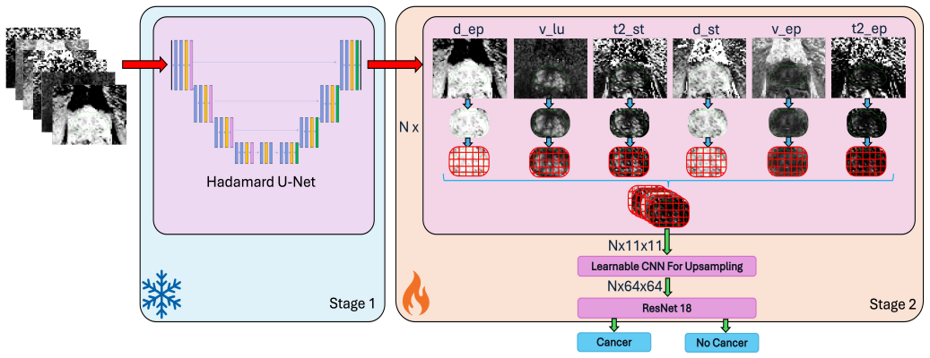
The authors state that the HBR-Net-18 framework, which uses a Hadamard U-Net to suppress bias fields across six HM-MRI parametric maps generated by a physics-informed autoencoder and then applies ResNet-18 to classify 11-by-11 overlapping patches that incorporate 2D and 3D context, delivers balanced sensitivity and specificity that exceed those of conventional radiomics methods and baseline CNN models.
What carries the argument
The Hadamard-Bias Network that applies a U-Net to remove intensity inhomogeneities from the parametric maps before ResNet-18 performs patch-level classification.
If this is right
- The corrected parametric maps enable more consistent patch classification than uncorrected images.
- Incorporation of adjacent-slice information improves spatial coherence of the detection output.
- The overall pipeline supports direct clinical use by balancing sensitivity and specificity without additional manual steps.
Where Pith is reading between the lines
- The same bias-correction step could be tested on quantitative maps from other organs or MRI protocols.
- Patch-level output could be aggregated into full-volume segmentations with limited additional training.
- Performance on multi-center data with varying scanner protocols would indicate robustness beyond the current experiments.
Load-bearing premise
The Hadamard U-Net reliably removes intensity inhomogeneities from the parametric maps and the 2D-plus-3D patch classification supplies enough spatial context for accurate cancer detection.
What would settle it
An independent test set where the framework shows lower sensitivity or specificity than the radiomics or baseline CNN comparators would disprove the performance advantage.
Figures

read the original abstract
Magnetic Resonance Imaging (MRI) is vital for prostate cancer (PCa) diagnosis. While advanced techniques such as Hybrid Multi-dimensional MRI (HM-MRI) have enhanced diagnostic capabilities, the significant need remains for robust, automated Artificial Intelligence (AI)-based detection methods. In this study, we combine quantitative HM-MRI of tissue composition with an AI-based neural network. We propose the Hadamard-Bias Network plus ResNet18 (HBR-Net-18), a two-stage AI framework for PCa detection. In the first stage, a Hadamard U-Net-based algorithm suppresses intensity inhomogeneities (bias fields) across six parametric HM-MRI maps generated via a Physics-Informed Autoencoder (PIA). In the second stage, a Residual Network (ResNet-18) performs patch-level classification. The framework utilizes overlapping 11-by-11 patches, incorporating both 2D intra-slice and 3D inter-slice (adjacent-slice) information to improve spatial consistency. Our experimental results demonstrate that HB-Net achieves balanced sensitivity and specificity, significantly outperforming conventional radiomics-based approaches and baseline CNN models, highlighting its potential for clinical deployment.
Editorial analysis
A structured set of objections, weighed in public.
Referee Report
Summary. The manuscript proposes a two-stage framework (HBR-Net-18 or HB-Net) for prostate cancer detection on Hybrid Multi-dimensional MRI (HM-MRI). Stage 1 applies a Hadamard U-Net to suppress intensity inhomogeneities across six parametric maps produced by a Physics-Informed Autoencoder (PIA). Stage 2 feeds overlapping 11×11 patches (incorporating 2D intra-slice and 3D adjacent-slice information) into a ResNet-18 for patch-level classification. The central claim is that the resulting model achieves balanced sensitivity and specificity while significantly outperforming conventional radiomics approaches and baseline CNN models, with potential for clinical deployment.
Significance. If substantiated with rigorous validation, the work could contribute to more robust automated PCa detection by combining quantitative HM-MRI tissue maps with targeted bias-field correction and multi-dimensional patch classification. Mitigating MRI intensity inhomogeneities remains a practical barrier in clinical imaging, and the hybrid 2D/3D strategy may improve spatial consistency; the overall pipeline offers a concrete example of physics-informed preprocessing paired with residual networks.
major comments (2)
- [Results] Results section: the headline claim that HB-Net 'significantly outperforming conventional radiomics-based approaches and baseline CNN models' is unsupported by any reported numerical values for sensitivity, specificity, AUC, or statistical tests (p-values, confidence intervals). This omission is load-bearing because the abstract and introduction position outperformance as the primary evidence of the framework's value.
- [Methods] Methods and Results sections: no quantitative metrics (coefficient of variation, NMI, or bias-field residual error) or ablation studies are presented to demonstrate that the Hadamard U-Net actually suppresses inhomogeneities or that the 3D adjacent-slice information in the ResNet-18 improves spatial consistency. Without these controls, it is impossible to attribute any performance gains to the proposed components rather than dataset-specific factors.
minor comments (2)
- [Abstract] The acronyms HB-Net and HBR-Net-18 are used without explicit definition of their relationship; consistent terminology would reduce ambiguity.
- A summary table comparing sensitivity, specificity, and other metrics across all methods (radiomics, baseline CNNs, and HB-Net) with error bars would substantially improve clarity of the performance claims.
Simulated Author's Rebuttal
We thank the referee for the detailed and constructive feedback on our manuscript. We appreciate the emphasis on rigorous validation and have addressed each major comment below. The suggested revisions will improve the clarity and substantiation of our claims.
read point-by-point responses
-
Referee: [Results] Results section: the headline claim that HB-Net 'significantly outperforming conventional radiomics-based approaches and baseline CNN models' is unsupported by any reported numerical values for sensitivity, specificity, AUC, or statistical tests (p-values, confidence intervals). This omission is load-bearing because the abstract and introduction position outperformance as the primary evidence of the framework's value.
Authors: We agree that the Results section would benefit from a more explicit and self-contained presentation of the quantitative performance metrics to directly support the outperformance claim. Although the abstract summarizes the balanced sensitivity and specificity and the figures/tables contain the detailed comparisons, we will revise the Results section to include a dedicated paragraph explicitly reporting the sensitivity, specificity, AUC values for HB-Net versus the radiomics and baseline CNN models, along with the associated statistical tests (p-values and confidence intervals). This addition will ensure the headline claim is fully substantiated within the main text without relying on cross-references. revision: yes
-
Referee: [Methods] Methods and Results sections: no quantitative metrics (coefficient of variation, NMI, or bias-field residual error) or ablation studies are presented to demonstrate that the Hadamard U-Net actually suppresses inhomogeneities or that the 3D adjacent-slice information in the ResNet-18 improves spatial consistency. Without these controls, it is impossible to attribute any performance gains to the proposed components rather than dataset-specific factors.
Authors: We acknowledge that quantitative controls would strengthen attribution of performance gains to the individual components. We will add to the Results section quantitative metrics evaluating the Hadamard U-Net bias correction, including coefficient of variation and normalized mutual information (NMI) computed on the parametric maps before and after correction. We will also include ablation experiments comparing the full HBR-Net-18 model against variants that omit the bias-correction stage and that use only 2D (intra-slice) patches without adjacent-slice information. These additions will provide direct evidence for the contribution of each proposed element. revision: yes
Circularity Check
No circularity: standard empirical ML pipeline with no self-referential derivations
full rationale
The paper describes a two-stage empirical framework (Hadamard U-Net bias correction on PIA-derived parametric maps followed by ResNet-18 patch classification) evaluated on MRI datasets. No equations, predictions, or first-principles results are presented that reduce to the inputs by construction. There are no self-definitional loops, fitted parameters renamed as predictions, load-bearing self-citations of uniqueness theorems, or ansatzes smuggled via prior work. All performance claims rest on experimental comparisons to radiomics and baseline CNNs rather than tautological identities. This is a conventional applied computer-vision study whose validity hinges on data and ablations, not on circular reasoning.
Axiom & Free-Parameter Ledger
Reference graph
Works this paper leans on
-
[1]
The diagnosis and treatment of prostate cancer: a review,
Mark S Litwin and Hung-Jui Tan, “The diagnosis and treatment of prostate cancer: a review,”Jama, vol. 317, no. 24, pp. 2532–2542, 2017
work page 2017
-
[2]
Hybrid multi-dimensional mri of prostate cancer,
Aritrick Chatterjee and Aytekin Oto, “Hybrid multi-dimensional mri of prostate cancer,” inCancer Detection and Diagnosis, pp. 155–162. CRC Press, 2025
work page 2025
-
[3]
Radiomics in prostate cancer: An up-to-date review,
Matteo Ferro, Ottavio de Cobelli, Gennaro Musi, Francesco Del Giu- dice, Giuseppe Carrieri, Gian Maria Busetto, Ugo Giovanni Falagario, Alessandro Sciarra, Martina Maggi, Felice Crocetto, et al., “Radiomics in prostate cancer: An up-to-date review,”Therapeutic Advances in Urology, vol. 14, pp. 17562872221109020, 2022
work page 2022
-
[4]
Multipara- metric mri and radiomics in prostate cancer: a review,
Yu Sun, Hayley M Reynolds, Bimal Parameswaran, Darren Wraith, Mary E Finnegan, Scott Williams, and Annette Haworth, “Multipara- metric mri and radiomics in prostate cancer: a review,”Australasian physical & engineering sciences in medicine, vol. 42, no. 1, pp. 3–25, 2019
work page 2019
-
[5]
Anindo Saha, Joeran S Bosma, Jasper J Twilt, Bram van Ginneken, Anders Bjartell, Anwar R Padhani, David Bonekamp, Geert Villeirs, Georg Salomon, Gianluca Giannarini, et al., “Artificial intelligence and radiologists in prostate cancer detection on mri (pi-cai): an international, paired, non-inferiority, confirmatory study,”The Lancet Oncology, vol. 25, no...
work page 2024
-
[6]
Ai-powered prostate cancer detection: a multi-centre, multi-scanner validation study,
Francesco Giganti, Nadia Moreira da Silva, Michael Yeung, Lucy Davies, Amy Frary, Mirjana Ferrer Rodriguez, Nikita Sushentsev, Nicholas Ashley, Adrian Andreou, Alison Bradley, et al., “Ai-powered prostate cancer detection: a multi-centre, multi-scanner validation study,” European Radiology, pp. 1–10, 2025
work page 2025
-
[7]
Feng Liu, Yuanshen Zhao, Jukun Song, Guilan Tu, Yadong Liu, Yunsong Peng, Jiahui Mao, Chongzhe Yan, and Rongpin Wang, “A hybrid classification model with radiomics and cnn for high and low grading of prostate cancer gleason score on mp-mri,”Displays, vol. 83, pp. 102703, 2024
work page 2024
-
[8]
Md Shakhawat Hossain, Md Sahilur Rahman, Munim Ahmed, Anowar Hussen, Zahid Ullah, and Mona Jamjoom, “Automated gleason grading of prostate cancer from low-resolution histopathology images using an ensemble network of cnn and transformer models.,”Computers, Materials & Continua, vol. 84, no. 2, 2025
work page 2025
-
[9]
Batuhan Gundogdu, Aritrick Chatterjee, Milica Medved, Ulas Bagci, Gregory S Karczmar, and Aytekin Oto, “Physics-informed autoencoder for prostate tissue microstructure profiling with hybrid multidimensional mri,”Radiology: Artificial Intelligence, vol. 7, no. 2, pp. e240167, 2025
work page 2025
-
[10]
A probabilistic hadamard u-net for mri bias field correction,
Xin Zhu, Hongyi Pan, Batuhan Gundogdu, Debesh Jha, Yury Velichko, Adam B Murphy, Ashley Ross, Baris Turkbey, Ahmet Enis Cetin, and Ulas Bagci, “A probabilistic hadamard u-net for mri bias field correction,” inInternational Workshop on Machine Learning in Medical Imaging. Springer, 2024, pp. 208–217
work page 2024
-
[11]
Deep residual learning for image recognition,
Kaiming He, Xiangyu Zhang, Shaoqing Ren, and Jian Sun, “Deep residual learning for image recognition,” inProceedings of the IEEE conference on computer vision and pattern recognition, 2016, pp. 770– 778
work page 2016
-
[12]
Aritrick Chatterjee, Roger M Bourne, Shiyang Wang, Ajit Devaraj, Alexander J Gallan, Tatjana Antic, Gregory S Karczmar, and Aytekin Oto, “Diagnosis of prostate cancer with noninvasive estimation of prostate tissue composition by using hybrid multidimensional mr imag- ing: a feasibility study,”Radiology, vol. 287, no. 3, pp. 864–873, 2018
work page 2018
-
[13]
Aritrick Chatterjee, Ambereen N Yousuf, Roger Engelmann, Carla Har- math, Grace Lee, Milica Medved, Ernest B Jamison, Abel Lorente Cam- pos, Batuhan Gundogdu, Glenn Gerber, et al., “Prospective validation of an automated hybrid multidimensional mri tool for prostate cancer detection using targeted biopsy: Comparison with pi-rads-based assess- ment,”Radiol...
work page 2025
-
[14]
N4itk: Improved n3 bias correction,
Nicholas J. Tustison, Brian B. Avants, Philip A. Cook, Yuanjie Zheng, Alexander Egan, Paul A. Yushkevich, and James C. Gee, “N4itk: Improved n3 bias correction,”IEEE Transactions on Medical Imaging, vol. 29, no. 6, pp. 1310–1320, 2010
work page 2010
-
[15]
Focal loss for dense object detection,
Tsung-Yi Lin, Priya Goyal, Ross Girshick, Kaiming He, and Piotr Doll´ar, “Focal loss for dense object detection,”IEEE Transactions on Pattern Analysis and Machine Intelligence, vol. 42, no. 2, pp. 318–327, 2020
work page 2020
-
[16]
Abel L. Campos, Aritrick Chatterjee, Roger Engelmann, Gregory S. Karczmar, and Aytek Oto, “Multi-voxel, spatially aware radiomics analysis for prostate cancer diagnosis using pia-hybrid mri biomarkers,” Manuscript submitted for publication, 2025
work page 2025
-
[17]
Zhen Kang, Enhua Xiao, Zhen Li, and Liang Wang, “Deep learning based on resnet-18 for classification of prostate imaging-reporting and data system category 3 lesions,”Academic Radiology, vol. 31, no. 6, pp. 2412–2423, 2024
work page 2024
-
[18]
Improved prostate cancer diagnosis using a modified resnet50- based deep learning architecture,
Fatma M Talaat, Shaker El-Sappagh, Khaled Alnowaiser, and Esraa Hassan, “Improved prostate cancer diagnosis using a modified resnet50- based deep learning architecture,”BMC Medical Informatics and Decision Making, vol. 24, no. 1, pp. 23, 2024
work page 2024
-
[19]
Sanjay Yadav and Sanyam Shukla, “Analysis of k-fold cross-validation over hold-out validation on colossal datasets for quality classification,” in 2016 IEEE 6th International conference on advanced computing (IACC). IEEE, 2016, pp. 78–83
work page 2016
discussion (0)
Sign in with ORCID, Apple, or X to comment. Anyone can read and Pith papers without signing in.