Recognition: unknown
Pyramid Self-contrastive Learning Framework for Test-time Ultrasound Image Denoising
Pith reviewed 2026-05-14 20:57 UTC · model grok-4.3
The pith
Self-contrastive learning on sub-aperture signals produces denoised ultrasound images from a single test sample.
A machine-rendered reading of the paper's core claim, the machinery that carries it, and where it could break.
Core claim
The Aperture-to-Aperture (A2A) framework disentangles anatomical similarity and noise randomness from shuffled sub-apertures through self-contrastive learning in pyramid latent spaces. The clean image is then decoded from the anatomy space, while discarding the noise space. A2A is trained at test time on one noisy sample of SAU signals, so it fundamentally eliminates the domain shift and pretraining costs.
What carries the argument
Aperture-to-Aperture (A2A) self-contrastive learning in pyramid latent spaces that contrasts shuffled sub-aperture signals to separate shared anatomy from differing noise.
If this is right
- Denoising requires no large labeled datasets or explicit noise models.
- The approach avoids domain shift because training occurs on the exact test sample.
- Only two aperture signals suffice for the reported in vivo quality gains.
- Clearer images support improved anatomical visualization and functional assessment.
- The method handles both electronic noise from 0 to 30 dB and varying inclusion geometries.
Where Pith is reading between the lines
- The same sub-aperture contrastive principle could apply to other multi-transmission ultrasound modes.
- One-shot adaptation may enable patient-specific denoising directly on clinical scanners.
- Pyramid levels likely help preserve fine structural details that single-scale methods blur.
Load-bearing premise
Sub-aperture signals share enough common anatomical content while their noise differs independently enough for contrastive learning to separate the two reliably.
What would settle it
Running the method on sub-aperture data where noise is strongly correlated across apertures or where anatomy varies markedly between them produces no measurable SNR or CNR improvement.
Figures


read the original abstract
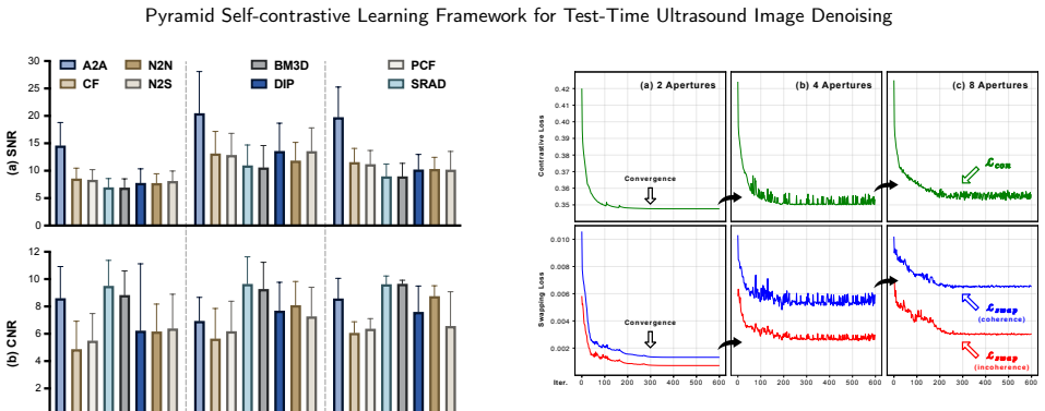
The inherent electronic and speckle noise complicates clinical interpretation of ultrasound images. Conventional denoising methods rely on explicit noise assumptions whose validity diminishes under composite noise conditions. Learning-based methods require massive labeled data and model parameters. These pre-defined and pre-trained manners entail an inevitable domain shift in complex in vivo environments, so they are limited to a specific noise type and often blur structural details. In this study, we propose a pure test-time training framework for one-shot ultrasound image denoising and apply it to synthetic aperture ultrasound (SAU), which synthesizes transmit focus from sub-aperture transmissions. Our Aperture-to-Aperture (A2A) framework disentangles anatomical similarity and noise randomness from shuffled sub-apertures through self-contrastive learning in pyramid latent spaces. The clean image is then decoded from the anatomy space, while discarding the noise space. A2A is trained at test time on one noisy sample of SAU signals, so it fundamentally eliminates the domain shift and pretraining costs. Simulation experiments, including electronic noise levels of 0 to 30 dB and different inclusion geometries, demonstrated an improvement of 69.3% SNR and 34.4% CNR by A2A. The in vivo results showed 84.8% SNR and 25.7% CNR gains using only two aperture data of the heart in six echocardiographic views, liver, and kidney. A2A delivers clear images/signals across diverse imaging targets and configurations, paving the way for more reliable anatomical visualization and functional assessment by ultrasound.
Editorial analysis
A structured set of objections, weighed in public.
Referee Report
Summary. The manuscript proposes the Aperture-to-Aperture (A2A) framework, a pure test-time training method for one-shot denoising of synthetic aperture ultrasound (SAU) images. It applies self-contrastive learning in pyramid latent spaces to shuffled sub-aperture transmissions in order to disentangle shared anatomical content from noise randomness; the clean image is decoded from the anatomy latent space while the noise space is discarded. Simulation experiments report 69.3% SNR and 34.4% CNR gains across electronic noise levels and inclusion geometries; in-vivo results on heart, liver, and kidney data using only two apertures report 84.8% SNR and 25.7% CNR gains.
Significance. If the disentanglement is reliable, the approach would be significant for clinical ultrasound by enabling domain-shift-free denoising without pre-training or large labeled datasets, directly addressing composite electronic-plus-speckle noise while preserving structural detail.
major comments (2)
- [A2A framework description] The core claim in the A2A framework description rests on the assumption that sub-aperture signals share identical anatomical content and differ solely by independent noise realizations. Speckle, however, is multiplicative and arises from coherent interference within the resolution cell, making it statistically dependent on local tissue structure rather than additive and independent across apertures. With only two apertures in the in-vivo experiments, the contrastive objective has limited negative samples and no explicit speckle model, raising the risk that structural speckle leaks into the anatomy space and undermines the one-shot decoding step that discards the noise space.
- [Experimental results] Table or figure reporting quantitative results (simulation and in-vivo SNR/CNR gains) provides no error bars, statistical tests, or detailed baseline comparisons, and the abstract supplies no equations, loss formulations, or architecture diagrams. This weakens support for the reported 69.3% SNR and 84.8% SNR improvements and makes it difficult to evaluate robustness against overfitting in one-shot training on complex noise.
minor comments (1)
- [Method] Pyramid depth and contrastive hyperparameters are free parameters but are not specified or ablated in the method or experiments.
Simulated Author's Rebuttal
We thank the referee for the constructive and insightful comments on our manuscript. We address each major comment point by point below and indicate the planned revisions.
read point-by-point responses
-
Referee: [A2A framework description] The core claim in the A2A framework description rests on the assumption that sub-aperture signals share identical anatomical content and differ solely by independent noise realizations. Speckle, however, is multiplicative and arises from coherent interference within the resolution cell, making it statistically dependent on local tissue structure rather than additive and independent across apertures. With only two apertures in the in-vivo experiments, the contrastive objective has limited negative samples and no explicit speckle model, raising the risk that structural speckle leaks into the anatomy space and undermines the one-shot decoding step that discards the noise space.
Authors: We appreciate the referee highlighting the structure-dependent nature of speckle. In SAU sub-aperture transmissions, different apertures provide angular diversity that decorrelates speckle realizations for the same underlying anatomy, which our pyramid self-contrastive objective exploits by contrasting multi-scale features across shuffled apertures to isolate shared content. We acknowledge that this is an approximation without an explicit speckle model and that two apertures limit negative sample diversity, potentially allowing some leakage. In revision we will expand the method section with a dedicated discussion of these assumptions, their validity in SAU, and risks of leakage, supported by additional qualitative visualizations of the disentangled spaces. The core framework remains unchanged. revision: partial
-
Referee: [Experimental results] Table or figure reporting quantitative results (simulation and in-vivo SNR/CNR gains) provides no error bars, statistical tests, or detailed baseline comparisons, and the abstract supplies no equations, loss formulations, or architecture diagrams. This weakens support for the reported 69.3% SNR and 84.8% SNR improvements and makes it difficult to evaluate robustness against overfitting in one-shot training on complex noise.
Authors: We agree that stronger statistical reporting and presentation are needed. In the revised manuscript we will add error bars (standard deviation across repeated noise realizations and training runs) to all SNR/CNR tables and figures, include paired statistical tests (e.g., Wilcoxon signed-rank) for significance, and expand baseline comparisons with additional methods plus ablation studies on pyramid depth and contrastive terms to assess one-shot robustness. Key loss equations and a compact architecture diagram will be added to the main text (or supplementary), and the abstract will be updated to reference the loss formulation. These changes directly address the concerns about support and overfitting evaluation. revision: yes
Circularity Check
No circularity in A2A framework derivation
full rationale
The paper introduces a test-time self-contrastive learning method on shuffled sub-aperture signals to separate anatomy and noise in pyramid latent spaces, with the clean image decoded from the anatomy space. This relies on the stated premise that sub-apertures share anatomical content while differing in noise realizations, applied via standard contrastive objectives without any fitted parameters being renamed as predictions, self-definitional loops, or load-bearing self-citations. Reported SNR/CNR gains are empirical outcomes from simulation and in vivo tests on single samples, not quantities forced by construction from the inputs. The derivation chain is self-contained and independent of the target results.
Axiom & Free-Parameter Ledger
free parameters (1)
- pyramid depth and contrastive hyperparameters
axioms (1)
- domain assumption Sub-aperture signals share identical anatomical content while noise realizations are statistically independent.
Reference graph
Works this paper leans on
-
[1]
J. A. Jensen, S. I. Nikolov, K. L. Gammelmark, M. H. Pedersen, Syntheticapertureultrasoundimaging,Ultrasonics44(2006)e5–e15
2006
-
[2]
Papadacci, M
C. Papadacci, M. Pernot, M. Couade, M. Fink, M. Tanter, High- contrastultrafastimagingoftheheart,IEEEtransactionsonultrason- ics, ferroelectrics, and frequency control 61 (2) (2014) 288–301
2014
-
[3]
Krissian, R
K. Krissian, R. Kikinis, C.-F. Westin, K. Vosburgh, Speckle- constrained filtering of ultrasound images, in: 2005 IEEE Computer Society Conference on Computer Vision and Pattern Recognition (CVPR’05), Vol. 2, IEEE, 2005, pp. 547–552
2005
-
[4]
H. Xie, L. E. Pierce, F. T. Ulaby, Statistical properties of logarith- mically transformed speckle, IEEE transactions on geoscience and remote sensing 40 (3) (2002) 721–727
2002
-
[5]
Loupas, W
T. Loupas, W. McDicken, P. Allan, Noise reduction in ultrasonic images by digital filtering, The British journal of radiology 60 (712) (1987) 389–392
1987
-
[6]
M.Gupta,H.Taneja,L.Chand,Performanceenhancementandanaly- sisoffiltersinultrasoundimagedenoising,Procediacomputerscience 132 (2018) 643–652
2018
-
[7]
Dabov, A
K. Dabov, A. Foi, V. Katkovnik, K. Egiazarian, Image denoising by sparse 3-d transform-domain collaborative filtering, IEEE Transac- tions on image processing 16 (8) (2007) 2080–2095
2007
-
[8]
Y. Yu, S. T. Acton, Speckle reducing anisotropic diffusion, IEEE Transactions on image processing 11 (11) (2002) 1260–1270
2002
-
[9]
Buades, B
A. Buades, B. Coll, J.-M. Morel, Non-local means denoising, Image processing on line 1 (2011) 208–212
2011
-
[10]
Coupé, P
P. Coupé, P. Hellier, C. Kervrann, C. Barillot, Nonlocal means-based speckle filtering for ultrasound images, IEEE transactions on image processing 18 (10) (2009) 2221–2229
2009
-
[11]
J. Yang, J. Fan, D. Ai, X. Wang, Y. Zheng, S. Tang, Y. Wang, Local statistics and non-local mean filter for speckle noise reduction in medical ultrasound image, Neurocomputing 195 (2016) 88–95
2016
-
[12]
X.Li,T.Wang,Researchonspinalultrasoundimagedenoisingbased on an improved bm3d algorithm, in: Fifth International Conference onImageProcessingandIntelligentControl(IPIC2025),Vol.13782, SPIE, 2025, pp. 152–156
2025
-
[13]
Y. Gan, E. Angelini, A. Laine, C. Hendon, Bm3d-based ultrasound image denoising via brushlet thresholding, in: 2015 IEEE 12th Inter- nationalSymposiumonBiomedicalImaging(ISBI),IEEE,2015,pp. 667–670
2015
-
[14]
Hollman, K
K. Hollman, K. Rigby, M. O’donnell, Coherence factor of speckle fromamulti-rowprobe,in:1999IEEEUltrasonicsSymposium.Pro- ceedings. International Symposium (Cat. No. 99CH37027), Vol. 2, IEEE, 1999, pp. 1257–1260
1999
-
[15]
P.-C.Li,M.-L.Li,Adaptiveimagingusingthegeneralizedcoherence factor,IEEEtransactionsonultrasonics,ferroelectrics,andfrequency control 50 (2) (2003) 128–141
2003
-
[16]
Camacho, M
J. Camacho, M. Parrilla, C. Fritsch, Phase coherence imaging, IEEE transactions on ultrasonics, ferroelectrics, and frequency control 56 (5) (2009) 958–974
2009
-
[17]
Wang, C.-H
S.-L. Wang, C.-H. Chang, H.-C. Yang, Y.-H. Chou, P.-C. Li, Perfor- mance evaluation of coherence-based adaptive imaging using clini- cal breast data, IEEE transactions on ultrasonics, ferroelectrics, and frequency control 54 (8) (2007) 1669–1679
2007
-
[18]
B. M. Asl, A. Mahloojifar, Minimum variance beamforming com- bined with adaptive coherence weighting applied to medical ultra- sound imaging, IEEE transactions on ultrasonics, ferroelectrics, and frequency control 56 (9) (2009) 1923–1931
2009
-
[19]
O’Donnell, Y
M. O’Donnell, Y. Wang, Coded excitation for synthetic aperture ultrasound imaging, IEEE transactions on ultrasonics, ferroelectrics, and frequency control 52 (2) (2005) 171–176
2005
-
[20]
X. Li, N. Navab, Z. Jiang, Speckle2self: Self-supervised ultrasound specklereductionwithoutcleandata,MedicalImageAnalysis(2025) 103755
2025
-
[21]
K. J. Parker, Ultrasonic attenuation and absorption in liver tissue, Ultrasound in medicine & biology 9 (4) (1983) 363–369
1983
-
[22]
P.C.Li,M.ODonnell,Evaluationalspatialcompounding,Ultrasonic imaging 16 (3) (1994) 176–189
1994
-
[23]
M. A. L. Bell, R. Goswami, J. A. Kisslo, J. J. Dahl, G. E. Trahey, Short-lagspatialcoherenceimagingofcardiacultrasounddata:Initial clinical results, Ultrasound in medicine & biology 39 (10) (2013) 1861–1874
2013
-
[24]
Y. Wang, C. Zheng, H. Peng, X. Chen, Short-lag spatial coherence combined with eigenspace-based minimum variance beamformer for synthetic aperture ultrasound imaging, Computers in Biology and Medicine 91 (2017) 267–276
2017
-
[25]
R. B. Kuc, Application of kalman filtering techniques to diagnostic ultrasound, Ultrasonic Imaging 1 (2) (1979) 105–120
1979
-
[26]
3917–3920
F.Y.Rizi,H.A.Noubari,S.K.Setarehdan,Wavelet-basedultrasound imagedenoising:Performanceanalysisandcomparison,in:2011An- nual International Conference of the IEEE Engineering in Medicine and Biology Society, IEEE, 2011, pp. 3917–3920
2011
-
[27]
Rabbani, M
H. Rabbani, M. Vafadust, P. Abolmaesumi, S. Gazor, Speckle noise reduction of medical ultrasound images in complex wavelet domain using mixture priors, IEEE transactions on biomedical engineering 55 (9) (2008) 2152–2160
2008
-
[28]
Khare, P
S. Khare, P. Kaushik, Speckle filtering of ultrasonic images using weighted nuclear norm minimization in wavelet domain, Biomedical Signal Processing and Control 70 (2021) 102997
2021
-
[29]
Zhu, C.-W
L. Zhu, C.-W. Fu, M. S. Brown, P.-A. Heng, A non-local low-rank framework for ultrasound speckle reduction, in: Proceedings of the IEEE conference on computer vision and pattern recognition, 2017, pp. 5650–5658
2017
-
[30]
M. A. Lediju, G. E. Trahey, B. C. Byram, J. J. Dahl, Short-lag spatial coherence of backscattered echoes: Imaging characteristics, IEEEtransactionsonultrasonics,ferroelectrics,andfrequencycontrol 58 (7) (2011) 1377–1388
2011
-
[31]
Kokil, S
P. Kokil, S. Sudharson, Despeckling of clinical ultrasound images using deep residual learning, Computer Methods and Programs in Biomedicine 194 (2020) 105477. J. Zhang et al.:Preprint submitted to ElsevierPage 13 of 14 Pyramid Self-contrastive Learning Framework for Test-Time Ultrasound Image Denoising
2020
-
[32]
K. M. Mohamed, M. H. Ali, Ultrasound images enhancement using unet-deep learning according to resolution and speckle noise, Inter- national Journal of Mechanical Engineering 7 (5)
-
[33]
Zhang, J
L. Zhang, J. Zhang, Ultrasound image denoising using generative adversarial networks with residual dense connectivity and weighted joint loss, PeerJ Computer Science 8 (2022) e873
2022
-
[34]
Karaoğlu, H
O. Karaoğlu, H. Ş. Bilge, I. Uluer, Removal of speckle noises from ultrasound images using five different deep learning networks, Engi- neering Science and Technology, an International Journal 29 (2022) 101030
2022
-
[35]
Singh, A
H. Singh, A. S. Ahmed, F. Melandsø, A. Habib, Ultrasonic image denoising using machine learning in point contact excitation and detection method, Ultrasonics 127 (2023) 106834
2023
-
[36]
Asgariandehkordi, S
H. Asgariandehkordi, S. Goudarzi, M. Sharifzadeh, A. Basarab, H. Rivaz, Denoising plane wave ultrasound images using diffu- sion probabilistic models, IEEE Transactions on Ultrasonics, Ferro- electrics, and Frequency Control
-
[37]
Zhang, C
Y. Zhang, C. Huneau, J. Idier, D. Mateus, Ultrasound image re- construction with denoising diffusion restoration models, in: Inter- national Conference on Medical Image Computing and Computer- Assisted Intervention, Springer, 2023, pp. 193–203
2023
-
[38]
Yancheng, X
L. Yancheng, X. Zeng, Q. Dong, X. Wang, Red-mam: A residual encoder-decoder network based on multi-attention fusion for ultra- sound image denoising, Biomedical Signal Processing and Control 79 (2023) 104062
2023
-
[39]
D. Hyun, L. L. Brickson, K. T. Looby, J. J. Dahl, Beamforming and speckle reduction using neural networks, IEEE transactions on ultrasonics, ferroelectrics, and frequency control 66 (5) (2019) 898– 910
2019
-
[40]
X. Yu, S. Luan, S. Lei, J. Huang, Z. Liu, X. Xue, T. Ma, Y. Ding, B. Zhu, Deep learning for fast denoising filtering in ultrasound local- ization microscopy, Physics in Medicine & Biology 68 (20) (2023) 205002
2023
-
[41]
Cammarasana, P
S. Cammarasana, P. Nicolardi, G. Patanè, Real-time denoising of ultrasound images based on deep learning, Medical & Biological Engineering & Computing 60 (8) (2022) 2229–2244
2022
-
[42]
T.-T.Zhang,H.Shu,K.-Y.Lam,C.-Y.Chow,A.Li,Featuredecompo- sition and enhancement for unsupervised medical ultrasound image denoising and instance segmentation, Applied Intelligence 53 (8) (2023) 9548–9561
2023
-
[43]
H. Wu, T. T. Huynh, R. Souvenir, Echocardiogram enhancement using supervised manifold denoising, Medical image analysis 24 (1) (2015) 41–51
2015
-
[44]
Jarosik, M
P. Jarosik, M. Lewandowski, Z. Klimonda, M. Byra, Pixel-wise deep reinforcement learning approach for ultrasound image denoising, in: 2021 IEEE International Ultrasonics Symposium (IUS), IEEE, 2021, pp. 1–4
2021
-
[45]
Jiang, C
M. Jiang, C. You, M. Wang, H. Zhang, Z. Gao, D. Wu, T. Tan, Controllable deep learning denoising model for ultrasound images using synthetic noisy image, in: Computer Graphics International Conference, Springer, 2023, pp. 297–308
2023
-
[46]
Noise2Noise: Learning Image Restoration without Clean Data
J. Lehtinen, J. Munkberg, J. Hasselgren, S. Laine, T. Karras, M. Ait- tala, T. Aila, Noise2noise: Learning image restoration without clean data, arXiv preprint arXiv:1803.04189
work page internal anchor Pith review Pith/arXiv arXiv
- [47]
- [48]
-
[49]
D. Jung, M. Kang, S. H. Park, N. Guezzi, J. Yu, Unsupervised speckle noise reduction technique for clinical ultrasound imaging, Ultrasonography 43 (5) (2024) 327–344
2024
-
[50]
C.Yu,F.Ren,S.Bao,Y.Yang,X.Xu,Self-supervisedultrasoundim- agedenoisingbasedonweightedjointloss,DigitalSignalProcessing 162 (2025) 105151
2025
- [51]
-
[52]
J. Huh, S. Khan, S. Choi, D. Shin, J. E. Lee, E. S. Lee, J. C. Ye, Tunable image quality control of 3-d ultrasound using switchable cyclegan, Medical Image Analysis 83 (2023) 102651
2023
-
[53]
S. Muth, S. Dort, I. A. Sebag, M.-J. Blais, D. Garcia, Unsupervised dealiasing and denoising of color-doppler data, Medical image anal- ysis 15 (4) (2011) 577–588
2011
-
[54]
Ulyanov, A
D. Ulyanov, A. Vedaldi, V. Lempitsky, Deep image prior, in: Pro- ceedings of the IEEE conference on computer vision and pattern recognition, 2018, pp. 9446–9454
2018
-
[55]
Batson, L
J. Batson, L. Royer, Noise2self: Blind denoising by self-supervision, in: International conference on machine learning, PMLR, 2019, pp. 524–533
2019
-
[56]
A. M. Christensen, I. M. Rosado-Mendez, T. J. Hall, A systematized review of quantitative ultrasound based on first-order speckle statis- tics,IEEETransactionsonUltrasonics,Ferroelectrics,andFrequency Control 71 (7) (2024) 872–886
2024
-
[57]
Zhang, Y
Y. Zhang, Y. Guo, W.-N. Lee, Ultrafast ultrasound imaging with cascadeddual-polaritywaves,IEEETransactionsonMedicalImaging 37 (4) (2017) 906–917
2017
-
[58]
T. D. Mast, Empirical relationships between acoustic parameters in human soft tissues, Acoustics Research Letters Online 1 (2) (2000) 37–42
2000
-
[59]
Y. Sun, X. Wang, Z. Liu, J. Miller, A. Efros, M. Hardt, Test-time training with self-supervision for generalization under distribution shifts,in:Internationalconferenceonmachinelearning,PMLR,2020, pp. 9229–9248
2020
-
[60]
Jaiswal, A
A. Jaiswal, A. R. Babu, M. Z. Zadeh, D. Banerjee, F. Makedon, A survey on contrastive self-supervised learning, Technologies 9 (1) (2020) 2
2020
-
[61]
T. Chen, S. Kornblith, M. Norouzi, G. Hinton, A simple framework for contrastive learning of visual representations, in: International conference on machine learning, PmLR, 2020, pp. 1597–1607
2020
-
[62]
M.Gutmann,A.Hyvärinen,Noise-contrastiveestimation:Anewesti- mationprincipleforunnormalizedstatisticalmodels,in:Proceedings ofthethirteenthinternationalconferenceonartificialintelligenceand statistics, JMLR Workshop and Conference Proceedings, 2010, pp. 297–304
2010
-
[63]
A. v. d. Oord, Y. Li, O. Vinyals, Representation learning with con- trastive predictive coding, arXiv preprint arXiv:1807.03748
work page internal anchor Pith review Pith/arXiv arXiv
-
[64]
X. Wang, R. Zhang, C. Shen, T. Kong, L. Li, Dense contrastive learningforself-supervisedvisualpre-training,in:Proceedingsofthe IEEE/CVF conference on computer vision and pattern recognition, 2021, pp. 3024–3033
2021
-
[65]
Zhang, B
J. Zhang, B. Dai, H. Guan, W.-N. Lee, Msaf: A multi-level sensing adversarial framework for signal recovery in synthetic aperture ultra- sound, IEEE Journal of Biomedical and Health Informatics
-
[66]
B. E. Treeby, J. Jaros, A. P. Rendell, B. T. Cox, Modeling nonlinear ultrasound propagation in heterogeneous media with power law ab- sorption using a k-space pseudospectral method, The Journal of the Acoustical Society of America 131 (6) (2012) 4324–4336
2012
-
[67]
Mitchell, P
C. Mitchell, P. S. Rahko, L. A. Blauwet, B. Canaday, J. A. Finstuen, M. C. Foster, K. Horton, K. O. Ogunyankin, R. A. Palma, E. J. Velazquez, Guidelines for performing a comprehensive transthoracic echocardiographic examination in adults: recommendations from the americansocietyofechocardiography,JournaloftheAmericanSoci- ety of Echocardiography 32 (1) (2...
2019
-
[68]
F. Wang, H. Liu, Understanding the behaviour of contrastive loss, in:ProceedingsoftheIEEE/CVFconferenceoncomputervisionand pattern recognition, 2021, pp. 2495–2504. J. Zhang et al.:Preprint submitted to ElsevierPage 14 of 14
2021
discussion (0)
Sign in with ORCID, Apple, or X to comment. Anyone can read and Pith papers without signing in.