REVIEW 2 major objections 1 minor 63 references
Quantifying the biophysical properties of stomatocytes in health and disease
T0 review · 2 major / 1 minor · reviewed 2026-06-28 · grok-4.3
Pith's one-line read Stomatocyte volume controls spleen slit passage while membrane motion controls blood viscosity.
desk verdict The simulations tie fixed-area volume reduction to an order-of-magnitude IES pressure difference that matches the OHS/DHS splenectomy split, but the fixed-moduli choice is the load-bearing assumption. read the letter →
The pith
A machine-rendered reading of the paper's core claim, the machinery that carries it, and where it could break.
The reading
What carries the argument
Three stomatocyte models at fixed membrane area and decreasing volumes simulated with dissipative particle dynamics and run through microfluidic slit and flow assays.
What would settle it
Direct measurement of the critical pressure needed for actual patient stomatocytes of known hydration state to cross slits of interendothelial size, or measurement of low-shear viscosity in their blood at physiological hematocrit.
Extended reading notes
Core claim
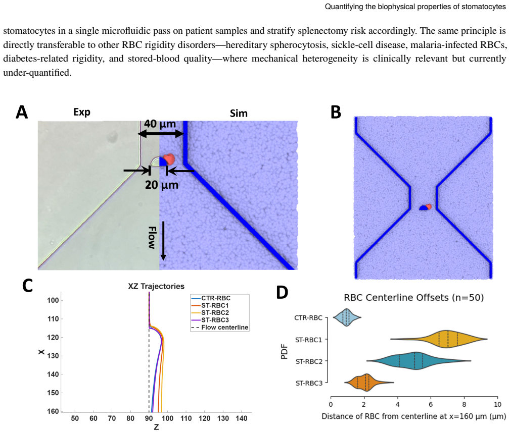
Tracing this parameter set through five mechanically orthogonal assays, we find that interendothelial-slit (IES) traversal is geometry-dominated: overhydrated ST-RBC1 requires an order of magnitude higher critical pressure than healthy RBCs, whereas dehydrated ST-RBC3 passes freely. ST-RBC3 nonetheless suppresses membrane tank-treading and raises low-shear whole-blood viscosity by ~29% at physiological haematocrit. A funnel-obstacle chip amplifies these differences into a label-free centerline-offset signal predicted to separate all four RBC types.
Load-bearing premise
The three stomatocyte models at fixed membrane area and decreasing volumes accurately represent the biophysical range from overhydrated to dehydrated hereditary stomatocytosis.
Editorial extensions
If this is right
- Overhydrated stomatocytes require an order of magnitude higher critical pressure to traverse interendothelial slits than healthy red blood cells.
- Dehydrated stomatocytes pass interendothelial slits freely but suppress membrane tank-treading during flow.
- Dehydrated stomatocytes raise low-shear whole-blood viscosity by approximately 29 percent at physiological haematocrit.
- A funnel-obstacle microfluidic chip produces a label-free centerline-offset signal that separates healthy, overhydrated, and dehydrated red blood cell types.
Reading between the lines
- The same geometry-versus-viscosity separation may appear in other red-cell shape disorders that alter surface-to-volume ratio.
- The funnel-obstacle chip layout could be miniaturized for point-of-care testing of red-cell disorders before splenectomy decisions.
- Cytoplasmic viscosity changes could interact with the volume effects in patient samples in ways the fixed-parameter models do not yet capture.
Editorial analysis
A structured set of objections, weighed in public.
Referee Report
Summary. The manuscript uses dissipative particle dynamics (DPD) simulations to build a healthy discocyte control and three stomatocyte models (ST-RBC1-3) at fixed membrane area with volumes 109.7, 101.5, and 89.8 fL, intended to span overhydrated to dehydrated hereditary stomatocytosis. These models are traced through five mechanically orthogonal assays (IES traversal, tank-treading, whole-blood viscosity, etc.), yielding the claims that IES passage is geometry-dominated (ST-RBC1 requires ~10 imes higher critical pressure than healthy RBCs while ST-RBC3 passes freely) and that ST-RBC3 suppresses tank-treading while elevating low-shear viscosity by ~29% at physiological haematocrit, comparable to Gaucher disease; a funnel-obstacle microfluidic chip is predicted to separate all four phenotypes by ~4.5 SD via centerline offset.
Significance. If the models are faithful biophysical proxies, the work supplies a unified, multi-assay framework that mechanistically resolves the splenectomy paradox in hereditary stomatocytosis and identifies a label-free microfluidic signature for pre-operative risk stratification. The use of five orthogonal readouts and the explicit volume series constitute a strength in linking single-cell geometry to macroscopic filtration and rheology outcomes.
major comments (2)
- [Abstract and model-construction description] Abstract and model-construction description: ST-RBC1-3 are constructed at fixed membrane area with only volume reduction (109.7/101.5/89.8 fL). The central claim that 'IES traversal is geometry-dominated' (order-of-magnitude pressure difference) is load-bearing on the assumption that shear modulus and bending rigidity remain identical to healthy RBCs. Hereditary stomatocytosis commonly involves membrane-protein defects that alter these moduli; without a sensitivity analysis or explicit justification for holding them fixed, the geometry-only interpretation cannot be isolated from possible modulus effects.
- [Viscosity and tank-treading results (Abstract)] Viscosity and tank-treading results (Abstract): The ~29% low-shear viscosity elevation for ST-RBC3 and the suppression of tank-treading are reported without error bars, number of independent runs, or direct comparison to experimental stomatocyte rheology data. Because these quantities underpin the claim that dehydrated stomatocytes produce Gaucher-like hyperviscosity despite free IES passage, the quantitative robustness of the prediction requires explicit validation or uncertainty quantification.
minor comments (1)
- [Abstract] Abstract: the abbreviation 'HS' for hereditary stomatocytosis risks confusion with the far more common hereditary spherocytosis; consider 'HSt' or full spelling on first use.
Simulated Author's Rebuttal
We thank the referee for their detailed and constructive review. We address each major comment below and will revise the manuscript accordingly to strengthen the presentation of our modeling assumptions and quantitative results.
read point-by-point responses
-
Referee: [Abstract and model-construction description] Abstract and model-construction description: ST-RBC1-3 are constructed at fixed membrane area with only volume reduction (109.7/101.5/89.8 fL). The central claim that 'IES traversal is geometry-dominated' (order-of-magnitude pressure difference) is load-bearing on the assumption that shear modulus and bending rigidity remain identical to healthy RBCs. Hereditary stomatocytosis commonly involves membrane-protein defects that alter these moduli; without a sensitivity analysis or explicit justification for holding them fixed, the geometry-only interpretation cannot be isolated from possible modulus effects.
Authors: We agree that the assumption of fixed shear modulus and bending rigidity requires explicit justification to support the geometry-dominated interpretation. In the revised manuscript we will add a dedicated paragraph in the Methods section explaining that the primary defect in hereditary stomatocytosis is ion-transport dysregulation leading to volume change, with literature support indicating that membrane mechanical properties are not the dominant altered parameter in the stomatocyte phenotypes modeled here. We will also include a limited sensitivity analysis in which bending rigidity is varied by ±20% around the baseline value, confirming that the order-of-magnitude difference in critical IES pressure between ST-RBC1 and healthy RBCs persists. revision: yes
-
Referee: [Viscosity and tank-treading results (Abstract)] Viscosity and tank-treading results (Abstract): The ~29% low-shear viscosity elevation for ST-RBC3 and the suppression of tank-treading are reported without error bars, number of independent runs, or direct comparison to experimental stomatocyte rheology data. Because these quantities underpin the claim that dehydrated stomatocytes produce Gaucher-like hyperviscosity despite free IES passage, the quantitative robustness of the prediction requires explicit validation or uncertainty quantification.
Authors: We acknowledge that the viscosity and tank-treading results would be strengthened by reporting uncertainty and additional validation. In the revision we will add error bars (standard deviation across n=5 independent runs per condition), explicitly state the number of runs performed, and include a direct comparison to published experimental rheology data on dehydrated or stomatocytic RBCs (or the closest available analogs) to support the ~29% elevation claim relative to Gaucher disease. revision: yes
Circularity Check
No significant circularity; derivation uses independent simulation methods on explicitly constructed models
full rationale
The paper constructs stomatocyte models at fixed membrane area with prescribed volumes (109.7/101.5/89.8 fL) and applies established DPD methods plus microfluidic imaging across five assays. No equations or claims reduce a prediction to a fitted parameter by construction, nor does any load-bearing step rely on a self-citation chain that itself lacks independent verification. The central results (geometry-dominated IES traversal, viscosity increase) follow from the chosen parameter set rather than re-deriving that set from the outputs. This is the normal case of a self-contained modeling study.
Assumptions & free parameters
free parameters (1)
- ST-RBC volumes =
109.7, 101.5, 89.8 fL
assumptions (1)
- domain assumption DPD simulations faithfully reproduce RBC membrane and cytoplasmic mechanics under the stated conditions
invented entities (1)
-
ST-RBC1-3 stomatocyte models
Cite this review
Pith. "Pith review of Quantifying the biophysical properties of stomatocytes in health and disease." pith.science (2026). https://pith.science/paper/VQTA73GK
@misc{pith2026260605227,
  author       = {Pith},
  title        = {Pith review of: Quantifying the biophysical properties of stomatocytes in health and disease},
  year         = {2026},
  howpublished = {\url{https://pith.science/paper/VQTA73GK}},
  note         = {Machine review of arXiv:2606.05227}
}
read the original abstract
Hereditary stomatocytosis (HS) comprises red blood cell (RBC) disorders characterized by cup-shaped erythrocytes that respond oppositely to splenectomy: curative in overhydrated HS (OHS) but potentially thrombogenic in dehydrated HS (DHS/xerocytosis). This paradox persists because RBC biomechanics is governed by partly independent parameters--shear modulus, bending rigidity, surface-to-volume ratio (S/V), and cytoplasmic viscosity--that existing assays capture only piecemeal. Here we combine dissipative particle dynamics (DPD) simulations with microfluidic imaging to construct a control discocyte and three stomatocyte models (ST-RBC1-3) at fixed membrane area and decreasing volume (109.7, 101.5, 89.8 fL), spanning the OHS-to-DHS range. Tracing this parameter set through five mechanically orthogonal assays, we find that interendothelial-slit (IES) traversal is geometry-dominated: overhydrated ST-RBC1 requires an order of magnitude higher critical pressure than healthy RBCs, whereas dehydrated ST-RBC3 passes freely. ST-RBC3 nonetheless suppresses membrane tank-treading and raises low-shear whole-blood viscosity by ~29% at physiological haematocrit, comparable to Gaucher-disease hyperviscosity. A funnel-obstacle chip amplifies these differences into a label-free centerline-offset signal predicted to separate all four RBC types (~4.5 standard deviations between extreme phenotypes). These results unite single-cell mechanics, splenic filtration, and hemorheology in one framework, resolve the splenectomy paradox, and point toward microfluidic pre-operative risk stratification in HS.
Figures


Figures from the paper (6 more)


Reference graph
Works this paper leans on
-
[1]
Structure and function of the spleen
R. E. Mebius and G. Kraal. “Structure and function of the spleen”. In:Nature Reviews Immunology5 (2005), pp. 606–616. Manuscript of paper23 Chai et al
2005
-
[2]
Mechanics of diseased red blood cells in human spleen and consequences for hereditary blood disorders
H. Li et al. “Mechanics of diseased red blood cells in human spleen and consequences for hereditary blood disorders”. In: Proceedings of the National Academy of Sciences115 (2018), pp. 9574–9579
2018
-
[3]
Biomechanical properties of red blood cells in health and disease towards microfluidics
Giovanna Tomaiuolo. “Biomechanical properties of red blood cells in health and disease towards microfluidics”. In: Biomicrofluidics8.5 (2014)
2014
-
[4]
Shear Dependence of Effective Cell Volume as a Determinant of Blood Viscosity
Shu Chien. “Shear Dependence of Effective Cell Volume as a Determinant of Blood Viscosity”. In:Science168.3934 (1970), pp. 977–979
1970
-
[5]
Blood rheology and hemodynamics
Oguz K Baskurt and Herbert J Meiselman. “Blood rheology and hemodynamics”. In:Seminars in thrombosis and hemostasis. Vol. 29. 05. Copyright©2003 by Thieme Medical Publishers, Inc., 333 Seventh Avenue, New ... 2003, pp. 435–450
2003
-
[6]
Guidelines for the diagnosis and management of hereditary spherocytosis—2011 update
Paula H B Bolton-Maggs et al. “Guidelines for the diagnosis and management of hereditary spherocytosis—2011 update”. In:British Journal of Haematology156.1 (2012), pp. 37–49
2011
-
[7]
A novel red blood cell deformability biomarker is associated with hemolysis and vaso-occlusive crises in sickle cell disease
Mallorie Sahun, Emmanuelle Bernit, Samuel Atwell, et al. “A novel red blood cell deformability biomarker is associated with hemolysis and vaso-occlusive crises in sickle cell disease”. In:Scientific Reports15 (2025), p. 15864
2025
-
[8]
Disorders of erythrocyte hydration
Patrick G Gallagher. “Disorders of erythrocyte hydration”. In:Blood, The Journal of the American Society of Hematology 130.25 (2017), pp. 2699–2708
2017
Show all 63 references
-
[9]
The evolving landscape of hereditary stomatocytosis
Immacolata Andolfo, Achille Iolascon, and Roberta Russo. “The evolving landscape of hereditary stomatocytosis”. In: Blood145.26 (2025), pp. 3089–3100
2025
-
[10]
Abnormal properties of red blood cells suggest a role in the pathophysiology of Gaucher disease
Melanie Franco et al. “Abnormal properties of red blood cells suggest a role in the pathophysiology of Gaucher disease”. In:Blood, The Journal of the American Society of Hematology121.3 (2013), pp. 546–555
2013
-
[11]
The hereditary stomatocytoses
Joanna F Flatt and Lesley J Bruce. “The hereditary stomatocytoses”. In:Haematologica94.8 (2009), pp. 1039–1041
2009
-
[12]
Hereditary dehydrated and overhydrated stomatocytosis: recent advances
Jean Delaunay. “Hereditary dehydrated and overhydrated stomatocytosis: recent advances”. In:Current Opinion in Hematology6.2 (1999), pp. 110–114
1999
-
[13]
Erythrocyte flow through the interendothelial slits of the splenic venous sinus
Ming Dao, Ian MacDonald, and RJ Asaro. “Erythrocyte flow through the interendothelial slits of the splenic venous sinus”. In:Biomechanics and modeling in mechanobiology20.6 (2021), pp. 2227–2245
2021
-
[14]
Physical mechanisms of red blood cell splenic filtration
Alexis Moreau et al. “Physical mechanisms of red blood cell splenic filtration”. In:Proceedings of the National Academy of Sciences120.44 (2023), e2300095120
2023
-
[15]
Blood viscosity in tube flow: dependence on diameter and hematocrit
A R Pries, D Neuhaus, and P Gaehtgens. “Blood viscosity in tube flow: dependence on diameter and hematocrit”. In: American Journal of Physiology–Heart and Circulatory Physiology263.6 (1992), H1770–H1778
1992
-
[16]
Microfluidic study of retention and elimination of abnormal red blood cells by human spleen with implications for sickle cell disease
Yuhao Qiang et al. “Microfluidic study of retention and elimination of abnormal red blood cells by human spleen with implications for sickle cell disease”. In:Proceedings of the National Academy of Sciences120.6 (2023), e2217607120
2023
-
[17]
Kinetics of red blood cell passage through interendothelial slits into venous sinuses in rat spleen, analyzed by in vivo microscopy
IC MacDonald et al. “Kinetics of red blood cell passage through interendothelial slits into venous sinuses in rat spleen, analyzed by in vivo microscopy”. In:Microvascular research33.1 (1987), pp. 118–134
1987
-
[18]
Biomechanics of red blood cells in human spleen and consequences for physiology and disease
Igor V Pivkin et al. “Biomechanics of red blood cells in human spleen and consequences for physiology and disease”. In: Proceedings of the National Academy of Sciences113.28 (2016), pp. 7804–7809
2016
-
[19]
A functional microengineered model of the human splenon-on-a-chip
L G Rigat-Brugarolas et al. “A functional microengineered model of the human splenon-on-a-chip”. In:Lab on a Chip 14.10 (2014), pp. 1715–1724
2014
-
[20]
A coarse-grained red blood cell membrane model to study stomatocyte- discocyte-echinocyte morphologies
Nadeeshani Maheshika Geekiyanage et al. “A coarse-grained red blood cell membrane model to study stomatocyte- discocyte-echinocyte morphologies”. In:PLoS one14.4 (2019), e0215447
2019
-
[21]
Stomatocyte–discocyte–echinocytesequenceofthehuman red blood cell: Evidence for the bilayer–couple hypothesis from membrane mechanics
GeraldHWLim,MichaelWortis,andRanjanMukhopadhyay.“Stomatocyte–discocyte–echinocytesequenceofthehuman red blood cell: Evidence for the bilayer–couple hypothesis from membrane mechanics”. In:Proceedings of the National Academy of Sciences99.26 (2002), pp. 16766–16769
2002
-
[22]
Piezo1 links mechanical forces to red blood cell volume
Stuart M Cahalan et al. “Piezo1 links mechanical forces to red blood cell volume”. In:eLife4 (2015), e07370. 24Manuscript of paper Quantifying the biophysical properties of stomatocytes
2015
-
[23]
Genotype-phenotype correlation and risk stratification in a cohort of 123 hereditary stomatocy- tosis patients
Immacolata Andolfo et al. “Genotype-phenotype correlation and risk stratification in a cohort of 123 hereditary stomatocy- tosis patients”. In:American Journal of Hematology93.12 (2018), pp. 1509–1517
2018
-
[24]
Clinical and biological features in PIEZO1-hereditary xerocytosis and Gardos channelopathy: a retrospective series of 126 patients
Véronique Picard et al. “Clinical and biological features in PIEZO1-hereditary xerocytosis and Gardos channelopathy: a retrospective series of 126 patients”. In:Haematologica104.8 (2019), pp. 1554–1564
2019
-
[25]
Dehydrated hereditary stomatocytosis: clinical perspectives
Henrik Frederiksen. “Dehydrated hereditary stomatocytosis: clinical perspectives”. In:Journal of Blood Medicine10 (2019), pp. 183–195
2019
-
[26]
Hereditary spherocytosis
Silverio Perrotta, Patrick G Gallagher, and Narla Mohandas. “Hereditary spherocytosis”. In:The Lancet372.9647 (2008), pp. 1411–1426
2008
-
[27]
An overview of hereditary spherocytosis and the curative effects of splenectomy
Kyril Turpaev et al. “An overview of hereditary spherocytosis and the curative effects of splenectomy”. In:Frontiers in Physiology16 (2025), p. 1497588
2025
-
[28]
OpenRBC: A fast simulator of red blood cells at protein resolution
Yu-Hang Tang et al. “OpenRBC: A fast simulator of red blood cells at protein resolution”. In:Biophysical Journal112.10 (2017), pp. 2030–2037
2017
-
[29]
A multiscale biomechanical model of platelets: Correlating with in-vitro results
Peng Zhang et al. “A multiscale biomechanical model of platelets: Correlating with in-vitro results”. In:Journal of biomechanics50 (2017), pp. 26–33
2017
-
[30]
Dynamics of the axon plasma membrane skeleton
Zhaojie Chai, Shiju Gu, and George Lykotrafitis. “Dynamics of the axon plasma membrane skeleton”. In:Soft Matter 19.14 (2023), pp. 2514–2528
2023
-
[31]
The periodic axon membrane skeleton leads to Na nanodomains but does not impact action potentials
Zhaojie Chai, Anastasios V Tzingounis, and George Lykotrafitis. “The periodic axon membrane skeleton leads to Na nanodomains but does not impact action potentials”. In:Biophysical Journal121.18 (2022), pp. 3334–3344
2022
-
[32]
MD/DPD multiscale framework for predicting morphology and stresses of red blood cells in health and disease
Hung-Yu Chang et al. “MD/DPD multiscale framework for predicting morphology and stresses of red blood cells in health and disease”. In:PLoS computational biology12.10 (2016), e1005173
2016
-
[33]
Deep reinforcement learning with a particle dynamics environment applied to emergency evacuation of a room with obstacles
Yihao Zhang, Zhaojie Chai, and George Lykotrafitis. “Deep reinforcement learning with a particle dynamics environment applied to emergency evacuation of a room with obstacles”. In:Physica A: Statistical Mechanics and its Applications571 (2021), p. 125845
2021
-
[34]
A deep reinforcement learning model based on deterministic policy gradient for collective neural crest cell migration
Yihao Zhang et al. “A deep reinforcement learning model based on deterministic policy gradient for collective neural crest cell migration”. In:arXiv preprint arXiv:2007.03190(2020)
2007
-
[35]
A multiscale red blood cell model with accurate mechanics, rheology, and dynamics
D.A. Fedosov, B. Caswell, and G.E. Karniadakis. “A multiscale red blood cell model with accurate mechanics, rheology, and dynamics”. In:Biophysical Journal98.10 (2010), pp. 2215–2225
2010
-
[36]
Multiscale Modeling of Red Blood Cell Mechanics and Blood Flow in Malaria
Dmitry A. Fedosov et al. “Multiscale Modeling of Red Blood Cell Mechanics and Blood Flow in Malaria”. In:PLOS Computational Biology7 (2011), pp. 1–13
2011
-
[37]
Dissipative particle dynamics: Bridging the gap between atomistic and mesoscopic simulation
Robert D Groot and Patrick B Warren. “Dissipative particle dynamics: Bridging the gap between atomistic and mesoscopic simulation”. In:The Journal of chemical physics107.11 (1997), pp. 4423–4435
1997
-
[38]
Simulating microscopic hydrodynamic phenomena with dissipative particle dynamics
PJ Hoogerbrugge and JMVA Koelman. “Simulating microscopic hydrodynamic phenomena with dissipative particle dynamics”. In:Europhysics Letters19.3 (1992), p. 155
1992
-
[39]
GRAFT-ATHENA: Self-Improving Agentic Teams for Autonomous Discovery and Evolutionary Numerical Algorithms
Juan Diego Toscano, Zhaojie Chai, and George Em Karniadakis. “GRAFT-ATHENA: Self-Improving Agentic Teams for Autonomous Discovery and Evolutionary Numerical Algorithms”. In:arXiv preprint arXiv:2605.11117(2026).doi: 10.48550/arXiv.2605.11117.url:https://arxiv.org/abs/2605.11117
-
[40]
Mechanics of the human red blood cell deformed by optical tweezers
Ming Dao, Chwee Teck Lim, and Subra Suresh. “Mechanics of the human red blood cell deformed by optical tweezers”. In: Journal of the Mechanics and Physics of Solids51.11–12 (2003), pp. 2259–2280
2003
-
[41]
Spectrin-level modeling of the cytoskeleton and optical tweezers stretching of the erythrocyte
Ju Li et al. “Spectrin-level modeling of the cytoskeleton and optical tweezers stretching of the erythrocyte”. In:Biophysical Journal88.5 (2005), pp. 3707–3719
2005
-
[42]
Determination of red blood cell membrane viscosity from rheoscopic observations of tank-treading motion
Roger Tran-Son-Tay, SP Sutera, and PR Rao. “Determination of red blood cell membrane viscosity from rheoscopic observations of tank-treading motion”. In:Biophysical Journal46.1 (1984), pp. 65–72. Manuscript of paper25 Chai et al
1984
-
[43]
Microrheologic investigation of erythrocyte deformability in diabetes mellitus
JR Williamson et al. “Microrheologic investigation of erythrocyte deformability in diabetes mellitus”. In:Blood65.6 (1985), pp. 1493–1499
1985
-
[44]
Tank-tread frequency of the red cell membrane: dependence on the viscosity of the suspending medium
Thomas M Fischer. “Tank-tread frequency of the red cell membrane: dependence on the viscosity of the suspending medium”. In:Biophysical Journal93.7 (2007), pp. 2553–2561
2007
-
[45]
In silico biophysics and rheology of blood and red blood cells in Gaucher Disease
Zhaojie Chai et al. “In silico biophysics and rheology of blood and red blood cells in Gaucher Disease”. In:PLOS Computational Biology21.9 (2025), e1012705
2025
-
[46]
Stomatocyte–discocyte–echinocyte transformations of erythrocyte modulated by membrane– cytoskeleton mechanical properties
Haizhou Wen et al. “Stomatocyte–discocyte–echinocyte transformations of erythrocyte modulated by membrane– cytoskeleton mechanical properties”. In:Biophysical Journal124.2 (2025), pp. 267–283
2025
-
[47]
Stomatocyte–discocyte–echinocytetransformationonapparentcapillaryviscosity
XinyueLiuetal.“Stomatocyte–discocyte–echinocytetransformationonapparentcapillaryviscosity”.In:PhysicsofFluids 37.5 (2025), p. 051910
2025
-
[48]
Quantifying the biophysical characteristics of <i>Plasmodium-falciparum</i>-parasitized red blood cells in microcirculation
D. A. Fedosov et al. “Quantifying the biophysical characteristics of <i>Plasmodium-falciparum</i>-parasitized red blood cells in microcirculation”. In:Proceedings of the National Academy of Sciences108.1 (2011), pp. 35–39
2011
-
[49]
Predictinghumanbloodviscosityinsilico
DmitryA.Fedosovetal.“Predictinghumanbloodviscosityinsilico”.In:ProceedingsoftheNationalAcademyofSciences 108.29 (2011), pp. 11772–11777
2011
-
[50]
Quantifying Fibrinogen-Dependent Aggregation of Red Blood Cells in Type 2 Diabetes Mellitus
Yixiang Deng et al. “Quantifying Fibrinogen-Dependent Aggregation of Red Blood Cells in Type 2 Diabetes Mellitus”. In: Biophysical Journal119.5 (2020), pp. 900–912
2020
-
[51]
Kinetics of sickle cell biorheology and implications for painful vasoocclusive crisis
E Du et al. “Kinetics of sickle cell biorheology and implications for painful vasoocclusive crisis”. In:Proceedings of the National Academy of Sciences112.5 (2015), pp. 1422–1427
2015
-
[52]
Mechanical fatigue of human red blood cells
Yuhao Qiang et al. “Mechanical fatigue of human red blood cells”. In:Proceedings of the National Academy of Sciences 116.40 (2019), pp. 19828–19834
2019
-
[53]
Measuring red blood cell deformability and its heterogeneity using a fast microfluidic device
Savita Kumari et al. “Measuring red blood cell deformability and its heterogeneity using a fast microfluidic device”. In: Cell Reports Physical Science5.8 (2024)
2024
-
[54]
A simplified method for calculating surface area of mammalian erythrocytes
Ion Udroiu. “A simplified method for calculating surface area of mammalian erythrocytes”. In:Methods and Protocols7.1 (2024), p. 11
2024
-
[55]
Membrane assembly and remodeling during reticulocyte maturation
JA Chasis et al. “Membrane assembly and remodeling during reticulocyte maturation”. In:Blood74.3 (1989), pp. 1112– 1120
1989
-
[56]
Motion of a tank-treading ellipsoidal particle in a shear flow
S R Keller and R Skalak. “Motion of a tank-treading ellipsoidal particle in a shear flow”. In:Journal of Fluid Mechanics 120 (1982), pp. 27–47
1982
-
[57]
Swinging of red blood cells under shear flow
Manouk Abkarian, Magalie Faivre, and Annie Viallat. “Swinging of red blood cells under shear flow”. In:Physical Review Letters98.18 (2007), p. 188302
2007
-
[58]
Full dynamics of a red blood cell in shear flow
Jules Dupire, Marius Socol, and Annie Viallat. “Full dynamics of a red blood cell in shear flow”. In:Proceedings of the National Academy of Sciences109.51 (2012), pp. 20808–20813
2012
-
[59]
BLOOD-VISCOSITYINDIABETICPATIENTS
F.Skovborgetal.“BLOOD-VISCOSITYINDIABETICPATIENTS”.In:TheLancet287.7429(1966).Originallypublished as Volume 1, Issue 7429, pp. 129–131.issn: 0140-6736.doi:https://doi.org/10.1016/S0140-6736(66)91264-5
1966 doi
-
[60]
Red cell rheology in stomatocyte–echinocyte transformation: roles of cell geometry and cell shape
W H Reinhart and S Chien. “Red cell rheology in stomatocyte–echinocyte transformation: roles of cell geometry and cell shape”. In:Blood67.4 (1986), pp. 1110–1118
1986
-
[61]
AMultiscaleSignaling-BiophysicalFramework Reveals Mechanisms of Macrophage-Mediated RBC Clearance in Sickle Cell and Gaucher Disease
ZhaojieChai,NazaninAhmadiDaryakenari,andGeorgeEmKarniadakis.“AMultiscaleSignaling-BiophysicalFramework Reveals Mechanisms of Macrophage-Mediated RBC Clearance in Sickle Cell and Gaucher Disease”. In:bioRxiv(2026), p. 2026.04.20.719505.doi:10.1101/2026.04.20.719505
2026 doi
-
[62]
Real-time deformability cytometry: on-the-fly cell mechanical phenotyping
Oliver Otto et al. “Real-time deformability cytometry: on-the-fly cell mechanical phenotyping”. In:Nature Methods12.3 (2015), pp. 199–202
2015
-
[63]
Unbiased morphometric assessment of red blood cell storage lesion in the presence of shear-induced stomatocytes
C Boecker et al. “Unbiased morphometric assessment of red blood cell storage lesion in the presence of shear-induced stomatocytes”. In:Transfusion Medicine and Hemotherapy52.3 (2025), pp. 190–203. 26Manuscript of paper
2025
Reviewed June 28, 2026 · model on record in the stance chip above.
Discussion (0). Sign in to comment.