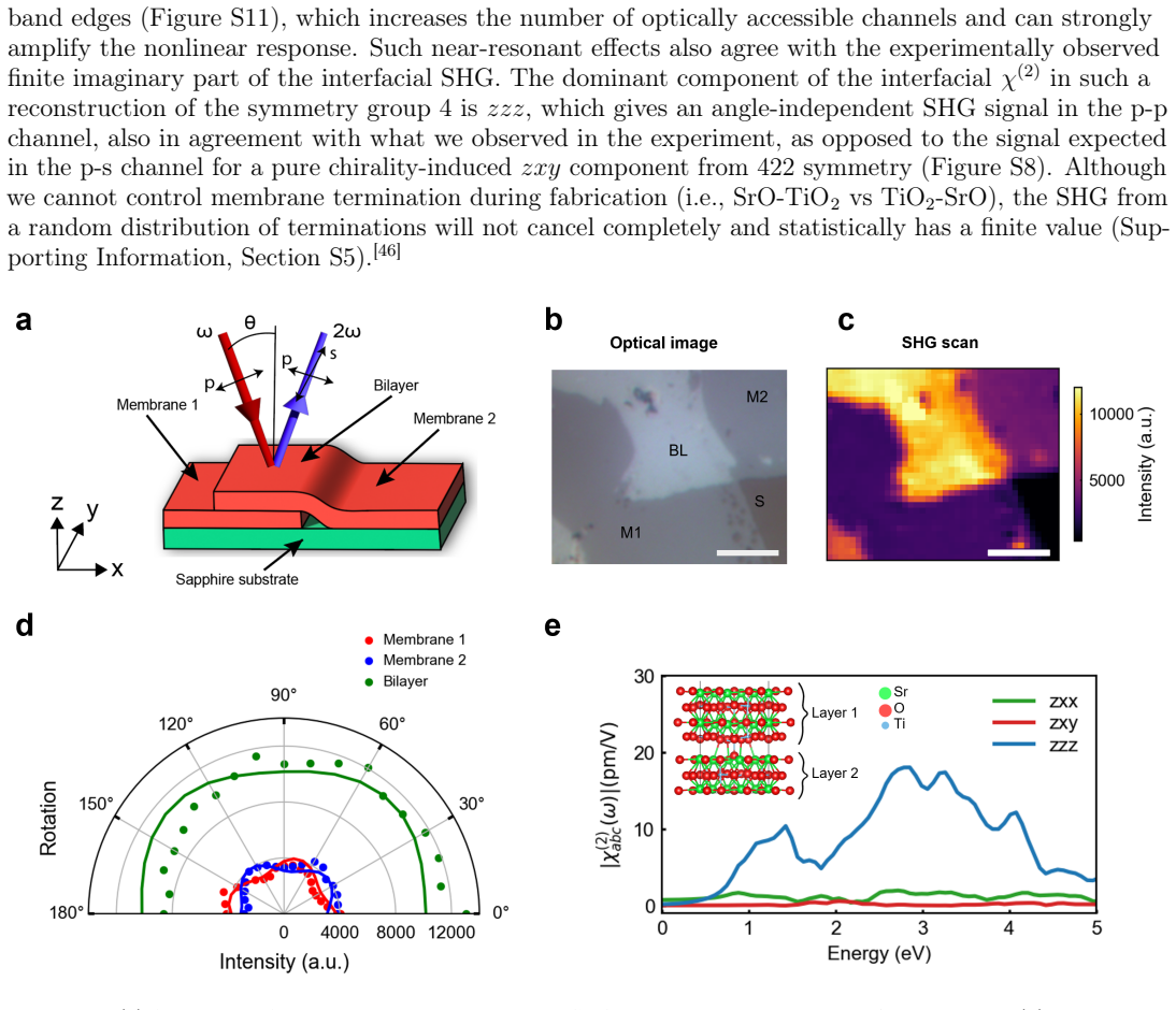
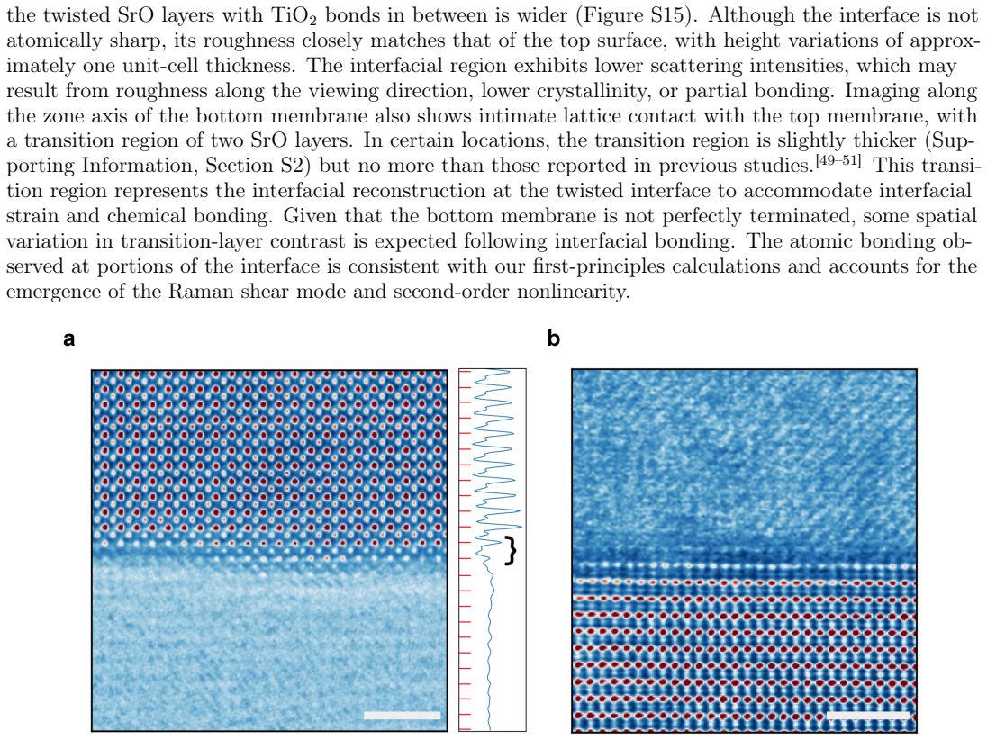
Optical Signature of Moir\'e Superlattices Formed by Twisted SrTiO₃ Membranes
Pith reviewed 2026-06-28 00:36 UTC · model grok-4.3
The pith
Twisted SrTiO3 bilayers at 36 degrees exhibit new low-frequency Raman modes and strong second harmonic generation due to an asymmetric interface.
A machine-rendered reading of the paper's core claim, the machinery that carries it, and where it could break.
Core claim
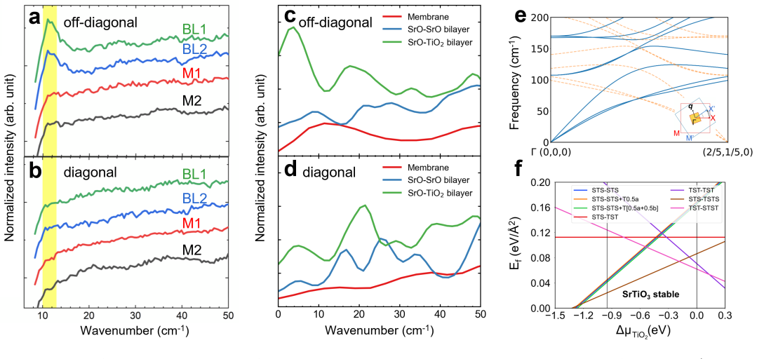
Millimeter-scale twisted SrTiO3 bilayers at 36 degrees, close to the Σ5 coincidence site lattice condition, display new low-frequency vibrational modes whose Raman activity is greatly enhanced by an asymmetric twisted interface between the SrO and TiO2 layers, as predicted by molecular dynamics simulations; this interface is energetically favorable and produces strong second harmonic generation comparable to the surface, consistent with enhanced interlayer coupling after annealing.
What carries the argument
The asymmetric twisted interface between SrO and TiO2 layers in the moiré superlattice, which enhances Raman activity of new modes and generates strong SHG.
Load-bearing premise
The observed new Raman modes and SHG arise specifically from the moiré-induced asymmetric interface rather than from fabrication defects or other contributions.
What would settle it
Observing the same new Raman modes and strong SHG in untwisted or symmetrically terminated SrTiO3 bilayers would falsify the claim that they are due to the twisted asymmetric interface.
Figures
read the original abstract
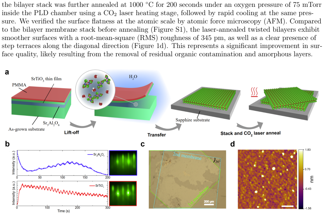
Moir\'e superlattices formed at the interfaces of mismatched lattices have attracted significant interest over the past decade due to their large tunability of band parameters and interactions among electrons, spins, and lattices. Superlattices made from twisted perovskite oxides may have strong structure and potential modulation, but evidence of such modulation over macroscopic areas, particularly at large twisting angles, has not been clearly demonstrated so far. Here, we fabricated millimeter-scale twisted oxide bilayers at $36^\circ$ angle, close to the simple coincidence site lattice condition $\Sigma5$, from freestanding SrTiO$_3$ membranes. We discovered new low-frequency vibrational modes whose Raman activity, according to molecular dynamics simulations, is greatly enhanced by an asymmetric, twisted interface between the SrO and TiO$_2$ layers. Such an interface is energetically favorable from first-principles calculations and is corroborated by the observation of strong second harmonic generation from the interface comparable to that from the SrTiO$_3$ surface throughout the bilayer region. The results are consistent with interlayer coupling enhanced by high-temperature annealing and confirmed by cross-sectional scanning transmission electron microscopy imaging. Our work sheds light on the structural behavior of twisted oxides and provides directions for tuning their phononic and nonlinear optical properties in future studies.
Editorial analysis
A structured set of objections, weighed in public.
Referee Report
Summary. The manuscript reports fabrication of millimeter-scale 36°-twisted SrTiO₃ bilayers from freestanding membranes and presents multi-technique evidence (Raman spectroscopy, SHG, cross-sectional STEM, MD simulations, and DFT) for new low-frequency vibrational modes whose Raman activity is enhanced by an energetically favored asymmetric SrO-TiO₂ interface at the moiré superlattice. The central claim is that these optical signatures arise specifically from the twisted, annealed interface rather than from generic fabrication artifacts.
Significance. If the attribution to the moiré-induced asymmetry holds, the work would provide the first macroscopic-scale demonstration of tunable phononic and nonlinear-optical responses in twisted perovskite membranes, extending moiré physics beyond van der Waals materials and offering a route to engineer interlayer coupling via high-temperature annealing. The combination of experiment with MD/DFT predictions is a strength, though the current data remain qualitative.
major comments (2)
- [Experimental methods and Results (Raman/SHG)] The central attribution of the new low-frequency Raman modes and enhanced SHG to the moiré superlattice and its SrO-TiO₂ asymmetry (Abstract; Results section on Raman and SHG) rests on the assumption that these features are absent in untwisted controls. No such control bilayers fabricated by the identical membrane-transfer and annealing protocol are reported, leaving open the possibility that the signals arise from surface reconstruction, residual strain, or defects introduced during the freestanding-membrane process rather than from the 36° twist itself.
- [MD simulations and Raman data] Quantitative support is limited: the manuscript states that Raman activity is 'greatly enhanced' by the asymmetric interface according to MD, yet no computed Raman intensities, error bars on experimental spectra, or statistical comparison across multiple twist angles or annealing conditions are provided (Abstract; MD/DFT section). This weakens the claim that the observed modes are a direct, falsifiable signature of the moiré asymmetry.
minor comments (2)
- [Fabrication] The coincidence-site-lattice angle is given as 36° (close to Σ5); a brief note on the exact angular tolerance achieved in the transfer process and its effect on the moiré period would clarify reproducibility.
- [Figures] Figure captions and axis labels for Raman spectra and SHG maps should explicitly state the number of independent samples measured and whether data are averaged or representative.
Simulated Author's Rebuttal
We thank the referee for their thorough review and valuable suggestions. We address each major comment below and indicate where revisions will be made to the manuscript.
read point-by-point responses
-
Referee: The central attribution of the new low-frequency Raman modes and enhanced SHG to the moiré superlattice and its SrO-TiO₂ asymmetry (Abstract; Results section on Raman and SHG) rests on the assumption that these features are absent in untwisted controls. No such control bilayers fabricated by the identical membrane-transfer and annealing protocol are reported, leaving open the possibility that the signals arise from surface reconstruction, residual strain, or defects introduced during the freestanding-membrane process rather than from the 36° twist itself.
Authors: We agree that untwisted control bilayers prepared with the identical protocol are important for ruling out fabrication artifacts. While our comparisons to single-layer membranes show the new modes only in twisted samples, we will add data from untwisted bilayers fabricated similarly in the revised manuscript to directly address this point. revision: yes
-
Referee: Quantitative support is limited: the manuscript states that Raman activity is 'greatly enhanced' by the asymmetric interface according to MD, yet no computed Raman intensities, error bars on experimental spectra, or statistical comparison across multiple twist angles or annealing conditions are provided (Abstract; MD/DFT section). This weakens the claim that the observed modes are a direct, falsifiable signature of the moiré asymmetry.
Authors: We acknowledge the lack of explicit computed Raman intensities in the presented MD results. In the revision, we will include the calculated Raman spectra and intensities from the simulations, as well as error bars on the experimental Raman data. A full statistical comparison across multiple twist angles and annealing conditions is not feasible within the scope of this work, as it would require a separate extensive study; however, the focus on the 36° twist near Σ5 is justified by the coincidence site lattice condition. revision: partial
Circularity Check
No circularity: experimental claims cross-validated by independent simulations and imaging
full rationale
The paper reports fabrication of twisted SrTiO3 bilayers, observation of new low-frequency Raman modes, SHG signal, and STEM imaging. These are corroborated by separate first-principles calculations (energetic favorability of asymmetry) and MD simulations (enhanced Raman activity), none of which reduce to a fitted parameter defined by the target result or to a self-citation chain. No derivation step equates a prediction to its input by construction; the central attribution rests on external corroboration rather than tautology.
Axiom & Free-Parameter Ledger
axioms (1)
- domain assumption Standard DFT and molecular-dynamics force fields accurately capture SrTiO3 interface energetics and vibrational modes.
Reference graph
Works this paper leans on
-
[1]
E. Y. Andrei, D. K. Efetov, P. Jarillo-Herrero, A. H. MacDonald, K. F. Mak, T. Senthil, E. Tutuc, A. Yazdani, A. F. Young,Nature Reviews Materials2021,6, 3 201
-
[2]
D. M. Kennes, M. Claassen, L. Xian, A. Georges, A. J. Millis, J. Hone, C. R. Dean, D. N. Basov, A. N. Pasupathy, A. Rubio,Nature Physics2021,17, 2 155
-
[3]
C. N. Lau, M. W. Bockrath, K. F. Mak, F. Zhang,Nature2022,602, 7895 41. 12
-
[4]
J. Cai, E. Anderson, C. Wang, X. Zhang, X. Liu, W. Holtzmann, Y. Zhang, F. Fan, T. Taniguchi, K. Watanabe, Y. Ran, T. Cao, L. Fu, D. Xiao, W. Yao, X. Xu,Nature2023,622, 7981 63, number: 7981
-
[5]
Y. Li, C. Xiang, F. M. Chiabrera, S. Yun, H. Zhang, D. J. Kelly, R. T. Dahm, C. K. Kirchert, T. E. L. Cozannet, F. Trier, et al.,Advanced Materials2022,34, 38 2203187
-
[6]
Pryds, D.-S
N. Pryds, D.-S. Park, T. S. Jespersen, S. Yun,APL Materials2024,12, 1 010901
-
[7]
S. Lee, D. J. P. de Sousa, B. Jalan, T. Low,Science Advances2024,10, 47 eadq0293
-
[8]
T. Xu, T. Qian, J. Pang, J. Zhang, S. Li, R. He, J. Wang, T. Shimada,Research2025,80621
-
[9]
N. A. Shahed, K. Samanta, M. Elekhtiar, K. Huang, C.-B. Eom, M. S. Rzchowski, K. D. Be- lashchenko, E. Y. Tsymbal,Physical Review B2025,111, 19 195420
-
[10]
D. Lu, D. J. Baek, S. S. Hong, L. F. Kourkoutis, Y. Hikita, H. Y. Hwang,Nature Materials2016
-
[11]
R. Xu, J. Huang, E. S. Barnard, S. S. Hong, P. Singh, E. K. Wong, T. Jansen, V. Harbola, J. Xiao, B. Y. Wang, S. Crossley, D. Lu, S. Liu, H. Y. Hwang,Nature Communications2020,11, 1 3141
-
[13]
M.-S. Kim, K. Lee, R. Ishikawa, K. Song, N. A. Shahed, K.-T. Eom, M. S. Rzchowski, E. Y. Tsym- bal, N. Shibata, T. Mizoguchi, C.-B. Eom, S.-Y. Choi,ACS Nano2025,19, 46 39714
-
[14]
Zubko, S
P. Zubko, S. Gariglio, M. Gabay, P. Ghosez, J.-M. Triscone,Annual Review of Condensed Matter Physics2011,2, Volume 2, 2011 141
2011
-
[15]
H. Kp, X. Wei, C.-H. Lee, D. Yoon, Y. Lee, K. J. Crust, Y.-T. Shao, R. Xu, J.-H. Kang, C. Liang, J. Park, H. Y. Hwang, D. A. Muller,arXiv:2510.230422025
-
[16]
H. Wang, V. Harbola, Y.-J. Wu, P. A. van Aken, J. Mannhart,Advanced Materials2024,36, 32 2405065
-
[17]
Huang, L
S. Huang, L. Liang, X. Ling, A. A. Puretzky, D. B. Geohegan, B. G. Sumpter, J. Kong, V. Meunier, M. S. Dresselhaus,Nano Letters2016,16, 2 1435
-
[18]
Lin, Q.-H
M.-L. Lin, Q.-H. Tan, J.-B. Wu, X.-S. Chen, J.-H. Wang, Y.-H. Pan, X. Zhang, X. Cong, J. Zhang, W. Ji, P.-A. Hu, K.-H. Liu, P.-H. Tan,ACS Nano2018,12, 8 8770
-
[19]
K.-Q. Lin, J. Holler, J. M. Bauer, P. Parzefall, M. Scheuck, B. Peng, T. Korn, S. Bange, J. M. Lup- ton, C. Schüller,Advanced Materials2021,33, 34 2008333
-
[20]
J. Quan, L. Linhart, M.-L. Lin, D. Lee, J. Zhu, C.-Y. Wang, W.-T. Hsu, J. Choi, J. Embley, C. Young, T. Taniguchi, K. Watanabe, C.-K. Shih, K. Lai, A. H. MacDonald, P.-H. Tan, F. Libisch, X. Li,Nature Materials2021,20, 8 1100
-
[21]
J. Quan, G. Chen, L. Linhart, Z. Liu, T. Taniguchi, K. Watanabe, F. Libisch, R. Huang, X. Li,Nano Letters2023,23, 24 11510
-
[22]
P. Ci, Y. Zhao, M. Sun, Y. Rho, Y. Chen, C. P. Grigoropoulos, S. Jin, X. Li, J. Wu,Nano Letters 2022,22, 22 9027
2022
-
[23]
Paradisanos, A
I. Paradisanos, A. M. S. Raven, T. Amand, C. Robert, P. Renucci, K. Watanabe, T. Taniguchi, I. C. Gerber, X. Marie, B. Urbaszek,Physical Review B2022,105, 11 115420
-
[24]
L. Du, Z. Huang, J. Zhang, F. Ye, Q. Dai, H. Deng, G. Zhang, Z. Sun,Nature Materials2024,23, 9 1179
-
[25]
H. Zhu, B. I. Yakobson,Nature Materials2024,23316. 13
-
[26]
Padilla, D
J. Padilla, D. Vanderbilt,Surface Science1998,418, 1 64
-
[27]
Kawasaki, K
M. Kawasaki, K. Takahashi, T. Maeda, R. Tsuchiya, M. Shinohara, O. Ishiyama, T. Yonezawa, M. Yoshimoto, H. Koinuma,Science1994,266, 5190 1540
-
[28]
Erdman, K
N. Erdman, K. R. Poeppelmeier, M. Asta, O. Warschkow, D. E. Ellis, L. D. Marks,Nature2002,419, 6902 55
-
[29]
W. G. Nilsen, J. G. Skinner,The Journal of Chemical Physics1968,48, 5 2240
-
[30]
C. H. Perry, J. H. Fertel, T. F. McNelly,The Journal of Chemical Physics1967,47, 5 1619
-
[31]
O’Shea, R
D. O’Shea, R. Kolluri, H. Cummins,Solid State Communications1967,5, 4 241
-
[32]
A. G. Razumnaya, Y. I. Yuzyuk, I. N. Zakharchenko, Y. A. Tikhonov, N. Ortega, A. Kumar, R. S. Katiyar, M. El Marssi, I. A. Lukyanchuk,Ferroelectrics2016,501, 1 61
-
[33]
Gupta, R
S. Gupta, R. Katiyar,Journal of Raman Spectroscopy2001,32, 10 885
-
[34]
A. A. Sirenko, I. A. Akimov, J. R. Fox, A. M. Clark, H.-C. Li, W. Si, X. X. Xi,Physical Review Letters1999,82, 22 4500
-
[35]
Shirane, Y
G. Shirane, Y. Yamada,Physical Review1969,177, 2 858
-
[36]
P. A. Fleury, J. F. Scott, J. M. Worlock,Physical Review Letters1968,21, 1 16
-
[37]
Tadano, S
T. Tadano, S. Tsuneyuki,Journal of the Ceramic Society of Japan2019,127, 6 404
-
[38]
Reuter, M
K. Reuter, M. Scheffler,Physical Review Letters2003,90, 4 046103
-
[39]
S. Qin, S. Banerjee, M. G. Sensoy, A. M. Rappe,ACS Catalysis2024,14, 23 17253
-
[40]
J.-L. Do, T. Friscic,ACS central science2017,3, 1 13
-
[41]
X. Liu, Y. Li, L. Zeng, X. Li, N. Chen, S. Bai, H. He, Q. Wang, C. Zhang,Advanced Materials2022, 34, 46 2108327
-
[42]
Cheng, M
Z. Cheng, M. R. Jones,Nature Communications2022,13, 1 4207
-
[43]
S. Zhou, J. Li, J. Lu, H. Liu, J.-Y. Kim, A. Kim, L. Yao, C. Liu, C. Qian, Z. D. Hood, et al.,Nature 2022,612, 7939 259
2022
-
[44]
Zhang, J
Z. Zhang, J. Zhang, Z.-J. Liu, N. S. Dahod, W. Paritmongkol, N. Brown, A. Stollmann, W. S. Lee, Y.-C. Chien, Z. Dai, et al.,Science Advances2023,9, 33 eadg4417
-
[45]
Yaffe, Y
O. Yaffe, Y. Guo, L. Z. Tan, D. A. Egger, T. Hull, C. C. Stoumpos, F. Zheng, T. F. Heinz, L. Kro- nik, M. G. Kanatzidis, et al.,Physical Review Letters2017,118, 13 136001
-
[46]
Biswas, R
A. Biswas, R. Xu, G. A. Alvarez, J. Zhang, J. Christiansen-Salameh, A. B. Puthirath, K. Burns, J. A. Hachtel, T. Li, S. A. Iyengar, T. Gray, C. Li, X. Zhang, H. Kannan, J. Elkins, T. S. Pieshkov, R. Vajtai, A. G. Birdwell, M. R. Neupane, E. J. Garratt, T. G. Ivanov, B. B. Pate, Y. Zhao, H. Zhu, Z. Tian, A. Rubio, P. M. Ajayan,Advanced Materials2023,34, 39 2304624
-
[47]
J. E. Sipe, D. J. Moss, H. M. van Driel,Physical Review B1987,35, 3 1129
-
[48]
R. Zhao, K. Jin, H. Guo, H. Lu, G. Yang,Science China Physics, Mechanics and Astronomy2013, 56, 12 2370
-
[49]
Sánchez-Santolino, V
G. Sánchez-Santolino, V. Rouco, S. Puebla, H. Aramberri, V. Zamora, M. Cabero, F. A. Cuellar, C. Munuera, F. Mompean, M. Garcia-Hernandez, A. Castellanos-Gomez, J. Íñiguez, C. Leon, J. San- tamaria,Nature2024,626, 7999 529
-
[50]
M.-S. Kim, K. Lee, R. Ishikawa, K. Song, N. A. Shahed, K.-T. Eom, M. S. Rzchowski, E. Y. Tsym- bal, N. Shibata, T. Mizoguchi, C.-B. Eom, S.-Y. Choi,ACS Nano2025,19, 46 39714. 14
-
[51]
Zhang, J
Y. Zhang, J. Ge, S. Su, Y. Li, W. Zhang, L. Lyu, J. Song, Y. Liu, Y. Lei, H. Du, G. Zhong, B. Huang, J. Li, C. Li,Adv. Mater.2025, e19300
2025
-
[52]
Giannozzi, S
P. Giannozzi, S. Baroni, N. Bonini, M. Calandra, R. Car, C. Cavazzoni, D. Ceresoli, G. L. Chiarotti, M. Cococcioni, I. Dabo, et al.,Journal of Physics: Condensed matter2009,21, 39 395502
-
[53]
Giannozzi, O
P. Giannozzi, O. Andreussi, T. Brumme, O. Bunau, M. B. Nardelli, M. Calandra, R. Car, C. Cavaz- zoni, D. Ceresoli, M. Cococcioni, et al.,Journal of Physics: Condensed matter2017,29, 46 465901
-
[54]
A. M. Rappe, K. M. Rabe, E. Kaxiras, J. Joannopoulos,Physical Review B1990,41, 2 1227
-
[55]
N. J. Ramer, A. M. Rappe,Physical Review B1999,59, 19 12471
-
[56]
J. P. Perdew, K. Burke, M. Ernzerhof,Physical Review Letters1996,77, 18 3865
-
[57]
Grimme,Journal of Computational Chemistry2006,27, 15 1787
S. Grimme,Journal of Computational Chemistry2006,27, 15 1787
-
[58]
Grimme, J
S. Grimme, J. Antony, S. Ehrlich, H. Krieg,The Journal of Chemical Physics2010,132, 15
-
[59]
Aversa, J
C. Aversa, J. E. Sipe,Physical Review B1995,52, 20 14636
-
[60]
J. E. Sipe, A. I. Shkrebtii,Physical Review B2000,61, 8 5337. Supporting Information Supporting Information is available from the Wiley Online Library or from the author. 15
discussion (0)
Sign in with ORCID, Apple, or X to comment. Anyone can read and Pith papers without signing in.