CSWinUNETR: Segmentation of Thin Anatomical Structures in Medical Images
Pith reviewed 2026-06-26 18:26 UTC · model grok-4.3
The pith
CSWinUNETR recovers fine branches in thin tortuous structures by using cross-shaped stripe attention and dynamic snake convolution.
A machine-rendered reading of the paper's core claim, the machinery that carries it, and where it could break.
Core claim
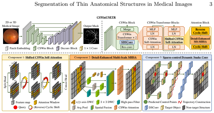
CSWinUNETR employs cross-shaped stripe self-attention to model long-range principal-axis context with cyclic shifts for better information exchange, a detail-enhanced multi-scale self-attention module to aggregate features from multi-resolution representations, and sparse-control dynamic snake convolution to build dense curvilinear kernels from sparse control points, resulting in superior segmentation of thin structures on four benchmarks across ophthalmology, neurovascular imaging, and dermatology without task-specific post-processing or topology-aware losses.
What carries the argument
CSWinUNETR architecture that integrates cross-shaped stripe self-attention, detail-enhanced multi-scale self-attention, and sparse-control dynamic snake convolution to handle thin tortuous geometry.
If this is right
- The model produces higher-accuracy segmentations of retinal vessels, cerebral vessels, and wrinkles on multiple public datasets.
- It supports both 2D and 3D inputs without requiring additional topology constraints.
- It avoids the need for specialized post-processing steps or custom loss functions common in prior thin-structure work.
- It shows consistent gains over both convolutional and Transformer baselines across ophthalmology, neurovascular, and dermatology tasks.
Where Pith is reading between the lines
- The sparse-control dynamic snake convolution mechanism could be tested on other curve-following tasks outside medicine, such as tracing roads or fibers in non-medical images.
- Combining the architecture with existing 3D vessel datasets might reveal whether the same modules scale to volumetric CT or MR angiography without retraining from scratch.
- If the cyclic-shift mechanism proves critical, similar stripe-based attention patterns could be adapted to other domains that require long-range linear context along principal axes.
Load-bearing premise
The specific choices of cross-shaped stripe self-attention, detail-enhanced multi-scale self-attention, and sparse-control dynamic snake convolution are responsible for the improved recovery of fine branches rather than other training or data factors.
What would settle it
Running the same four benchmarks and finding that CSWinUNETR does not exceed prior state-of-the-art methods in metrics that measure continuity of thin branches would falsify the performance claim.
Figures

read the original abstract
Accurate segmentation of thin, tortuous anatomical structures, such as retinal vessels, cerebral vasculature, and facial wrinkles, remains challenging due to low contrast, frequent discontinuities, and severe class imbalance. Although recent convolutional and Transformer-based models have improved performance, they often yield fragmented predictions and fail to recover fine branches. We propose CSWinUNETR, a general-purpose backbone for 2D and 3D thin-structure segmentation. It employs cross-shaped stripe self-attention to model long-range principal-axis context and incorporates cyclic shifts to enhance information exchange across stripes. To better preserve fine-grained details, we further introduce a detail-enhanced multi-scale self-attention module that aggregates contextual features from multi-resolution representations. In addition, we propose sparse-control dynamic snake convolution, which reconstructs reliable dense curvilinear kernels from sparsely predicted control points to better follow tortuous geometry. Extensive experiments on four benchmarks across ophthalmology, neurovascular imaging, and dermatology demonstrate that CSWinUNETR consistently outperforms state-of-the-art methods without task-specific post-processing or topology-aware losses. The code is available at https://github.com/labhai/CSWinUNETR.
Editorial analysis
A structured set of objections, weighed in public.
Referee Report
Summary. The manuscript proposes CSWinUNETR, a general-purpose backbone for 2D/3D segmentation of thin, tortuous anatomical structures (e.g., retinal vessels, cerebral vasculature, facial wrinkles). The architecture combines cross-shaped stripe self-attention with cyclic shifts for long-range context, a detail-enhanced multi-scale self-attention module for fine-grained features, and sparse-control dynamic snake convolution to follow curvilinear geometry. It reports consistent outperformance over prior convolutional and Transformer models on four benchmarks spanning ophthalmology, neurovascular imaging, and dermatology, without task-specific post-processing or topology-aware losses. Code is released at https://github.com/labhai/CSWinUNETR.
Significance. If the empirical superiority holds under rigorous evaluation, the work would offer a reusable backbone for a persistent challenge in medical image analysis where standard models produce fragmented outputs on low-contrast, imbalanced thin structures. The public code release is a clear strength that enables direct reproducibility and extension.
major comments (1)
- [Abstract] Abstract: the central claim that CSWinUNETR 'consistently outperforms state-of-the-art methods' on four benchmarks is presented without any quantitative metrics (Dice, sensitivity, etc.), ablation tables, statistical significance tests, dataset identifiers, or even summary performance numbers. This absence makes it impossible to evaluate whether the data support the stated superiority or the contribution of the three proposed modules.
Simulated Author's Rebuttal
We thank the referee for the detailed review and constructive comment on the abstract. We address the concern point-by-point below and will revise the manuscript accordingly.
read point-by-point responses
-
Referee: [Abstract] Abstract: the central claim that CSWinUNETR 'consistently outperforms state-of-the-art methods' on four benchmarks is presented without any quantitative metrics (Dice, sensitivity, etc.), ablation tables, statistical significance tests, dataset identifiers, or even summary performance numbers. This absence makes it impossible to evaluate whether the data support the stated superiority or the contribution of the three proposed modules.
Authors: We agree that the abstract would be strengthened by including concrete quantitative indicators. In the revised manuscript we will expand the abstract to report key summary metrics (e.g., mean Dice and sensitivity on the primary benchmarks), name the four datasets, and briefly note that detailed ablation studies and statistical tests appear in the main text. The full experimental tables, ablations, and significance results are already present in Sections 4 and 5; the revision will simply surface the most salient numbers in the abstract itself. revision: yes
Circularity Check
No significant circularity detected
full rationale
The paper is an empirical architecture proposal whose central claim is benchmark superiority of CSWinUNETR on four medical segmentation datasets. No equations, fitted parameters, or predictions appear that reduce by construction to the inputs; the three described modules (cross-shaped stripe attention, detail-enhanced multi-scale attention, sparse-control dynamic snake convolution) are presented as design choices whose value is tested externally rather than derived from the evaluation data itself. No self-citation chains or uniqueness theorems are invoked as load-bearing premises. The result is therefore self-contained against external benchmarks.
Axiom & Free-Parameter Ledger
Reference graph
Works this paper leans on
-
[1]
International Journal of Computer Vision92(2), 192–210 (2011)
Benmansour, F., Cohen, L.D.: Tubular structure segmentation based on minimal path method and anisotropic enhancement. International Journal of Computer Vision92(2), 192–210 (2011)
2011
-
[2]
Pattern Recognition60, 949–970 (2016)
Bibiloni, P., González-Hidalgo, M., Massanet, S.: A survey on curvilinear object segmentation in multiple applications. Pattern Recognition60, 949–970 (2016)
2016
-
[3]
IEEE transactions on pattern analysis and machine intelligence 44(12), 8766–8778 (2020)
Clough, J.R., Byrne, N., Oksuz, I., Zimmer, V.A., Schnabel, J.A., King, A.P.: A topological loss function for deep-learning based image segmentation using per- sistent homology. IEEE transactions on pattern analysis and machine intelligence 44(12), 8766–8778 (2020)
2020
-
[4]
Dice,L.R.:Measuresoftheamountofecologicassociationbetweenspecies.Ecology 26(3), 297–302 (1945)
1945
-
[5]
Dong, X., Bao, J., Chen, D., Zhang, W., Yu, N., Yuan, L., Chen, D., Guo, B.: Cswin transformer: A general vision transformer backbone with cross-shaped win- dows.In:ProceedingsoftheIEEE/CVFconferenceoncomputervisionandpattern recognition. pp. 12124–12134 (2022)
2022
-
[6]
In: International conference on medical image computing and computer-assisted intervention
Frangi, A.F., Niessen, W.J., Vincken, K.L., Viergever, M.A.: Multiscale vessel en- hancement filtering. In: International conference on medical image computing and computer-assisted intervention. pp. 130–137. Springer (1998)
1998
-
[7]
arXiv preprint arXiv:2212.03035 (2022)
Fu, L., Tian, H., Zhai, X.B., Gao, P., Peng, X.: Incepformer: efficient incep- tion transformer with pyramid pooling for semantic segmentation. arXiv preprint arXiv:2212.03035 (2022)
-
[8]
Tomography 11(5), 52 (2025)
Gao, Y., Jiang, Y., Peng, Y., Yuan, F., Zhang, X., Wang, J.: Medical image seg- mentation: A comprehensive review of deep learning-based methods. Tomography 11(5), 52 (2025)
2025
-
[9]
In: International MICCAI brainlesion workshop
Hatamizadeh, A., Nath, V., Tang, Y., Yang, D., Roth, H.R., Xu, D.: Swin unetr: Swin transformers for semantic segmentation of brain tumors in mri images. In: International MICCAI brainlesion workshop. pp. 272–284. Springer (2021)
2021
-
[10]
In: Proceedings of the IEEE/CVF winter conference on applications of computer vi- sion
Hatamizadeh, A., Tang, Y., Nath, V., Yang, D., Myronenko, A., Landman, B., Roth, H.R., Xu, D.: Unetr: Transformers for 3d medical image segmentation. In: Proceedings of the IEEE/CVF winter conference on applications of computer vi- sion. pp. 574–584 (2022)
2022
-
[11]
In: International Conference on Medical Image Computing and Computer-Assisted Intervention
He, Y., Nath, V., Yang, D., Tang, Y., Myronenko, A., Xu, D.: Swinunetr-v2: Stronger swin transformers with stagewise convolutions for 3d medical image segmentation. In: International Conference on Medical Image Computing and Computer-Assisted Intervention. pp. 416–426. Springer (2023) 10 J. Moon et al
2023
-
[12]
Advances in neural information processing systems32(2019)
Hu, X., Li, F., Samaras, D., Chen, C.: Topology-preserving deep image segmenta- tion. Advances in neural information processing systems32(2019)
2019
-
[13]
Nature methods18(2), 203–211 (2021)
Isensee, F., Jaeger, P.F., Kohl, S.A., Petersen, J., Maier-Hein, K.H.: nnu-net: a self-configuring method for deep learning-based biomedical image segmentation. Nature methods18(2), 203–211 (2021)
2021
-
[14]
Scientific data9(1), 475 (2022)
Jin, K., Huang, X., Zhou, J., Li, Y., Yan, Y., Sun, Y., Zhang, Q., Wang, Y., Ye, J.: Fives: A fundus image dataset for artificial intelligence based vessel segmentation. Scientific data9(1), 475 (2022)
2022
-
[15]
In: International conference on medical imaging with deep learning
Kervadec, H., Bouchtiba, J., Desrosiers, C., Granger, E., Dolz, J., Ayed, I.B.: Boundary loss for highly unbalanced segmentation. In: International conference on medical imaging with deep learning. pp. 285–296. PMLR (2019)
2019
-
[16]
In: European Conference on Computer Vision
Kirchhoff, Y., Rokuss, M.R., Roy, S., Kovacs, B., Ulrich, C., Wald, T., Zenk, M., Vollmuth, P., Kleesiek, J., Isensee, F., et al.: Skeleton recall loss for connectiv- ity conserving and resource efficient segmentation of thin tubular structures. In: European Conference on Computer Vision. pp. 218–234. Springer (2024)
2024
-
[17]
Medical Image Analysis89, 102929 (2023)
Lin, J., Huang, X., Zhou, H., Wang, Y., Zhang, Q.: Stimulus-guided adaptive trans- former network for retinal blood vessel segmentation in fundus images. Medical Image Analysis89, 102929 (2023)
2023
-
[18]
Information Fusion 113, 102634 (2025)
Liu, X., Gao, P., Yu, T., Wang, F., Yuan, R.Y.: Cswin-unet: Transformer unet with cross-shaped windows for medical image segmentation. Information Fusion 113, 102634 (2025)
2025
-
[19]
Liu, Z., Lin, Y., Cao, Y., Hu, H., Wei, Y., Zhang, Z., Lin, S., Guo, B.: Swin transformer:Hierarchicalvisiontransformerusingshiftedwindows.In:Proceedings of the IEEE/CVF international conference on computer vision. pp. 10012–10022 (2021)
2021
-
[20]
In: International Conference on Pattern Recognition
Moon, J., Chung, H., Jang, I.: Facial wrinkle segmentation for cosmetic derma- tology: Pretraining with texture map-based weak supervision. In: International Conference on Pattern Recognition. pp. 319–334. Springer (2024)
2024
-
[21]
Medical image analysis67, 101874 (2021)
Mou, L., Zhao, Y., Fu, H., Liu, Y., Cheng, J., Zheng, Y., Su, P., Yang, J., Chen, L., Frangi, A.F., et al.: Cs2-net: Deep learning segmentation of curvilinear structures in medical imaging. Medical image analysis67, 101874 (2021)
2021
-
[22]
In: Proceed- ings of the IEEE/CVF international conference on computer vision
Qi, Y., He, Y., Qi, X., Zhang, Y., Yang, G.: Dynamic snake convolution based on topological geometric constraints for tubular structure segmentation. In: Proceed- ings of the IEEE/CVF international conference on computer vision. pp. 6070–6079 (2023)
2023
-
[23]
In: International Conference on Medical Image Computing and Computer-Assisted Intervention
Shi,P.,Hu,J.,Yang,Y.,Gao,Z.,Liu,W.,Ma,T.:Centerlineboundarydicelossfor vascular segmentation. In: International Conference on Medical Image Computing and Computer-Assisted Intervention. pp. 46–56. Springer (2024)
2024
-
[24]
In: Proceedings of the IEEE/CVF conference on computer vision and pattern recognition
Shit, S., Paetzold, J.C., Sekuboyina, A., Ezhov, I., Unger, A., Zhylka, A., Pluim, J.P., Bauer, U., Menze, B.H.: cldice-a novel topology-preserving loss function for tubular structure segmentation. In: Proceedings of the IEEE/CVF conference on computer vision and pattern recognition. pp. 16560–16569 (2021)
2021
-
[25]
BMC medical imaging15(1), 29 (2015)
Taha, A.A., Hanbury, A.: Metrics for evaluating 3d medical image segmentation: analysis, selection, and tool. BMC medical imaging15(1), 29 (2015)
2015
-
[26]
In: Proceedings of the IEEE/CVF international conference on computer vision
Wu, H., Xiao, B., Codella, N., Liu, M., Dai, X., Yuan, L., Zhang, L.: Cvt: In- troducing convolutions to vision transformers. In: Proceedings of the IEEE/CVF international conference on computer vision. pp. 22–31 (2021)
2021
-
[27]
IEEE Journal of Biomedical and Health Informatics 28(3), 1472–1483 (2023) Segmentation of Thin Anatomical Structures in Medical Images 11
Xu, W., Yang, H., Shi, Y., Tan, T., Liu, W., Pan, X., Deng, Y., Gao, F., Su, R.: Ernet: edge regularization network for cerebral vessel segmentation in digital sub- traction angiography images. IEEE Journal of Biomedical and Health Informatics 28(3), 1472–1483 (2023) Segmentation of Thin Anatomical Structures in Medical Images 11
2023
-
[28]
ArXiv pp
Yang, K., Musio, F., Ma, Y., Juchler, N., Paetzold, J.C., Al-Maskari, R., Höher, L., Li, H.B., Hamamci, I.E., Sekuboyina, A., et al.: Benchmarking the cow with the topcow challenge: Topology-aware anatomical segmentation of the circle of willis for cta and mra. ArXiv pp. arXiv–2312 (2025)
2025
-
[29]
In: International Conference on Medical Image Computing and Computer-Assisted Intervention
Zhou, F., Gao, Z., Zhao, H., Xie, J., Meng, Y., Zhao, Y., Lip, G.Y., Zheng, Y.: Glcp: Global-to-local connectivity preservation for tubular structure segmentation. In: International Conference on Medical Image Computing and Computer-Assisted Intervention. pp. 237–247. Springer (2025)
2025
discussion (0)
Sign in with ORCID, Apple, or X to comment. Anyone can read and Pith papers without signing in.