HF Etching and Silanization: Evidence for the Role of Surface Hydroxyl Groups in Silicon Nitride Resonator Loss
Pith reviewed 2026-06-26 03:27 UTC · model grok-4.3
The pith
Surface hydroxyl groups drive mechanical energy loss in silicon nitride resonators, and silanization can cut this loss by up to half.
A machine-rendered reading of the paper's core claim, the machinery that carries it, and where it could break.
Core claim
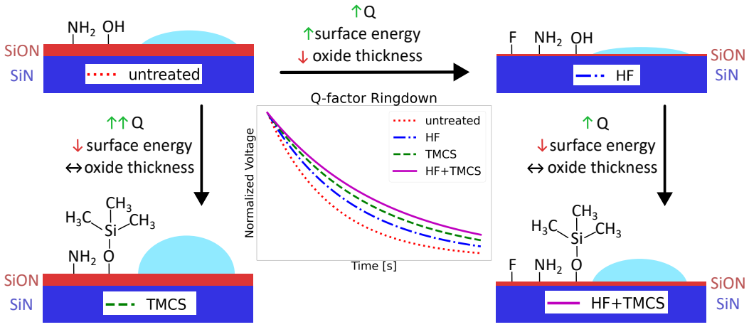
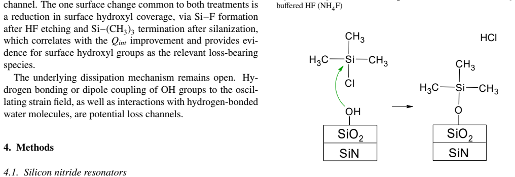
The authors show that TMCS silanization, which replaces surface hydroxyls with Si-(CH3)3 groups, yields substantially larger improvements in resonator Q_int than HF etching that removes the native oxide, demonstrating that hydroxyl groups rather than the oxide layer itself are the main source of surface-related mechanical dissipation in SiNx resonators.
What carries the argument
The differential effect of HF oxide removal versus TMCS hydroxyl passivation on measured Q_int, tracked by XPS, photothermal FTIR, and contact-angle data.
Load-bearing premise
The quality-factor gains come specifically from changes to surface hydroxyl groups and not from other side effects of the chemical treatments or from bulk material changes.
What would settle it
A surface treatment that removes or blocks hydroxyl groups without raising Q_int, or an unrelated treatment that raises Q_int without altering hydroxyl coverage, would falsify the link.
Figures
read the original abstract
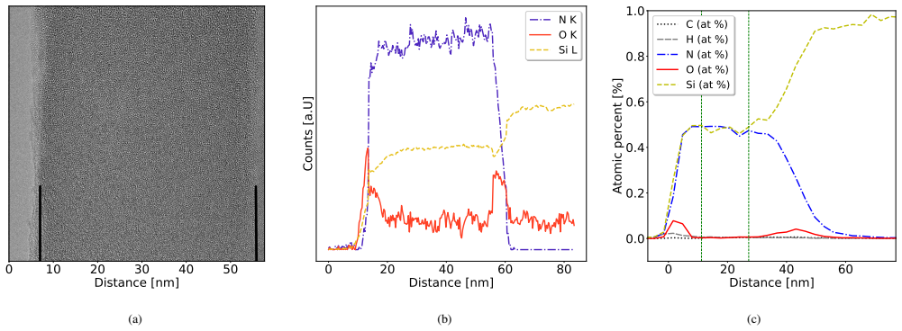
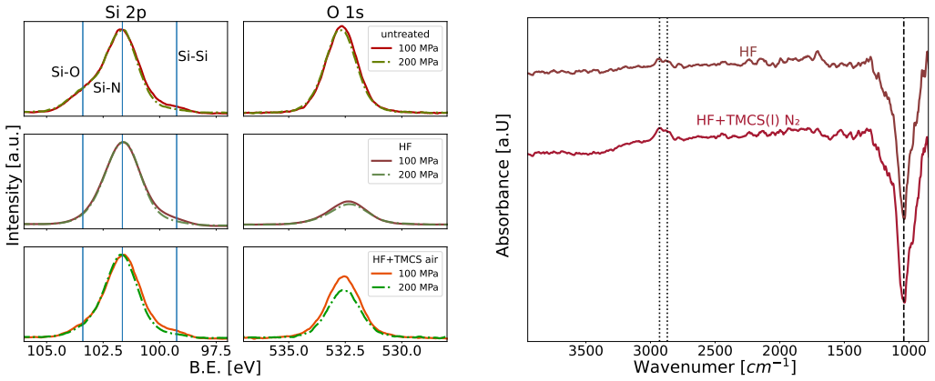
Silicon nitride $SiN_x$ nanomechanical resonators are central to sensing, quantum technologies, and fundamental physics experiments due to their exceptional mechanical quality factors (Q). However, as resonator thickness approaches the nano-scale, surface-related dissipation limits performance. Here, we investigate the role of surface chemistry in low-stress Si-rich SiNx membranes through a combination of hydrofluoric acid (HF) etching and trimethylchlorosilane (TMCS) silanization, correlated with surface characterization and mechanical measurements. Preliminary analysis by TEM-EELS, XPS, RBS/ERDA, and XRR reveals a native oxide surface layer (1-2 nm). Surface modification by HF and TMCS was subsequently evaluated using XPS, photothermal FTIR, contact-angle measurements, and intrinsic quality factor ($Q_{int}$) characterization. While HF etching effectively removes the native oxide and TMCS introduces hydrophobic $Si-(CH_3)_3$ termination, neither oxide thickness nor surface energy correlates with mechanical dissipation. TMCS treatments produce the largest enhancements, increasing $Q_{int}$ by up to 50%, whereas HF etching alone yields lower gains of 20-25%. These findings suggest surface hydroxyl groups as a key contributor to energy loss in $SiN_x$ resonators and demonstrate that chemical functionalization can substantially suppress surface dissipation.
Editorial analysis
A structured set of objections, weighed in public.
Referee Report
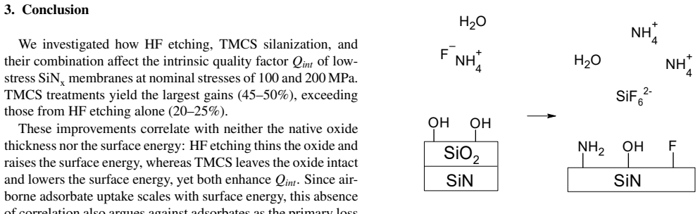
Summary. The manuscript reports experiments on low-stress Si-rich SiNx membranes where HF etching removes the native 1-2 nm oxide layer and TMCS silanization introduces Si-(CH3)3 termination. Surface characterization (XPS, photothermal FTIR, contact angle, TEM-EELS, RBS/ERDA, XRR) is correlated with intrinsic quality factor measurements. The key observations are that oxide thickness and surface energy show no correlation with dissipation, HF alone yields 20-25% Q_int gains, and TMCS yields up to 50% gains, from which the authors conclude that surface hydroxyl groups are a primary loss mechanism and that chemical functionalization can suppress surface dissipation.
Significance. If the attribution of Q_int gains specifically to hydroxyl passivation is substantiated, the result would be significant for resonator design in sensing, quantum technologies, and precision measurements, as it identifies a practical surface-chemistry route to reduce dissipation in thin SiNx devices without relying on oxide thickness or macroscopic surface energy. The combination of multiple surface probes with mechanical characterization is a positive aspect of the experimental design.
major comments (2)
- [Abstract] Abstract: the central inference that surface hydroxyl groups are the dominant loss contributor rests on the differential Q_int response to HF versus TMCS without a direct, quantitative correlation between photothermal FTIR OH signal intensity and Q_int across the measured samples; this leaves open the possibility that other uncharacterized treatment effects (bulk defects, stress changes, or additional surface species) drive the observed gains.
- [Abstract] Abstract: the reported Q_int increases (20-25% for HF, up to 50% for TMCS) are presented without error bars, sample counts, or statistical tests, so the claimed difference between treatments cannot be evaluated for robustness against measurement variability or confounding variables.
minor comments (1)
- [Abstract] The abstract refers to 'preliminary analysis' by TEM-EELS, XPS, RBS/ERDA, and XRR but does not indicate where the full datasets or error estimates for oxide thickness appear in the main text.
Simulated Author's Rebuttal
We thank the referee for their careful review and constructive comments on our manuscript. We address each major comment below.
read point-by-point responses
-
Referee: [Abstract] Abstract: the central inference that surface hydroxyl groups are the dominant loss contributor rests on the differential Q_int response to HF versus TMCS without a direct, quantitative correlation between photothermal FTIR OH signal intensity and Q_int across the measured samples; this leaves open the possibility that other uncharacterized treatment effects (bulk defects, stress changes, or additional surface species) drive the observed gains.
Authors: The inference is grounded in the specific surface chemistry: HF removes the native oxide (containing OH groups) while TMCS targets OH passivation via silanization, yielding larger Q gains. Photothermal FTIR confirms OH signal changes post-treatment, and complementary probes (XPS, contact angle, TEM-EELS, RBS/ERDA, XRR) show no evidence of bulk defects or stress changes. While a direct sample-by-sample quantitative correlation between FTIR OH intensity and Q_int is not presented, the differential response and ruling out of alternatives support the conclusion. We will expand the discussion section to address alternative explanations more explicitly. revision: partial
-
Referee: [Abstract] Abstract: the reported Q_int increases (20-25% for HF, up to 50% for TMCS) are presented without error bars, sample counts, or statistical tests, so the claimed difference between treatments cannot be evaluated for robustness against measurement variability or confounding variables.
Authors: We agree that these statistical details are needed for robustness assessment. The reported values are based on measurements from multiple devices across several samples per treatment. In the revised manuscript we will add error bars (standard deviation), specify sample and device counts, and include notes on variability in both the abstract and main text. revision: yes
Circularity Check
No circularity: purely experimental correlations with no fitted predictions or self-referential derivations
full rationale
The manuscript reports surface treatments (HF etching, TMCS silanization), characterization (XPS, FTIR, contact angle, TEM-EELS, etc.), and direct Q_int measurements on SiNx resonators. No equations, parameters fitted to subsets of the same data, or theoretical derivations appear. The central inference—that hydroxyl groups contribute to dissipation—is drawn from observed Q_int changes after treatments that alter surface chemistry, but this remains an empirical correlation open to falsification by independent measurements rather than a quantity defined by the same inputs. No self-citation chains or ansatzes are invoked as load-bearing steps. The work is self-contained experimental evidence.
Axiom & Free-Parameter Ledger
axioms (1)
- domain assumption XPS, FTIR, and contact-angle measurements accurately report surface hydroxyl coverage and hydrophobicity after HF and TMCS treatments.
Reference graph
Works this paper leans on
-
[1]
K. Kanellopulos, R. G. West, S. Emminger, P. Martini, M. Sauer, A. Foelske, S. Schmid, Stress-dependent opti- cal extinction in low-pressure chemical vapor deposition silicon nitride measured by nanomechanical photother- mal sensing, Nano Letters 24 (36) (2024) 11262–11268. doi:10.1021/acs.nanolett.4c02902
-
[2]
Sementilli, E
L. Sementilli, E. Romero, W. P. Bowen, Nanomechani- cal dissipation and strain engineering, Advanced Func- tional Materials 32 (3) (Aug. 2021).doi:10.1002/ adfm.202105247
2021
-
[3]
Schmid, K
S. Schmid, K. Jensen, K. Nielsen, A. Boisen, Damping mechanisms in high-q micro and nanomechanical string resonators, Physical Review B—Condensed Matter and Materials Physics 84 (16) (2011) 165307
2011
-
[4]
R. Norte, J. Moura, S. Gröblacher, Mechanical resonators for quantum optomechanics experiments at room temper- ature, Physical Review Letters 116 (14) (2016) 147202. doi:10.1103/physrevlett.116.147202
-
[5]
Poggio, C
M. Poggio, C. L. Degen, Force-detected nuclear mag- netic resonance: recent advances and future challenges, Nanotechnology 21 (34) (2010) 342001.doi:10.1088/ 0957-4484/21/34/342001
2010
-
[6]
J. Košata, O. Zilberberg, C. L. Degen, R. Chitra, A. Eich- ler, Spin detection via parametric frequency conversion in a membrane resonator, Physical Review Applied 14 (1) (2020) 014042.doi:10.1103/physrevapplied.14. 014042
-
[7]
D. Hälg, T. Gisler, Y . Tsaturyan, L. Catalini, U. Grob, M.-D. Krass, M. Héritier, H. Mattiat, A.-K. Thamm, R. Schirhagl, E. C. Langman, A. Schliesser, C. L. Degen, A. Eichler, Membrane-based scanning force microscopy, Physical Review Applied 15 (2) (2021) l021001.doi: 10.1103/physrevapplied.15.l021001. 9
-
[8]
M. A. Page, M. Goryachev, H. Miao, Y . Chen, Y . Ma, D. Mason, M. Rossi, C. D. Blair, L. Ju, D. G. Blair, A. Schliesser, M. E. Tobar, C. Zhao, Gravitational wave detectors with broadband high frequency sensitiv- ity, Communications Physics 4 (1) (Feb. 2021).doi: 10.1038/s42005-021-00526-2
-
[10]
D. Carney, G. Krnjaic, D. C. Moore, C. A. Regal, G. Afek, S. Bhave, B. Brubaker, T. Corbitt, J. Cripe, N. Crisosto, A. Geraci, S. Ghosh, J. G. E. Harris, A. Hook, E. W. Kolb, J. Kunjummen, R. F. Lang, T. Li, T. Lin, Z. Liu, J. Lykken, L. Magrini, J. Manley, N. Matsumoto, A. Monte, F. Mon- teiro, T. Purdy, C. J. Riedel, R. Singh, S. Singh, K. Sinha, J. M. ...
-
[11]
Tsaturyan, A
Y . Tsaturyan, A. Barg, E. S. Polzik, A. Schliesser, Ul- tracoherent nanomechanical resonators via soft clamping and dissipation dilution, Nature nanotechnology 12 (8) (2017) 776–783
2017
-
[12]
A. H. Ghadimi, S. A. Fedorov, N. J. Engelsen, M. J. Bereyhi, R. Schilling, D. J. Wilson, T. J. Kippenberg, Elastic strain engineering for ultralow mechanical dissi- pation, Science 360 (6390) (2018) 764–768
2018
-
[13]
L. Villanueva, S. Schmid, Evidence of surface loss as ubiquitous limiting damping mechanism in sin micro- and nanomechanical resonators, Physical Review Letters 113 (22) (2014) 227201.doi:10.1103/physrevlett. 113.227201
-
[14]
S. Schmid, T. Bagci, E. Zeuthen, J. M. Taylor, P. K. Her- ring, M. C. Cassidy, C. M. Marcus, L. Guillermo Vil- lanueva, B. Amato, A. Boisen, Y . Cheol Shin, J. Kong, A. S. Sørensen, K. Usami, E. S. Polzik, Single-layer graphene on silicon nitride micromembrane resonators, Journal of Applied Physics 115 (5) (Feb. 2014).doi: 10.1063/1.4862296
-
[15]
T. Ono, M. Esashi, Effect of ion attachment on mechanical dissipation of a resonator, Applied Physics Letters 87 (4) (Jul. 2005).doi:10.1063/1.1993771
-
[16]
M. Héritier, R. Pachlatko, Y . Tao, J. M. Abendroth, C. L. Degen, A. Eichler, Spatial correlation between fluctuat- ing and static fields over metal and dielectric substrates, Physical Review Letters 127 (21) (2021) 216101.doi: 10.1103/physrevlett.127.216101
-
[17]
Luhmann, A
N. Luhmann, A. Jachimowicz, J. Schalko, P. Sadeghi, M. Sauer, A. Foelske-Schmitz, S. Schmid, Effect of oxy- gen plasma on nanomechanical silicon nitride resonators, Applied Physics Letters 111 (6) (Aug. 2017).doi:10. 1063/1.4989775
2017
-
[18]
Y . Tao, P. Navaretti, R. Hauert, U. Grob, M. Poggio, C. L. Degen, Permanent reduction of dissipation in nanome- chanical si resonators by chemical surface protection, Nanotechnology 26 (46) (2015) 465501.doi:10.1088/ 0957-4484/26/46/465501
2015
-
[19]
J. A. Henry, Y . Wang, M. A. Hines, Effect of surface chemistry on the quality factors of micromechanical res- onators, in: Micro- and Nanotechnology Sensors, Sys- tems, and Applications III, SPIE, 2011, p. 80311A.doi: 10.1117/12.883185
-
[20]
Kinkel, K
J. Kinkel, K. Unger, Role of solvent and base in the silanization reaction of silicas for reversed-phase high- performance liquid chromatography, Journal of Chro- matography A 316 (1984) 193–200.doi:10.1016/ s0021-9673(00)96151-x
1984
-
[21]
M. Szkop, B. Kliszcz, A. A. Kasprzak, A simple and re- producible protocol of glass surface silanization for tirf microscopy imaging, Analytical Biochemistry 549 (2018) 119–123.doi:10.1016/j.ab.2018.03.020
-
[22]
A. Maharanwar, J. J. Weimer, Analysis of the uptake of chlorotrimethylsilane on glass from toluene solution- phase depositions, Surfaces and Interfaces 7 (2017) 29– 38, aFM.doi:10.1016/j.surfin.2017.01.007
-
[23]
T. Rezayi, M. H. Entezari, Achieving to a superhydropho- bic glass with high transparency by a simple sol–gel-dip- coating method, Surface and Coatings Technology 276 (2015) 557–564.doi:10.1016/j.surfcoat.2015. 06.015
-
[24]
A. Y . Fadeev, T. J. McCarthy, Self-assembly is not the only reaction possible between alkyltrichlorosilanes and surfaces: Monomolecular and oligomeric covalently at- tached layers of dichloro- and trichloroalkylsilanes on sil- icon, Langmuir 16 (18) (2000) 7268–7274.doi:10. 1021/la000471z
2000
-
[25]
L.-H. Liu, D. J. Michalak, T. P. Chopra, S. P. Pujari, W. Cabrera, D. Dick, J.-F. Veyan, R. Hourani, M. D. Halls, H. Zuilhof, Y . J. Chabal, Surface etching, chemical modi- fication and characterization of silicon nitride and silicon oxide—selective functionalization of si3n4and sio2, Jour- nal of Physics: Condensed Matter 28 (9) (2016) 094014. doi:10.108...
-
[26]
Y . Coffinier, R. Boukherroub, Surface modification of sil- icon nanowires for biosensing, Elsevier, 2022, Ch. Three, pp. 25–68.doi:10.1016/b978-0-12-821351-3. 00017-3
-
[27]
S. I. Raider, R. Flitsch, J. A. Aboaf, W. A. Pliskin, Sur- face oxidation of silicon nitride films, Journal of The Electrochemical Society 123 (4) (1976) 560–565.doi: 10.1149/1.2132877. 10
-
[28]
J. A. Wurzbach, F. J. Grunthaner, Compositional depth profile of a native oxide lpcvd mnos structure using x-ray photoelectron spectroscopy and chemical etching, Journal of The Electrochemical Society 130 (3) (1983) 691–699. doi:10.1149/1.2119784
-
[29]
P. Greil, R. Nitzsche, H. Friedrich, W. Hermel, Evaluation of oxygen content on silicon nitride powder surface from the measurement of the isoelectric point, Journal of the European Ceramic Society 7 (6) (1991) 353–359.doi: 10.1016/0955-2219(91)90058-8
-
[30]
L. Lamagna, C. Wiemer, M. Perego, S. Spiga, J. Ro- dríguez, D. Santiago Coll, M. E. Grillo, S. Klejna, S. D. Elliott, Mechanisms for substrate-enhanced growth dur- ing the early stages of atomic layer deposition of alu- mina onto silicon nitride surfaces, Chemistry of Materials 24 (6) (2012) 1080–1090.doi:10.1021/cm203362d
-
[31]
Pezzotti, The surface chemistry of silicon nitride, in: Silicon Nitride Bioceramics, Springer, 2024, pp
G. Pezzotti, The surface chemistry of silicon nitride, in: Silicon Nitride Bioceramics, Springer, 2024, pp. 101–123
2024
-
[32]
M. Brunet, D. Aureau, P. Chantraine, F. Guillemot, A. Etcheberry, A. C. Gouget-Laemmel, F. Ozanam, Etch- ing and chemical control of the silicon nitride surface, ACS Applied Materials I& Interfaces 9 (3) (2017) 3075– 3084.doi:10.1021/acsami.6b12880
-
[33]
S. Schmid, L. G. Villanueva, M. L. Roukes, Fundamentals of Nanomechanical Resonators, Springer International Publishing, 2023.doi:10.1007/978-3-031-29628-4
-
[34]
P. Temple-Boyer, C. Rossi, E. Saint-Etienne, E. Scheid, Residual stress in low pressure chemical vapor deposition sinx films deposited from silane and ammonia, Journal of Vacuum Science and Technology A: Vacuum, Surfaces and Films 16 (4) (1998) 2003–2007.doi:10.1116/1. 581302
work page doi:10.1116/1 1998
-
[35]
S. Han, R. Yang, C. Li, L. Yang, The wettability and numerical model of different silicon microstructural sur- faces, Applied Sciences 9 (3) (2019) 566.doi:10.3390/ app9030566
2019
-
[36]
P. Bryk, E. Korczeniewski, G. S. Szyma ´nski, P. Kowal- czyk, K. Terpiłowski, A. P. Terzyk, What is the value of water contact angle on silicon?, Materials 13 (7) (2020) 1554.doi:10.3390/ma13071554
-
[37]
Bohling, W
C. Bohling, W. Sigmund, Self-limitation of native oxides explained, Silicon 8 (3) (2015) 339–343.doi:10.1007/ s12633-015-9366-8
2015
-
[38]
A. Giesriegl, J. Blaschke, S. Naghdi, D. Eder, Rate- limiting steps of dye degradation over titania-silica core- shell photocatalysts, Catalysts 9 (7) (2019) 583.doi: 10.3390/catal9070583
-
[39]
I. Lisovskyy, M. V oitovych, A. Sarikov, S. Zlobin, A. Lukianov, O. Oberemok, O. Dubikovsky, Infrared study of the structure of silicon oxynitride films pro- duced by plasma enhanced chemical vapor deposition, Journal of Non-Crystalline Solids 617 (2023) 122502. doi:10.1016/j.jnoncrysol.2023.122502
-
[40]
A. Laades, M. Burkhardt, M. Roczen, C. Klimm, M. Blech, A. Lawerenz, Detailed investigation of the structural and passivation properties of silicon oxynitrides for silicon solar cells, physica status solidi c 9 (10–11) (2012) 2124–2127.doi:10.1002/pssc.201200245
-
[41]
F. Rebib, E. Tomasella, E. Bêche, J. Cellier, M. Jacquet, Ftir and xps investigations of a-sioxny thin films struc- ture, Journal of Physics: Conference Series 100 (8) (2008) 082034.doi:10.1088/1742-6596/100/8/082034
-
[42]
Fubini, M
B. Fubini, M. V olante, V . Bolis, E. Giamello, Reactivity towards water of silicon nitride: Energy of interaction and hydration dehydration mechanism, Journal of Ma- terials Science 24 (2) (1989) 549–556.doi:10.1007/ bf01107440
1989
-
[43]
D. Harame, L. Bousse, J. Shott, J. Meindl, Ion-sensing devices with silicon nitride and borosilicate glass insula- tors, IEEE Transactions on Electron Devices 34 (8) (1987) 1700–1707.doi:10.1109/t-ed.1987.23140
-
[44]
J. Yang, T. Ono, M. Esashi, Energy dissipation in submi- crometer thick single-crystal silicon cantilevers, Journal of Microelectromechanical Systems 11 (6) (2002) 775– 783.doi:10.1109/jmems.2002.805208
-
[45]
D. Chen, A. Kovach, X. Shen, S. Poust, A. M. Armani, On-chip ultra-high-q silicon oxynitride optical resonators, ACS Photonics 4 (9) (2017) 2376–2381.doi:10.1021/ acsphotonics.7b00752
2017
-
[46]
H. Shimizu, J.-J. Delaunay, R. Kometani, S. Wari- sawa, S. Ishihara, Evaluation of resonance characteris- tics change of silicon resonators due to surface treat- ment, Japanese Journal of Applied Physics 49 (6S) (2010) 06GN13.doi:10.1143/jjap.49.06gn13
-
[47]
C. Wu, S. H. Zandavi, C. A. Ward, Prediction of the wet- ting condition from the zeta adsorption isotherm, Phys. Chem. Chem. Phys. 16 (46) (2014) 25564–25572.doi: 10.1039/c4cp03585b
-
[48]
L. J. M. Schlangen, L. K. Koopal, M. A. C. Stuart, J. Lyk- lema, M. Robin, H. Toulhoat, Thin hydrocarbon and wa- ter films on bare and methylated silica: Vapor adsorption, wettability, adhesion, and surface forces, Langmuir 11 (5) (1995) 1701–1710.doi:10.1021/la00005a045
-
[49]
M. Della Ciana, A. Kovtun, C. Summonte, A. Candini, D. Cavalcoli, D. Gentili, R. Nipoti, C. Albonetti, Na- tive silicon oxide properties determined by doping, Lang- muir 39 (35) (2023) 12430–12451.doi:10.1021/acs. langmuir.3c01652. 11
work page doi:10.1021/acs 2023
-
[50]
L. Chen, X. He, H. Liu, L. Qian, S. H. Kim, Water ad- sorption on hydrophilic and hydrophobic surfaces of sili- con, The Journal of Physical Chemistry C 122 (21) (2018) 11385–11391.doi:10.1021/acs.jpcc.8b01821
-
[51]
T. H. Muster, C. A. Prestidge, R. A. Hayes, Water ad- sorption kinetics and contact angles of silica particles, Colloids and Surfaces A: Physicochemical and Engineer- ing Aspects 176 (2-3) (2001) 253–266.doi:10.1016/ s0927-7757(00)00600-2
2001
-
[52]
Cavalleri, A
N. Cavalleri, A. Giesriegl, R. G. West, K. Kanellopulos, D. Nazzari, P. Sadeghi, E. C. Langman, A. Schliesser, S. Schmid, Reduction of surface losses in silicon nitride resonators through thermal annealing in ultrahigh vac- uum, [manuscript unpublished] (2026)
2026
-
[53]
D. K. Owens, R. C. Wendt, Estimation of the surface free energy of polymers, Journal of Applied Polymer Science 13 (8) (1969) 1741–1747.doi:10.1002/app.1969. 070130815
-
[54]
F. Schell, C. Zwahr, A. F. Lasagni, Surfalize: A python library for surface topography and roughness analysis designed for periodic surface structures, Nanomaterials 14 (13) (2024) 1076.doi:10.3390/nano14131076
-
[55]
J. Timarac-Popovi ´c, J. Hiesberger, E. Šesto, N. Luh- mann, A. Giesriegl, H. Beši ´c, J. P. Lafleur, S. Schmid, Picogram-level nanoplastic analysis with nanoelectrome- chanical system fourier transform infrared spectroscopy: Nems-ftir, ACS Nano 20 (14) (2026) 11193–11208.doi: 10.1021/acsnano.5c22099
-
[56]
M. P. Seah, S. J. Spencer, Ultrathin sio2 on si ii. issues in quantification of the oxide thickness, Surface and In- terface Analysis 33 (8) (2002) 640–652.doi:10.1002/ sia.1433
2002
-
[57]
R. Hesse, P. Streubel, R. Szargan, Improved accuracy of quantitative xps analysis using predetermined spec- trometer transmission functions with unifit 2004, Surface and Interface Analysis 37 (7) (2005) 589–607.doi: 10.1002/sia.2056
-
[58]
Müller, N
A. Müller, N. Cavalleri, A. Glavic, C. Cancellieri, P. Mar- sik, G. Benga, A. Giesriegl, L. Jeurgens, S. Schmid, A. Eichler, G. Jeschke, A. Armanious, In-depth character- ization of silicon nitride thin membranes: hydrogen and paramagnetic defects as candidate surface-related dissipa- tion channels, [unpublished manuscript] (2026)
2026
-
[59]
S. Tanuma, C. J. Powell, D. R. Penn, Calculations of elec- tron inelastic mean free paths. v. data for 14 organic com- pounds over the 50–2000 ev range, Surface and Interface Analysis 21 (3) (1994) 165–176.doi:10.1002/sia. 740210302
work page doi:10.1002/sia 2000
-
[60]
Glavic, M
A. Glavic, M. Björck, GenX 3: the latest genera- tion of an established tool, Journal of Applied Crys- tallography 55 (4) (2022) 1063–1071.doi:10.1107/ S1600576722006653. 12 Supplementary Information S1. Q andQ int plot Figure S1 shows the Qs and intrinsic Qs (Qint) for modesn 2 +j 2 ≤100. The lines in the top plot represent the envelope from the mean of ...
2022
discussion (0)
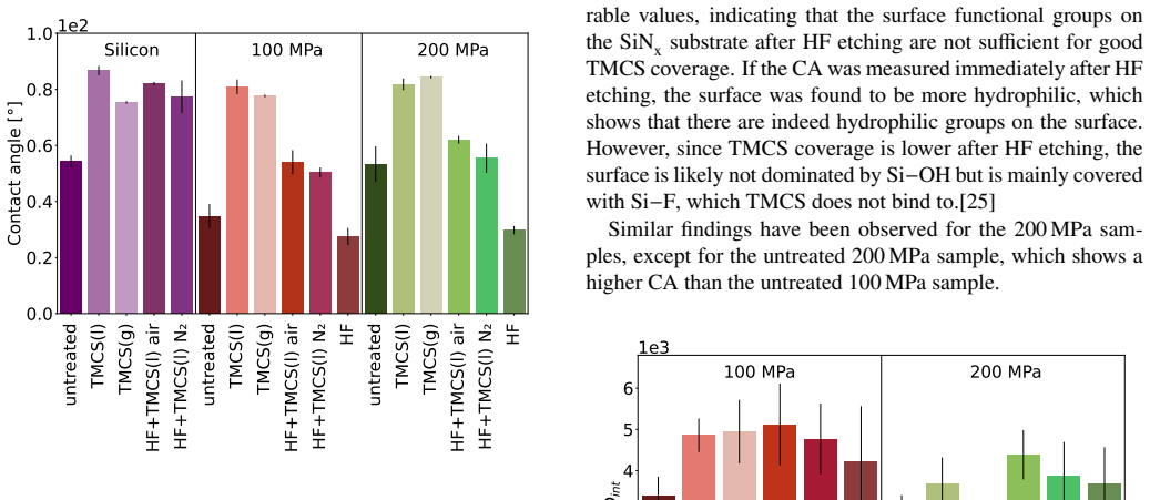
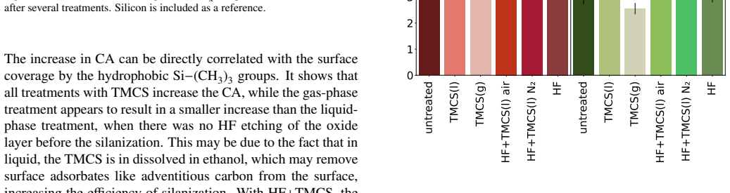
Sign in with ORCID, Apple, or X to comment. Anyone can read and Pith papers without signing in.