Tractography-Driven Synthetic Data Generation for Fiber Bundle Segmentation in Tracer Histology
Pith reviewed 2026-06-26 05:23 UTC · model grok-4.3
The pith
Synthetic patches from dMRI tractography let a U-Net match state-of-the-art fiber bundle segmentation in macaque histology using one-third the manual labels.
A machine-rendered reading of the paper's core claim, the machinery that carries it, and where it could break.
Core claim
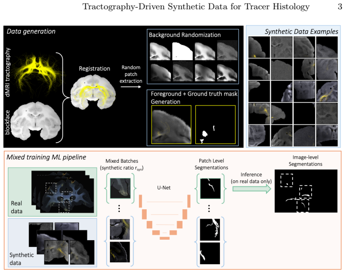
By using ex vivo dMRI tractography as a generative prior to synthesize 2D image patches for training, which provides sufficiently realistic foreground texture composed with blockface photo backgrounds and diversified via domain randomization, a 2D U-Net trained on mixed real and synthetic patches achieves performance comparable to the state-of-the-art while requiring 3x less manually annotated data, with improved generalization across brains and fiber bundle densities; training with synthetic data only leads to poor performance.
What carries the argument
ex vivo dMRI tractography as generative prior for synthesizing realistic 2D foreground patches of fiber bundles
If this is right
- Models generalize better to new brains and to bundles of varying densities.
- Comparable accuracy to current best methods is reached with only one-third the manual labels.
- Pure synthetic training is not enough and must be mixed with real examples.
- Annotation effort for tracer histology segmentation can be scaled down without losing performance.
Where Pith is reading between the lines
- The same generative-prior idea could lower labeling costs when validating tractography against histology in additional primate species.
- Domain randomization on composed patches might help close gaps between ex vivo and in vivo imaging data.
- Structural priors from one modality could be used to augment segmentation training in other medical imaging tasks where full annotation is expensive.
Load-bearing premise
That the texture produced by ex vivo dMRI tractography is close enough to real histology when placed on blockface backgrounds that the resulting synthetic patches improve a model's accuracy on actual sections.
What would settle it
A held-out brain test in which adding the synthetic patches to real training data fails to raise segmentation accuracy above the level achieved by real data alone.
Figures
read the original abstract
Diffusion MRI (dMRI) tractography enables non-invasive reconstruction of white-matter pathways, but its accuracy is fundamentally limited by indirect, low-resolution measurements of axonal organization. Tracer injection studies in non-human primates provide a gold standard for validating dMRI tractography. This, however, requires time-consuming manual annotation of fiber bundles in histology sections. We propose a synthetic-data augmented framework for automated fiber bundle segmentation in macaque tracer histology. Our approach uses ex vivo dMRI tractography as a generative prior to synthesize 2D image patches for training. This provides us with sufficiently realistic foreground texture, which we compose with backgrounds from blockface photos and diversify via domain randomization. A 2D U-Net is trained on mixed real and synthetic patches. Experiments on held-out brains demonstrate improved generalization across brains and fiber bundle densities compared to training with real data only. Training with synthetic data only leads to poor performance, underscoring the need for real supervision. Overall, our approach achieves performance comparable to the state-of-the-art while requiring 3x less manually annotated data.
Editorial analysis
A structured set of objections, weighed in public.
Referee Report
Summary. The manuscript proposes a synthetic-data-augmented framework for automated fiber bundle segmentation in macaque tracer histology. Ex vivo dMRI tractography supplies a generative prior for 2D foreground patches that are composed with blockface backgrounds and diversified by domain randomization; a 2D U-Net is trained on the resulting mixed real+synthetic patches. Experiments on held-out brains are reported to show improved generalization across brains and fiber-bundle densities relative to real-data-only training, with overall performance comparable to the state of the art while using 3× less manual annotation; synthetic-only training is shown to fail.
Significance. If the quantitative claims hold, the work would demonstrate a practical route to reducing the annotation burden that currently limits histology-based validation of dMRI tractography. The empirical finding that mixed training outperforms both real-only and synthetic-only regimes is potentially useful for other histology segmentation tasks that suffer from limited labeled data.
major comments (3)
- [Abstract] Abstract: the central claim that the method “achieves performance comparable to the state-of-the-art while requiring 3× less manually annotated data” is stated without any supporting numbers, tables, or error bars that quantify annotation volume versus Dice/IoU on the held-out brains.
- [Abstract] Abstract / Methods (generative prior): no quantitative fidelity metric (intensity histograms, Haralick features, embedding distances, etc.) is reported between tractography-derived foreground texture and real histology fiber appearance, yet this realism assumption is load-bearing for the claim that mixed training generalizes better than real data alone.
- [Abstract] Abstract: the statement that synthetic-only training “leads to poor performance” is given without the exact metrics, data-exclusion criteria, or cross-brain statistics that would allow the reader to assess how much the mixed-training gain depends on the real supervision component.
minor comments (1)
- [Abstract] The abstract would benefit from a brief parenthetical listing of the primary quantitative metrics (e.g., mean Dice on held-out brains) so that the “3× less annotation” claim can be evaluated at a glance.
Simulated Author's Rebuttal
We thank the referee for the constructive comments on our manuscript. We address each major point below and have revised the abstract (and, where needed, the methods) to supply the requested quantitative details from our existing experiments.
read point-by-point responses
-
Referee: [Abstract] Abstract: the central claim that the method “achieves performance comparable to the state-of-the-art while requiring 3× less manually annotated data” is stated without any supporting numbers, tables, or error bars that quantify annotation volume versus Dice/IoU on the held-out brains.
Authors: We agree that the abstract should contain explicit quantitative support. The experiments section already reports Dice/IoU scores, annotation volumes, and error bars for held-out brains under real-only, synthetic-only, and mixed regimes. We have revised the abstract to include the key numbers (e.g., performance at one-third annotation effort versus baselines) and cross-references to the relevant tables and figures. revision: yes
-
Referee: [Abstract] Abstract / Methods (generative prior): no quantitative fidelity metric (intensity histograms, Haralick features, embedding distances, etc.) is reported between tractography-derived foreground texture and real histology fiber appearance, yet this realism assumption is load-bearing for the claim that mixed training generalizes better than real data alone.
Authors: We acknowledge that direct fidelity metrics were not provided. The performance advantage of mixed over real-only training supplies indirect evidence that the tractography-derived patches are sufficiently realistic. To address the concern explicitly, we will add intensity-histogram and Haralick-feature comparisons between synthetic and real foreground patches to the revised Methods section. revision: yes
-
Referee: [Abstract] Abstract: the statement that synthetic-only training “leads to poor performance” is given without the exact metrics, data-exclusion criteria, or cross-brain statistics that would allow the reader to assess how much the mixed-training gain depends on the real supervision component.
Authors: We agree the abstract should supply these details. The results already contain the exact Dice/IoU values, held-out brain IDs, and cross-brain statistics for the synthetic-only condition. We have updated the abstract to include these numbers and criteria so readers can evaluate the contribution of the real-supervision component. revision: yes
Circularity Check
Empirical ML framework with external generative prior exhibits no circularity
full rationale
The paper describes a data-augmentation pipeline that takes ex vivo dMRI tractography (an independent imaging modality) as the source of foreground texture, composites it with blockface backgrounds, applies domain randomization, and trains a U-Net on the resulting mixed real+synthetic patches. Performance is measured by direct comparison against held-out real histology annotations. No equations, fitted parameters, or self-citations are presented that would make any reported metric equivalent to the input labels or to a prior result by the same authors. The realism assumption is an empirical premise, not a self-definitional or fitted-input reduction.
Axiom & Free-Parameter Ledger
axioms (1)
- domain assumption Ex vivo dMRI tractography can serve as a generative prior that produces sufficiently realistic foreground fiber texture for histology patch synthesis
Reference graph
Works this paper leans on
-
[1]
Insight j2(365), 1–35 (2009)
Avants, B.B., Tustison, N., Song, G., et al.: Advanced normalization tools (ants). Insight j2(365), 1–35 (2009)
2009
-
[2]
In: Interna- tional Workshop on Computational Diffusion MRI
Bintsi, K.M., Balbastre, Y., Wu, J., Lehman, J.F., Haber, S.N., Yendiki, A.: Fully automated segmentation of fiber bundles in anatomic tracing data. In: Interna- tional Workshop on Computational Diffusion MRI. pp. 81–92. Springer (2025)
2025
-
[3]
Current opinion in neurology19(6), 599–606 (2006)
Catani, M.: Diffusion tensor magnetic resonance imaging tractography in cognitive disorders. Current opinion in neurology19(6), 599–606 (2006)
2006
-
[4]
BMC evolutionary biology9, 1–19 (2009)
Chatterjee, H.J., Ho, S.Y., Barnes, I., Groves, C.: Estimating the phylogeny and divergence times of primates using a supermatrix approach. BMC evolutionary biology9, 1–19 (2009)
2009
-
[5]
arXiv preprint arXiv:2407.01419 (2024) 10 K
Chollet, E., Balbastre, Y., Mauri, C., Magnain, C., Fischl, B., Wang, H.: Neurovas- cular segmentation in soct with deep learning and synthetic training data. arXiv preprint arXiv:2407.01419 (2024) 10 K. M. Bintsi et al
-
[6]
In: Proceedings of the 26th annual meeting of the International Society of Magnetic Resonance in Medicine
Dhollander, T., Zanin, J., Nayagam, B.A., Rance, G., Connelly, A.: Feasibility and benefits of 3-tissue constrained spherical deconvolution for studying the brains of babies. In: Proceedings of the 26th annual meeting of the International Society of Magnetic Resonance in Medicine. vol. 3077 (2018)
2018
-
[7]
Proceedings of the National Academy of Sciences117(20), 11068–11075 (2020)
Friedmann, D., Pun, A., Adams, E.L., Lui, J.H., Kebschull, J.M., Grutzner, S.M., Castagnola, C., Tessier-Lavigne, M., Luo, L.: Mapping mesoscale axonal projec- tions in the mouse brain using a 3d convolutional network. Proceedings of the National Academy of Sciences117(20), 11068–11075 (2020)
2020
-
[8]
In: Generative Machine Learning Models in Medical Image Computing, pp
Friedrich, P., Frisch, Y., Cattin, P.C.: Deep generative models for 3d medical image synthesis. In: Generative Machine Learning Models in Medical Image Computing, pp. 255–278. Springer (2024)
2024
-
[9]
Imaging Neuroscience2, imag–2 (2024)
Gopinath, K., Hoopes, A., Alexander, D.C., Arnold, S.E., Balbastre, Y., Billot, B., Casamitjana, A., Cheng, Y., Chua, R.Y.Z., Edlow, B.L., et al.: Synthetic data in generalizable, learning-based neuroimaging. Imaging Neuroscience2, imag–2 (2024)
2024
-
[10]
Journal of Neuroscience33(11), 4804–4814 (2013)
Haynes, W.I., Haber, S.N.: The organization of prefrontal-subthalamic inputs in primates provides an anatomical substrate for both functional specificity and inte- gration: implications for basal ganglia models and deep brain stimulation. Journal of Neuroscience33(11), 4804–4814 (2013)
2013
-
[11]
Journal of Neuroscience33(7), 3190–3201 (2013)
Jbabdi, S., Lehman, J.F., Haber, S.N., Behrens, T.E.: Human and monkey ven- tral prefrontal fibers use the same organizational principles to reach their targets: tracing versus tractography. Journal of Neuroscience33(7), 3190–3201 (2013)
2013
-
[12]
Neu- roimage214, 116704 (2020)
Jones, R., Grisot, G., Augustinack, J., Magnain, C., Boas, D.A., Fischl, B., Wang, H., Yendiki, A.: Insight into the fundamental trade-offs of diffusion mri from polarization-sensitive optical coherence tomography in ex vivo human brain. Neu- roimage214, 116704 (2020)
2020
-
[13]
Journal of Neuroscience31(28), 10392–10402 (2011)
Lehman, J.F., Greenberg, B.D., McIntyre, C.C., Rasmussen, S.A., Haber, S.N.: Rules ventral prefrontal cortical axons use to reach their targets: implications for diffusion tensor imaging tractography and deep brain stimulation for psychiatric illness. Journal of Neuroscience31(28), 10392–10402 (2011)
2011
-
[14]
Decoupled Weight Decay Regularization
Loshchilov, I., Hutter, F., et al.: Fixing weight decay regularization in adam. arXiv preprint arXiv:1711.051015(5), 5 (2017)
work page internal anchor Pith review Pith/arXiv arXiv 2017
-
[15]
Journal of neuroscience methods291, 141–149 (2017)
Naito, T., Nagashima, Y., Taira, K., Uchio, N., Tsuji, S., Shimizu, J.: Identification and segmentation of myelinated nerve fibers in a cross-sectional optical microscopic image using a deep learning model. Journal of neuroscience methods291, 141–149 (2017)
2017
-
[16]
Computational and structural biotechnology journal23, 2892–2910 (2024)
Pezoulas, V.C., Zaridis, D.I., Mylona, E., Androutsos, C., Apostolidis, K., Tachos, N.S., Fotiadis, D.I.: Synthetic data generation methods in healthcare: A review on open-source tools and methods. Computational and structural biotechnology journal23, 2892–2910 (2024)
2024
-
[17]
SAM 2: Segment Anything in Images and Videos
Ravi, N., Gabeur, V., Hu, Y.T., Hu, R., Ryali, C., Ma, T., Khedr, H., Rädle, R., Rolland, C., Gustafson, L., et al.: Sam 2: Segment anything in images and videos. arXiv preprint arXiv:2408.00714 (2024)
work page internal anchor Pith review Pith/arXiv arXiv 2024
-
[18]
Reza, A.M.: Realization of the contrast limited adaptive histogram equalization (clahe)forreal-timeimageenhancement.JournalofVLSIsignalprocessingsystems for signal, image and video technology38(1), 35–44 (2004)
2004
-
[19]
In: Medical image computing and computer-assisted intervention–MICCAI 2015: 18th international conference, Munich, Germany, Oc- tober 5-9, 2015, proceedings, part III 18
Ronneberger, O., Fischer, P., Brox, T.: U-net: Convolutional networks for biomed- ical image segmentation. In: Medical image computing and computer-assisted intervention–MICCAI 2015: 18th international conference, Munich, Germany, Oc- tober 5-9, 2015, proceedings, part III 18. pp. 234–241. Springer (2015) Tractography-Driven Synthetic Data for Tracer Histology 11
2015
-
[20]
Journal of Neuroscience38(8), 2106–2117 (2018)
Safadi, Z., Grisot, G., Jbabdi, S., Behrens, T.E., Heilbronner, S.R., McLaughlin, N.C., Mandeville, J., Versace, A., Phillips, M.L., Lehman, J.F., et al.: Functional segmentation of the anterior limb of the internal capsule: linking white matter abnormalities to specific connections. Journal of Neuroscience38(8), 2106–2117 (2018)
2018
-
[21]
Neuroimage185, 1–11 (2019)
Schilling, K.G., Nath, V., Hansen, C., Parvathaneni, P., Blaber, J., Gao, Y., Neher, P., Aydogan, D.B., Shi, Y., Ocampo-Pineda, M., et al.: Limits to anatomical ac- curacy of diffusion tractography using modern approaches. Neuroimage185, 1–11 (2019)
2019
-
[22]
Imag- ing Neuroscience3, imag_a_00514 (2025)
Sundaresan, V., Lehman, J.F., Maffei, C., Haber, S.N., Yendiki, A.: Self-supervised segmentation and characterization of fiber bundles in anatomic tracing data. Imag- ing Neuroscience3, imag_a_00514 (2025)
2025
-
[23]
Wei, D., Lee, K., Li, H., Lu, R., Bae, J.A., Liu, Z., Zhang, L., dos Santos, M., Lin, Z., Uram, T., et al.: Axonem dataset: 3d axon instance segmentation of brain cor- tical regions. In: Medical Image Computing and Computer Assisted Intervention– MICCAI 2021: 24th International Conference, Strasbourg, France, September 27– October 1, 2021, Proceedings, Pa...
2021
-
[24]
Cell179(1), 268–281 (2019)
Winnubst, J., Bas, E., Ferreira, T.A., Wu, Z., Economo, M.N., Edson, P., Arthur, B.J.,Bruns,C.,Rokicki,K.,Schauder,D.,etal.:Reconstructionof1,000projection neurons reveals new cell types and organization of long-range connectivity in the mouse brain. Cell179(1), 268–281 (2019)
2019
-
[25]
Elife 11, e72534 (2022)
Yan, M., Yu, W., Lv, Q., Lv, Q., Bo, T., Chen, X., Liu, Y., Zhan, Y., Yan, S., Shen, X., et al.: Mapping brain-wide excitatory projectome of primate prefrontal cortex at submicron resolution and comparison with diffusion tractography. Elife 11, e72534 (2022)
2022
-
[26]
Physics in Medicine & Biology66(15), 15TR01 (2021)
Yang, J.Y.M., Yeh, C.H., Poupon, C., Calamante, F.: Diffusion mri tractogra- phy for neurosurgery: the basics, current state, technical reliability and challenges. Physics in Medicine & Biology66(15), 15TR01 (2021)
2021
-
[27]
Neuroimage256, 119146 (2022)
Yendiki, A., Aggarwal, M., Axer, M., Howard, A.F., van Walsum, A.M.v.C., Haber, S.N.: Post mortem mapping of connectional anatomy for the validation of diffusion mri. Neuroimage256, 119146 (2022)
2022
-
[28]
arXiv preprint arXiv:2408.00874 (2024)
Zhu, J., Hamdi, A., Qi, Y., Jin, Y., Wu, J.: Medical sam 2: Segment medical images as video via segment anything model 2. arXiv preprint arXiv:2408.00874 (2024)
discussion (0)
Sign in with ORCID, Apple, or X to comment. Anyone can read and Pith papers without signing in.