REVIEW 3 major objections 4 minor 29 references
A webcam-only system detects EEG cap electrodes in real time and checks they sit at the correct 10–20 head positions, reaching 0.94 mAP across held-out subjects.
Reviewed by Pith at T0; open to challenge. T0 means a machine referee read the full paper against a public rubric. the ladder, T0–T4 →
2026-08-01 10:37 UTC pith:KEC6KZ7Q
load-bearing objection The direct electrode-detection idea is real and the eval is mostly honest, but the headline mAP comes from the no-crop baseline while the deployed system always crops, so the system-level accuracy is never actually reported. the 3 major comments →
Real-Time EEG Cap Electrode Detection for Guided Point-of-Care Placement
The pith
A machine-rendered reading of the paper's core claim, the machinery that carries it, and where it could break.
Core claim
The paper's central claim is that electrode placement can be verified continuously during cap donning using only a single RGB camera, without depth sensors, markers, or post-hoc scanning. The key move is to separate detection from naming: a class-agnostic YOLO detector finds electrode-like blobs, and a deterministic geometric rule assigns each blob to a specific 10–20 role (FP1, FP2, Fz, F9, F10) based on facial landmarks. This design avoids needing per-role training data and lets the system check the electrodes' actual positions rather than assuming they match an atlas. The authors also show that a landmark-driven head crop is the decisive factor for maintaining accuracy at distance, liftin
What carries the argument
The load-bearing object is the geometric role-assignment stage (Stage 2), which converts anonymous detections into named anatomical roles. It uses the inter-eye distance from MediaPipe FaceLandmarker as a subject-specific reference scale (d_ref), then classifies each detection as a temporal electrode (F9/F10) if it lies beyond a horizontal margin past the outer eye corner and above a crown threshold, and assigns inner electrodes by greedy nearest-neighbour proximity to eyebrow landmarks. This deterministic rule is what turns a generic electrode detector into a placement validator, and it is evaluated directly against the detector's output.
Load-bearing premise
The whole pipeline assumes MediaPipe can locate the subject's face in the webcam frame; if face detection fails (poor lighting, extreme pose, occlusion, or distance beyond 0.3× scale), the adaptive crop is abandoned, the anatomical reference frame is lost, and the system cannot assign electrode roles.
What would settle it
Run the pipeline on a held-out subject wearing the Small cap under realistic dim lighting, where MediaPipe locates the face but the detector must rely on appearance alone; the paper predicts a sharp drop in mAP for the Small cap when subject familiarity is also removed, but if the drop vanishes with more subjects or if face detection succeeds and role assignment still fails, the geometric-stage assumption would be contradicted.
If this is right
- Non-specialist operators could receive real-time feedback on whether each electrode is in its correct 10–20 position during donning, potentially reducing the error rate that currently corrupts point-of-care EEG.
- The two-stage design (detection + geometric naming) can be ported to other electrode caps or extended to additional 10–20 sites without retraining the detector, since naming is rule-based.
- The landmark-driven head crop establishes a general principle: for small-object detection on the head, using facial landmarks to upscale the region of interest is more effective than simply increasing input resolution, and it works at no inference cost.
- A compact mobile backbone (YOLOv10n) reaches the real-time band on a CPU at 640 px with near-equal accuracy, suggesting the system can run on a phone or low-cost device in low-connectivity settings.
Where Pith is reading between the lines
- The 10–20 naming rule depends on a roughly frontal head pose and on the inter-eye distance being a reliable scale anchor; a natural extension would be to test pose robustness beyond the synthetic roll sweep, e.g., with real side or tilted head captures.
- The paper's evaluation focuses on five fixed electrode roles; extending to the full 10–20 system (e.g., C3/C4, O1/O2) would require either more landmark anchors or a richer geometric model, but the same architecture should transfer.
- If the system were deployed, a prospective clinical study comparing placement correctness (measured by a gold standard like optical scanning) with and without real-time guidance would be the decisive test of whether the vision loop actually improves EEG signal quality.
Editorial analysis
A structured set of objections, weighed in public.
Referee Report
Summary. The paper presents a real-time, webcam-only system that detects EEG cap electrodes and assigns them to named 10–20 roles. Stage 1 is a YOLO-based single-class electrode detector; Stage 2 is a deterministic geometric role-assignment stage using MediaPipe facial landmarks and an adaptive head crop. The evaluation uses subject-disjoint leave-one-subject-out (LOSO) cross-validation on 1,309 frames from 5 subjects wearing three cap sizes, reporting detector mAP@.5 = 0.94 ± 0.07, per-electrode miss rates, a leave-one-cap-out axis, synthetic robustness ablations (lighting, distance, roll), role-assignment accuracy (90.7% of matched detections), and throughput figures. The authors are transparent about confounds (Small-cap leave-cap-out is subject-confounded), the single-rater and fixed-box annotation convention, the synthetic nature of robustness tests, and the persistent Fz detection difficulty.
Significance. If the reported system-level performance matched the headline detector numbers, this would be a practically useful step toward automated EEG cap placement quality assurance in point-of-care settings. Strengths of the paper are the leakage-free LOSO protocol, the explicit leave-one-cap-out axis, the honest treatment of subject/cap confounding, the reproducible synthetic perturbation methodology, the deterministic and clearly specified geometric role-assignment rule, and the real-time throughput characterization. The main gap is that the reported headline metric appears to be detector-only, while the deployed system includes an adaptive crop whose effect at native scale is non-negligible; the end-to-end placement-validation accuracy, which is the clinically relevant quantity, is not reported.
major comments (3)
- [§5.2 / Figure 4 / Table 2] The headline detection metric is not the deployed system's metric. Figure 4 (left) reports that at 1.0× scale, with the adaptive head crop the mAP@.5 is 0.80, while the no-crop full-frame baseline is 0.94. Table 2's LOSO mAP@.5 = 0.94 ± 0.07 matches the no-crop baseline, and §5.2 states 'Without the crop, mAP@.5 falls from 0.94 ± 0.07 ...' — so Table 2 is evidently detector-only on full frames. However §4.3 states the deployed pipeline always applies the crop when a face is found. Thus the system as actually deployed has not been evaluated on the held-out LOSO folds. Please report system-level mAP@.5 and all-electrodes-found rate with the crop applied exactly as in §4.3, and state unambiguously which configuration produced Table 2.
- [§5.4 / Table 2] End-to-end placement-validation accuracy is missing. The paper's central claim is that the system 'validates their anatomical placement in real time,' but detection and role assignment are reported separately. Table 2 gives the all-electrodes-found detection rate (0.70 pooled; 0.07 on fold s4), and §5.4 gives role-assignment accuracy (90.7% of matched detections). No metric reports the fraction of frames in which the system finds all five electrodes and assigns all five roles correctly, under the deployed crop. This joint per-frame metric is the quantity a clinical user would care about; please add it.
- [§4.1 / §4.3] The training/evaluation protocol regarding the crop is unspecified. §4.1 says 'All results in this paper use this configuration,' but it is not clear whether training images were full-frame or cropped. If the detector was trained on full frames only, the inference-time crop is a train/test domain shift that may explain the 1.0× degradation in Figure 4. Please describe the training data transform precisely and, if the crop is used at inference, either include it in training or justify its absence.
minor comments (4)
- [§3 / Dataset] The dataset is small (5 subjects, 1,309 frames), annotations come from a single rater, and boxes are fixed-size squares. These limitations are acknowledged, but adding a brief statement on the absence of inter-rater reliability and the potential bias from a single rater would strengthen the reproducibility discussion.
- [§4.3 / §5.2] The fallback to the full frame when no face is found is stated, and the distance experiment shows that performance collapses at 0.3× when MediaPipe fails. It would be helpful to state explicitly in the limitations that the system cannot perform role assignment at all in such cases, and that the operational envelope therefore depends on reliable face detection.
- [Section 5.6 / Table 4] Table 4 reports YOLOv8s at 640 px with 0.86 ± 0.19 mAP@.5, while Table 2 reports YOLOv8s at 960 px with 0.943 ± 0.074. The resolution difference is clear from the text, but a sentence in the Table 4 caption restating that these are 640-px operating points would avoid confusion.
- [Data and code availability] The manuscript states that code and dataset are not publicly released. For a computer-vision methods paper, releasing at least the evaluation code and a small sample of anonymized data would substantially aid reproducibility; the current statement is a limitation for verification.
Circularity Check
No circularity: held-out LOSO evaluation and deterministic geometric stage; no prediction reduces to a fitted input or self-citation.
full rationale
Walking the claimed derivation chain, I find no step in which a reported result reduces by construction to its own inputs. The headline detection number (mAP@.5=0.94, Table 2) is a measured LOSO evaluation on held-out subjects; no test-set parameter fitting is described, and the paper explicitly reports per-fold results, a pooled value, and a subject-4 outlier. The geometric role-assignment stage (Section 4.2) is deterministic and is evaluated in Section 5.4 against manually annotated ground-truth labels ('reading each prediction’s true label off its IoU-matched ground-truth box'), so the role-accuracy figure is not defined in terms of the assignment rule being tested. The adaptive-crop/distance results in Section 5.2 are controlled ablations (with vs. without crop; with vs. without augmentation), not predictions derived from the fitted model. The one self-citation ([14], the BC-1 cap clinical validation) is background context and not load-bearing for the detection or role-assignment claims. The discrepancy between Table 2 (0.94) and Figure 4's 1.0× with-crop value (0.80) is an evaluation-protocol ambiguity about which configuration produced Table 2; it is a correctness/reporting concern, not a circularity, because neither number is obtained by fitting to the test set. The paper's own limitations (Sections 6(i)-(v): small subject count, synthetic perturbations, Fz miss rate, cap/subject confounding, role accuracy) are acknowledged weaknesses, not circular reductions. No self-citation chain, uniqueness import, or ansatz-by-citation is present. Score 0.
Axiom & Free-Parameter Ledger
free parameters (3)
- Temporal margin τ =
0.15·d_ref
- Head crop expansion factors =
1.8× horizontal, 2.5× vertical
- Geometric augmentation strengths =
rotation 10°, perspective 0.0005, mixup 0.1
axioms (4)
- domain assumption MediaPipe FaceLandmarker returns accurate facial landmarks in roughly frontal webcam views.
- domain assumption COCO-pretrained YOLO features transfer to EEG electrode detection.
- domain assumption The BC-1 cap's 5 electrodes follow a fixed 10-10/10-20 layout and appear as distinct visual objects.
- domain assumption Single-rater point-click annotation with fixed-size boxes is an adequate ground truth.
Cite this review
Pith. "Pith review of Real-Time EEG Cap Electrode Detection for Guided Point-of-Care Placement." pith.science (2026). https://pith.science/paper/KEC6KZ7Q
@misc{pith2026260720142,
  author       = {Pith},
  title        = {Pith review of: Real-Time EEG Cap Electrode Detection for Guided Point-of-Care Placement},
  year         = {2026},
  howpublished = {\url{https://pith.science/paper/KEC6KZ7Q}},
  note         = {Machine review of arXiv:2607.20142}
}
read the original abstract
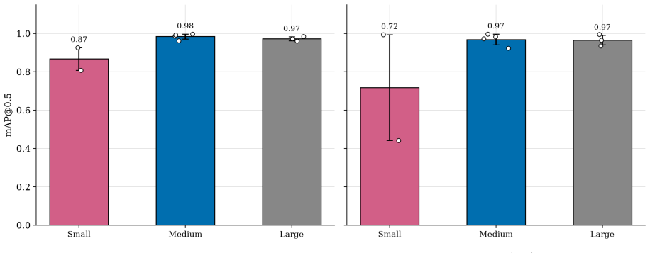
We present a two-stage vision system that detects EEG cap electrodes in a live webcam stream and validates their anatomical placement in real time. A single-class YOLO detector localises electrodes; a geometric stage assigns each detection to a named 10-20 role from facial landmarks. Evaluating under subject-disjoint leave-one-subject-out (LOSO) cross-validation across five subjects wearing the clinically-validated Small/Medium/Large caps, the detector attains mAP@.5 = 0.94 +/- 0.07 across five held-out folds (0.96 pooled). A dedicated leave-one-cap-out axis, holding out every frame of a cap regardless of subject, leaves Medium and Large mAP@.5 within 0.01 of LOSO (0.97, 0.97) while Small drops to 0.72 +/- 0.28, a gap confounded with subject familiarity rather than cap style. Geometric augmentation (rotation, perspective, mixup) improves in-plane-roll robustness and temporal-electrode recall at no inference cost, and a landmark-driven head crop extends the usable distance range, lifting mAP@.5 from 0.23 to 0.45 at 0.6 x apparent scale. A compact mobile-candidate backbone (YOLOv10n) keeps the detector at real-time throughput (19 FPS) on a commodity CPU at 640 px.
Figures




Reference graph
Works this paper leans on
-
[1]
Intersectoral global action plan on epilepsy and other neurological disorders 2022–2031
World Health Organization, “Intersectoral global action plan on epilepsy and other neurological disorders 2022–2031.” World Health Organization, Geneva, 2023. [Online]. Available: https:// www.who.int/publications/b/74388
2022
-
[2]
Epilepsy
World Health Organization, “Epilepsy.” [Online]. Available: https://www.who.int/news-room/ fact-sheets/detail/epilepsy
-
[3]
Epilepsy in low- to middle-income countries,
A. Sen, C. R. Newton, and G. Ngwende, “Epilepsy in low- to middle-income countries,” Current Opinion in Neurology, vol. 38, no. 2, pp. 121–127, 2025, doi: 10.1097/WCO.0000000000001350
-
[4]
Epilepsy care and outcome in low- and middle-income countries: A scoping review,
N. D. A. Bankole et al., “Epilepsy care and outcome in low- and middle-income countries: A scoping review,” Journal of Neurosciences in Rural Practice, vol. 15, no. 1, pp. 8–15, 2024, doi: 10.25259/JNRP_502_2023
-
[5]
Rapid response EEG: current state and future directions,
Z. Davey, P. B. Gupta, D. R. Li, R. U. Nayak, and P. Govindarajan, “Rapid response EEG: current state and future directions,” Current Neurology and Neuroscience Reports, vol. 22, no. 12, pp. 839–846, 2022, doi: 10.1007/s11910-022-01243-1
-
[6]
How much time is enough? Establishing an optimal duration of recording for ambulatory video EEG,
H. Klein, T. Pang, J. Slater, and R. E. Ramsay, “How much time is enough? Establishing an optimal duration of recording for ambulatory video EEG,” Epilepsia Open, vol. 6, no. 3, pp. 569–578, 2021, doi: 10.1002/epi4.12517
-
[7]
Automated interpretation of clinical electroencephalograms using artificial intel- ligence,
J. Tveit et al., “Automated interpretation of clinical electroencephalograms using artificial intel- ligence,” JAMA Neurology, vol. 80, no. 8, pp. 805–812, 2023, doi: 10.1001/jamaneurol.2023.1645
arXiv 2023
-
[8]
Large brain model for learning generic representations with tremendous EEG data in BCI,
W.-B. Jiang, L.-M. Zhao, and B.-L. Lu, “Large brain model for learning generic representations with tremendous EEG data in BCI,” in The Twelfth International Conference on Learning Representations, 2024. [Online]. Available: https://openreview.net/forum?id=QzTpTRVtrP
2024
-
[9]
REVE: a foundation model for EEG — adapting to any setup with large- scale pretraining on 25,000 subjects,
Y. El Ouahidi et al., “REVE: a foundation model for EEG — adapting to any setup with large- scale pretraining on 25,000 subjects,” in Advances in Neural Information Processing Systems, 2025
2025
-
[10]
Assistive artificial intelligence in epilepsy and its impact on epilepsy care in low- and middle-income countries,
N. Koirala et al., “Assistive artificial intelligence in epilepsy and its impact on epilepsy care in low- and middle-income countries,” Brain Sciences, vol. 15, no. 5, p. 481, 2025, doi: 10.3390/ brainsci15050481
2025
-
[11]
The ten-twenty electrode system of the International Federation,
H. H. Jasper, “The ten-twenty electrode system of the International Federation,” Electroen cephalography and Clinical Neurophysiology, vol. 10, pp. 371–375, 1958
1958
-
[12]
Y. Wang and J. Gotman, “The influence of electrode location errors on EEG dipole source localization with a realistic head model,” Clinical Neurophysiology, vol. 112, no. 9, pp. 1777– 1780, 2001, doi: 10.1016/S1388-2457(01)00594-6
-
[13]
S. S. Dalal, S. Rampp, F. Willomitzer, and S. Ettl, “Consequences of EEG electrode position error on ultimate beamformer source reconstruction performance,” Frontiers in Neuroscience, vol. 8, p. 42, 2014, doi: 10.3389/fnins.2014.00042
arXiv 2014
-
[14]
Low-cost portable EEG device for bridging the diagnostic gap in resource-limited areas,
S. Armand Larsen, L. Klok, W. Lehn-Schiøler, R. Gatej, and S. Beniczky, “Low-cost portable EEG device for bridging the diagnostic gap in resource-limited areas,” Epileptic Disorders, vol. 26, no. 5, pp. 694–700, 2024, doi: 10.1002/epd2.20266
-
[15]
T. L. Rich, B. T. Gillick, M. Chen, E. Bhatt, and J. R. Carey, “Electrode placement in transcranial direct current stimulation — How reliable is the determination of C3/C4?,” Brain Sciences, vol. 9, no. 3, p. 69, 2019, doi: 10.3390/brainsci9030069. page 12
-
[16]
O. Györfi et al., “Accuracy of high-density EEG electrode position measurement using an optical scanner compared with the photogrammetry method,” Clinical Neurophysiology Practice, vol. 7, pp. 135–138, 2022, doi: 10.1016/j.cnp.2022.04.002
-
[17]
Augmented reality-based electrode guidance system for reliable electroencephalography,
C. Song, S. Jeon, S. Lee, H.-G. Ha, J. Kim, and J. Hong, “Augmented reality-based electrode guidance system for reliable electroencephalography,” BioMedical Engineering OnLine, vol. 17, p. 64, 2018, doi: 10.1186/s12938-018-0500-x
-
[18]
C. Kaewrat and Y. Punsawad, “Utilizing augmented reality technology for teaching fundamen- tals of the human brain and EEG electrode placement,” International Journal on Smart Sensing and Intelligent Systems, vol. 15, no. 1, p. 20220005, 2022, doi: 10.2478/ijssis-2022-0005
-
[19]
Improving neuroimaging headgear placement robustness using facial-landmark-guided augmented reality,
F.-Y. Yen, Y.-A. Lin, and Q. Fang, “Improving neuroimaging headgear placement robustness using facial-landmark-guided augmented reality,” Neurophotonics, vol. 12, no. 4, p. 45005, 2025, doi: 10.1117/1.NPh.12.4.045005
-
[20]
You Only Look Once: Unified, Real-Time Object Detection,
J. Redmon, S. Divvala, R. Girshick, and A. Farhadi, “You Only Look Once: Unified, Real-Time Object Detection,” in IEEE Conference on Computer Vision and Pattern Recognition (CVPR), 2016, pp. 779–788
2016
-
[21]
Microsoft COCO: Common Objects in Context,
T.-Y. Lin, M. Maire, S. Belongie, and others, “Microsoft COCO: Common Objects in Context,” in European Conference on Computer Vision (ECCV), 2014, pp. 740–755
2014
-
[22]
Ultralytics YOLO
G. Jocher, A. Chaurasia, and J. Qiu, “Ultralytics YOLO.” [Online]. Available: https://github. com/ultralytics/ultralytics
-
[23]
YOLOv10: Real-Time End-to-End Object Detection,
A. Wang et al., “YOLOv10: Real-Time End-to-End Object Detection,” in Advances in Neural Information Processing Systems, 2024
2024
-
[24]
Ultralytics YOLO11
G. Jocher and J. Qiu, “Ultralytics YOLO11.” [Online]. Available: https://github.com/ ultralytics/ultralytics
-
[25]
MediaPipe: A Framework for Building Perception Pipelines,
C. Lugaresi, J. Tang, H. Nash, and others, “MediaPipe: A Framework for Building Perception Pipelines,” arXiv preprint arXiv:1906.08172, 2019
Pith/arXiv arXiv 1906
-
[26]
mixup: Beyond Empirical Risk Minimization,
H. Zhang, M. Cissé, Y. N. Dauphin, and D. Lopez-Paz, “mixup: Beyond Empirical Risk Minimization,” in International Conference on Learning Representations (ICLR), 2018
2018
-
[27]
American Clinical Neurophysiology Society Guideline 2: Guidelines for Standard Electrode Position Nomencla - ture,
J. N. Acharya, A. J. Hani, J. Cheek, P. Thirumala, and T. N. Tsuchida, “American Clinical Neurophysiology Society Guideline 2: Guidelines for Standard Electrode Position Nomencla - ture,” Journal of Clinical Neurophysiology , vol. 33, no. 4, pp. 308–311, 2016, doi: 10.1097/ WNP.0000000000000316
2016
-
[28]
NanoDet-Plus: Super Fast and High Accuracy Lightweight Anchor-Free Object Detection Model
RangiLyu, “NanoDet-Plus: Super Fast and High Accuracy Lightweight Anchor-Free Object Detection Model.” 2021
2021
-
[29]
PP-PicoDet: A Better Real-Time Object Detector on Mobile Devices,
G. Yu et al., “PP-PicoDet: A Better Real-Time Object Detector on Mobile Devices,” in arXiv preprint, 2021. doi: 10.48550/arXiv.2111.00902. page 13
This paper was first reviewed by deepseek-v4-flash on August 1, 2026.
discussion (0)
Sign in with ORCID, Apple, or X to comment. Anyone can read and Pith papers without signing in.